

The DOI matches its Crossref record; 2 of the 2 references it cites were found in a registry or an index.
Inhibition of TLR4 Induces M2 Microglial Polarization and Provides Neuroprotection via the NLRP3 Inflammasome in Alzheimer’s Disease
Overview
The claims and statistics of this paper haven’t been checked yet.
Run the check — free, no account needed. It reads the open-access text and checks the sources it cites.
Integrity signals
Registry
Cites
Statistics
Figures
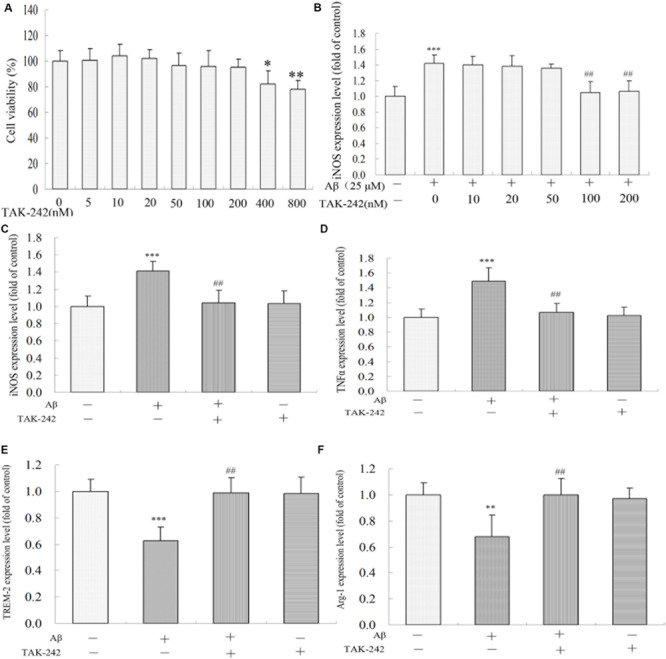
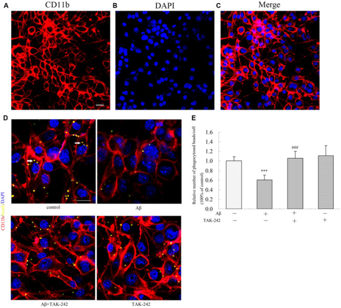
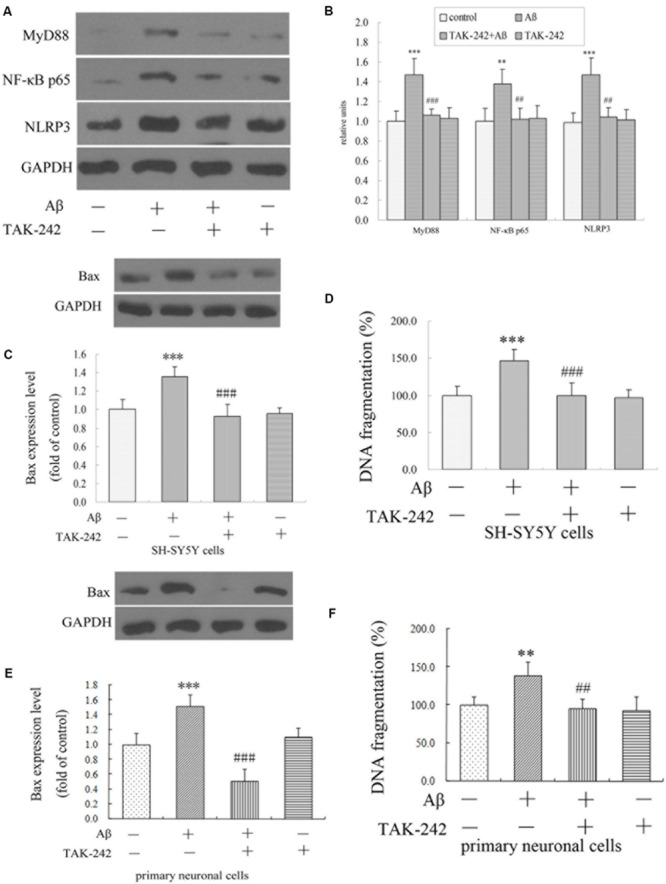
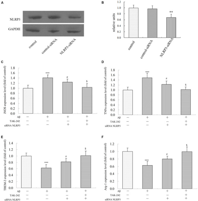
Verification
Claims the paper makes
The claims and statistics of this paper haven’t been checked yet.
Run the check — free, no account needed. It reads the open-access text and checks the sources it cites.
Claims other papers make about this one
Each is checked against this paper’s own text: does it say what the citing paper says it does?
The cited source says what the claim attributes to it.
In the citing paper
“In addition, TLR4 signaling has been shown to shift microglia toward an inflammatory state that is more disease-associated, ultimately compromising neuroprotection and cognitive function [139–143].”
From microRNA-132 attenuates inflammation in induced pluripotent stem cell-derived microglia from Alzheimer’s disease patients (2026) · cites this paper as [139]
Self-check
Percentages, sums and reported differences re-computed from the paper’s own numbers.
The self-check needs the paper’s full text.
Citations
Cites
Arcuri, C., Mecca, C., Bianchi, R., Giambanco, I., & Donato, R. (2017) · Frontiers in Molecular Neuroscience · doi:10.3389/fnmol.2017.00191 · Cited by 279
Zhou, J., Yu, W., Zhang, M., Tian, X., Li, Y., & Lü, Y. (2019) · Neurochemical Research · doi:10.1007/s11064-019-02748-x · Cited by 139
Research works printed but not linked: 45
- Al, 2018, Interferon regulatory factor 4/5 signaling impacts on microglial activation after ischemic stroke in mice., Eur. J. Neurosci., 47, 140, 10.1111/ejn.13778 · DOI
- Aryanpour, 2017, Progesterone therapy induces an M1 to M2 switch in microglia phenotype and suppresses NLRP3 inflammasome in a cuprizone-induced demyelination mouse model., Int. Immunopharmacol., 51, 131, 10.1016/j.intimp.2017.08.007 · DOI
- Oliveira, 2018, Inflammation as a possible link between dyslipidemia and Alzheimer’s disease., Neuroscience, 376, 127, 10.1016/j.neuroscience.2018.02.012 · DOI
- Businaro, 2018, Modulation of inflammation as a way of delaying Alzheimer’s disease progression: the Diet’s role., Curr Alzheimer Res, 15, 363, 10.2174/1567205014666170829100100 · DOI
- Cui, 2012, Liver X receptor activation attenuates inflammatory response and protects cholinergic neurons in APP/PS1 transgenic mice., Neuroscience, 210, 200, 10.1016/j.neuroscience.2012.02.047 · DOI
- Cui, 2013, Neuregulin1beta1 antagonizes apoptosis via ErbB4-dependent activation of PI3-kinase/Akt in APP/PS1 transgenic mice., Neurochem. Res., 38, 2237, 10.1007/s11064-013-1131-z · DOI
- Cui, 2017, Inhibition of PTEN attenuates endoplasmic reticulum stress and apoptosis via activation of PI3K/AKT pathway in Alzheimer’s disease., Neurochem. Res., 42, 3052, 10.1007/s11064-017-2338-1 · DOI
- Fahnestock, 2019, ProNGF and neurodegeneration in Alzheimer’s disease., Front. Neurosci., 13, 10.3389/fnins.2019.00129 · DOI
- Fiala, 2018, Immunotherapy of mild cognitive impairment by omega-3 supplementation: why are Amyloid-beta Antibodies and omega-3 not working in clinical trials., J. Alzheimers Dis., 62, 1013, 10.3233/JAD-170579 · DOI · Paper page
- Henn, 2009, The suitability of BV2 cells as alternative model system for primary microglia cultures or for animal experiments examining brain inflammation., ALTEX, 26, 83, 10.1016/j.jmpt.2008.11.018 · DOI
- Hu, 2017, Inhibition of TLR4 alleviates the inflammation and apoptosis of retinal ganglion cells in high glucose., Graefes Arch. Clin. Exp. Ophthalmol., 255, 2199, 10.1007/s00417-017-3772-0 · DOI
- Hu, 2012, Microglia/macrophage polarization dynamics reveal novel mechanism of injury expansion after focal cerebral ischemia., Stroke, 43, 3063, 10.1161/STROKEAHA.112.659656 · DOI
- Hua, 2015, TAK-242, an antagonist for Toll-like receptor 4, protects against acute cerebral ischemia/reperfusion injury in mice., J. Cereb. Blood Flow Metab., 35, 536, 10.1038/jcbfm.2014.240 · DOI
- Huang, 2017, TLR4 is a link between diabetes and Alzheimer’s disease., Behav. Brain Res., 316, 234, 10.1016/j.bbr.2016.08.047 · DOI
- Jankowsky, 2004, Mutant presenilins specifically elevate the levels of the 42 residue beta-amyloid peptide in vivo: evidence for augmentation of a 42-specific gamma secretase., Hum. Mol. Genet., 13, 159, 10.1093/hmg/ddh019 · DOI · Paper page
- Latta-Mahieu, 2018, Systemic immune-checkpoint blockade with anti-PD1 antibodies does not alter cerebral amyloid-beta burden in several amyloid transgenic mouse models., Glia, 66, 492, 10.1002/glia.23260 · DOI
- Li, 2014, Role of the TLR4 pathway in blood-spinal cord barrier dysfunction during the bimodal stage after ischemia/reperfusion injury in rats., J. Neuroinflam., 11, 10.1186/1742-2094-11-62 · DOI
- Lu, 2018, Quercetin ameliorates kidney injury and fibrosis by modulating M1/M2 macrophage polarization., Biochem. Pharmacol., 154, 203, 10.1016/j.bcp.2018.05.007 · DOI
- Ma, 2018, LncRNA XIST mediates bovine mammary epithelial cell inflammatory response via NF-kappaB/NLRP3 inflammasome pathway., Cell Prolif., 52, 10.1111/cpr.12525 · DOI
- Maezawa, 2018, Kv1.3 inhibition as a potential microglia-targeted therapy for Alzheimer’s disease: preclinical proof of concept., Brain, 141, 596, 10.1093/brain/awx346 · DOI · Paper page
- Matsunaga, 2011, TAK-242 (resatorvid), a small-molecule inhibitor of Toll-like receptor (TLR) 4 signaling, binds selectively to TLR4 and interferes with interactions between TLR4 and its adaptor molecules., Mol. Pharmacol., 79, 34, 10.1124/mol.110.068064 · DOI · Paper page
- McCarthy, 2016, Characterization of a novel adult murine immortalized microglial cell line and its activation by amyloid-beta., J. Neuroinflam., 13, 10.1186/s12974-016-0484-z · DOI
- McClain, 2011, Adolescent binge alcohol exposure induces long-lasting partial activation of microglia., Brain Behav. Immun., S120, 10.1016/j.bbi.2011.01.006 · DOI
- Miron, 2018, TLR4 gene expression and pro-inflammatory cytokines in Alzheimer’s disease and in response to hippocampal deafferentation in rodents., J. Alzheimers Dis., 63, 1547, 10.3233/JAD-171160 · DOI · Paper page
- Mohammad, 2018, Reno-protective effects of TAK-242 on acute kidney injury in a rat model., Biochem. Biophys. Res. Commun., 503, 304, 10.1016/j.bbrc.2018.06.020 · DOI
- Moser, 2018, TLR4 inhibitor TAK-242 attenuates the adverse neural effects of diet-induced obesity., J. Neuroinflam., 15, 10.1186/s12974-018-1340-0 · DOI
- O’Halloran, 2014, MyD88 acts as an adaptor protein for inflammatory signalling induced by amyloid-beta in macrophages., Immunol. Lett., 162, 109, 10.1016/j.imlet.2014.08.001 · DOI
- Rosciszewski, 2018, Toll-Like Receptor 4 (TLR4) AND TRIGGERING RECEPTOR EXPRESSED ON Myeloid Cells-2 (TREM-2) activation balance astrocyte polarization into a proinflammatory phenotype., Mol. Neurobiol., 55, 3875
- Saresella, 2014, A complex proinflammatory role for peripheral monocytes in Alzheimer’s disease., J. Alzheimers Dis., 38, 403, 10.3233/JAD-131160 · DOI
- Shi, 2013, Prion protein participates in the regulation of classical and alternative activation of BV2 microglia., J. Neurochem., 124, 168, 10.1111/jnc.12053 · DOI
- Shibata, 2017, Toll-like receptor 4 antagonist TAK-242 inhibits autoinflammatory symptoms in DITRA., J. Autoimmun., 80, 28, 10.1016/j.jaut.2017.01.007 · DOI
- Song, 2018, Knockdown of NLRP3 alleviates high glucose or TGFB1-induced EMT in human renal tubular cells., J. Mol. Endocrinol., 61, 101, 10.1530/JME-18-0069 · DOI
- Su, 2018, Inhibition of toll-like receptor 4 alleviates hyperalgesia induced by acute dural inflammation in experimental migraine., Mol. Pain, 14, 1, 10.1177/1744806918754612 · DOI
- Su, 2018, Effects of the TLR4/Myd88/NF-kappaB Signaling Pathway on NLRP3 inflammasome in coronary microembolization-induced myocardial injury., Cell. Physiol. Biochem., 47, 1497, 10.1159/000490866 · DOI
- Tai, 2018, Magnesium Lithospermate B suppresses lipopolysaccharide-induced neuroinflammation in BV2 Microglial cells and attenuates neurodegeneration in Lipopolysaccharide-injected mice., J. Mol. Neurosci., 64, 80, 10.1007/s12031-017-1007-9 · DOI
- Tan, 2013, The NLRP3 inflammasome in Alzheimer’s disease., Mol. Neurobiol., 48, 875, 10.1007/s12035-013-8475-x · DOI
- Tao, 2018, Andrographolide alleviates acute brain injury in a rat model of traumatic brain injury: possible involvement of inflammatory signaling., Front. Neurosci., 12, 10.3389/fnins.2018.00657 · DOI
- Tramullas, 2016, Obesity takes its toll on Visceral pain: high-Fat diet induces toll-like receptor 4-Dependent Visceral hypersensitivity., PLoS One, 11, 10.1371/journal.pone.0155367 · DOI
- Wang, 2017, TAK-242 protects against apoptosis in Coronary Microembolization-INDUCED MYOCARDIAL INJURY IN RATS BY SUPPRESSING TLR4/NF-kappaB signaling pathway., Cell. Physiol. Biochem., 41, 1675, 10.1159/000471248 · DOI
- Xie, 2016, Protective Effect of 2-Dodecyl-6-Methoxycyclohexa-2, 5-Diene-1, 4-Dione, isolated from Averrhoa Carambola L., Against Palmitic Acid-Induced Inflammation and Apoptosis in Min6 Cells by Inhibiting the TLR4-MyD88-NF-kappaB Signaling Pathway., Cell Physiol. Biochem., 39, 1705, 10.1159/000447871 · DOI
- Yang, 2018, Toll-like receptor 4-induced ryanodine receptor 2 oxidation and sarcoplasmic reticulum Ca(2+) leakage promote cardiac contractile dysfunction in sepsis., J. Biol. Chem., 293, 794, 10.1074/jbc.M117.812289 · DOI
- Zhang, 2017, Acute Hypoxia Induced an Imbalanced M1/M2 Activation of Microglia through NF-kappaB Signaling in Alzheimer’s Disease Mice and Wild-Type Littermates., Front. Aging Neurosci, 9, 10.3389/fnagi.2017.00282 · DOI
- Zhang, 2015, TAK-242, a Toll-Like receptor 4 antagonist, protects against Aldosterone-Induced cardiac and renal injury., PLoS One, 10, 10.1371/journal.pone.0142456 · DOI
- Zhao, 2016, Inhibition of TLR4 signalling-induced inflammation attenuates secondary injury after diffuse axonal injury in rats., Mediators Inflamm., 2016, 10.1155/2016/4706915 · DOI
- Zhou, 2017, Astragalus polysaccharides exerts immunomodulatory effects via TLR4-mediated MyD88-dependent signaling pathway in vitro and in vivo., Sci. Rep., 7, 10.1038/srep44822 · DOI
Cited by
Sorted by citations
Microglia polarization from M1 to M2 in neurodegenerative diseases
Guo, S., Wang, H., & Yin, Y. (2022) · Frontiers in Aging Neuroscience · doi:10.3389/fnagi.2022.815347 · Cited by 920
Cai, Y., Liu, J., Wang, B., Sun, M., & Yang, H. (2022) · Frontiers in Immunology · doi:10.3389/fimmu.2022.856376 · Cited by 351
Microglia Phenotypes in Aging and Neurodegenerative Diseases
Wendimu, M. Y., & Hooks, S. B. (2022) · Cells · doi:10.3390/cells11132091 · Cited by 336
Reich, N., & Hölscher, C. (2022) · Frontiers in Neuroscience · doi:10.3389/fnins.2022.970925 · Cited by 248
Di Benedetto, G., Burgaletto, C., Bellanca, C. M., Munafò, A., Bernardini, R., & Cantarella, G. (2022) · Cells · doi:10.3390/cells11172728 · Cited by 189
Metadata
- Authors
- Cui, W., Sun, C., Ma, Y., Wang, S., Wang, X., & Zhang, Y.
- Venue
- Frontiers in Neuroscience
- Publisher
- Frontiers Media SA
- Published
- 2020-05-20
- Volume
- 14
- ISSN
- 1662-453X
- PMC
- PMC7251077
- Cited by
- 220
- References
- 2
- Funders
- National Natural Science Foundation of China (U1804185, 81873459, U1804166)
- Science and Technology Department of Henan Province (192102310346)
About these checks
Registry, retraction, Cites and statistics results are computed, not judged, with one exception: a printed reference that no index lists is read once by a model before it counts as a citation error. Claims are extracted by a model and kept only when their quote appears verbatim in the text; each cited source is then checked, with a model’s help, for whether it says what the claim relies on. Those verdicts can be wrong — read the claim’s quote and the cited source before relying on one.
Statistics and arithmetic are recomputed, not judged.
Confidence bands are shown only for fields calibrated on our annotated test set. None is calibrated yet, so no band is shown.
What counts as a claim ›How we measure confidence ›How we check this ›
Are you an author, or do you see a problem with this page? Request a review
