

The DOI matches its Crossref record; 223 of the 224 references it cites were found in a registry or an index.
Glial functions in the blood-brain communication at the circumventricular organs
Open accessAbstract
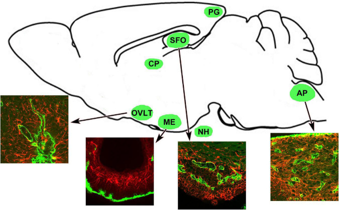
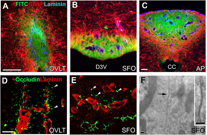
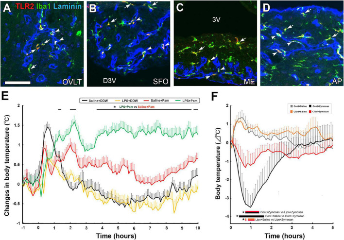
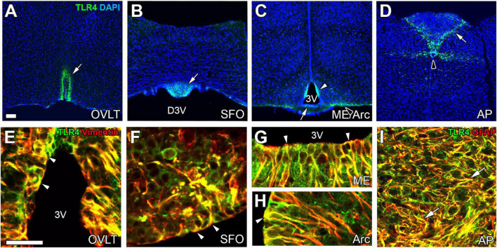
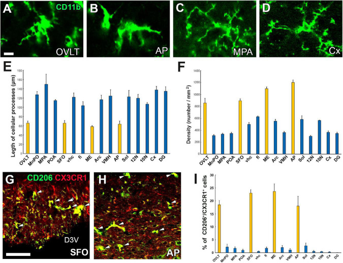
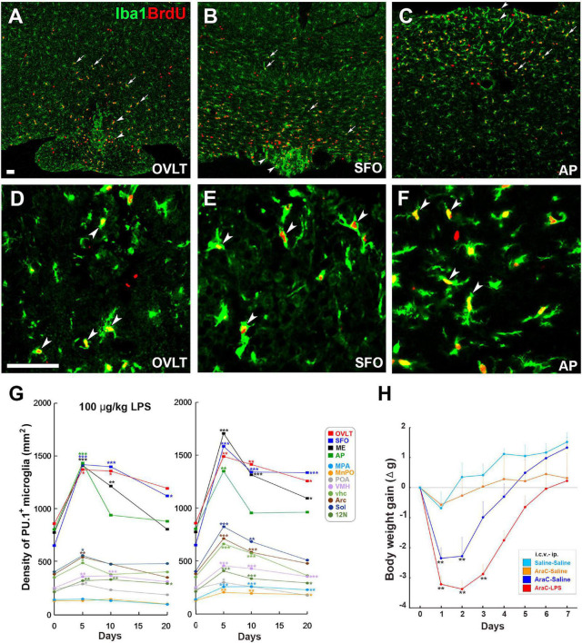
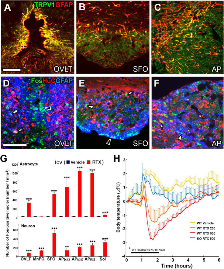
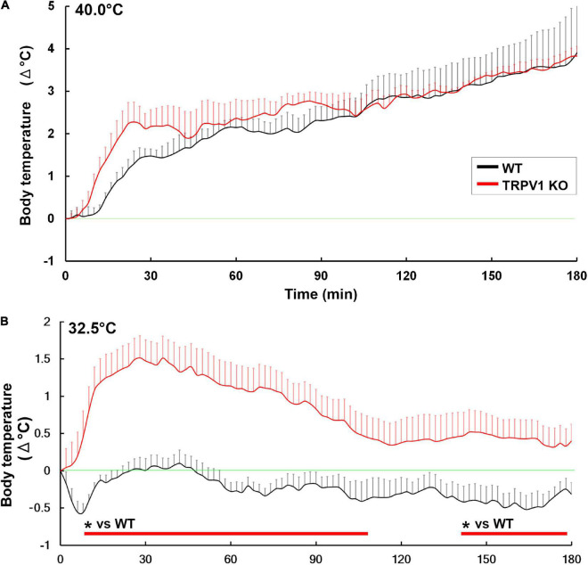
The circumventricular organs (CVOs) are located around the brain ventricles, lack a blood-brain barrier (BBB) and sense blood-derived molecules. This review discusses recent advances in the importance of CVO functions, especially glial cells transferring periphery inflammation signals to the brain. The CVOs show size-limited vascular permeability, allowing the passage of molecules with molecular weight <10,000. This indicates that the lack of an endothelial cell barrier does not mean the free movement of blood-derived molecules into the CVO parenchyma. Astrocytes and tanycytes constitute a dense barrier at the distal CVO subdivision, preventing the free diffusion of blood-derived molecules into neighboring brain regions. Tanycytes in the CVOs mediate communication between cerebrospinal fluid and brain parenchyma via transcytosis. Microglia and macrophages of the CVOs are essential for transmitting peripheral information to other brain regions via toll-like receptor 2 (TLR2). Inhibition of TLR2 signaling or depletion of microglia and macrophages in the brain eliminates TLR2-dependent inflammatory responses. In contrast to TLR2, astrocytes and tanycytes in the CVOs of the brain are crucial for initiating lipopolysaccharide (LPS)-induced inflammatory responses via TLR4. Depletion of microglia and macrophages augments LPS-induced fever and chronic sickness responses. Microglia and macrophages in the CVOs are continuously activated, even under normal physiological conditions, as they exhibit activated morphology and express the M1/M2 marker proteins. Moreover, the microglial proliferation occurs in various regions, such as the hypothalamus, medulla oblongata, and telencephalon, with a marked increase in the CVOs, due to low-dose LPS administration, and after high-dose LPS administration, proliferation is seen in most brain regions, except for the cerebral cortex and hippocampus. A transient increase in the microglial population is beneficial during LPS-induced inflammation for attenuating sickness response. Transient receptor potential receptor vanilloid 1 expressed in astrocytes and tanycytes of the CVOs is responsible for thermoregulation upon exposure to a warm environment less than 37°C. Alternatively, Na x expressed in astrocytes and tanycytes of the CVOs is crucial for maintaining body fluid homeostasis. Thus, recent findings indicate that glial cells in the brain CVOs are essential for initiating neuroinflammatory responses and maintaining body fluid and thermal homeostasis.
Overview
The claims and statistics of this paper haven’t been checked yet.
Run the check — free, no account needed. It reads the open-access text and checks the sources it cites.
Integrity signals
Registry
Cites
Statistics
Figures
Verification
Claims the paper makes
The claims and statistics of this paper haven’t been checked yet.
Run the check — free, no account needed. It reads the open-access text and checks the sources it cites.
Claims other papers make about this one
Each is checked against this paper’s own text: does it say what the citing paper says it does?
The source backs part of the claim, not all of it — or backs it for a different population, condition or comparison.
In the citing paper
“Circumventricular organs (CVOs) are specialized structures located around the third and fourth ventricles that mediate communications between brain, blood and cerebrospinal fluid74,75 (CSF).”
From A high-resolution transcriptomic and spatial atlas of cell types in the whole mouse brain (2023) · cites this paper as [75]
Experimental — accuracy not yet measured
Self-check
Percentages, sums and reported differences re-computed from the paper’s own numbers.
The self-check needs the paper’s full text.
Citations
Cites
Items 201–223 of 223
Kasuga, R., Shiraki, C., Horikawa, R., YOSHIMURA, R., Kurganov, E., & Miyata, S. (2022) · Physiology & Behavior · doi:10.1016/j.physbeh.2022.113729 · Cited by 21
Self-citationMuneoka, S., Murayama, S., Nakano, Y., & Miyata, S. (2018) · Journal of Neuroimmunology · doi:10.1016/j.jneuroim.2018.04.017 · Cited by 20
Self-citationOliveira, G. R., Franci, C. R., Rodovalho, G. V., Franci, J. A. A., Morris, M., & Rocha, M. J. A. (2004) · Brain Research Bulletin · doi:10.1016/j.brainresbull.2004.04.009 · Cited by 19
Inagaki, H., Kurganov, E., Park, Y., Furube, E., & Miyata, S. (2019) · Physiology & Behavior · doi:10.1016/j.physbeh.2019.04.015 · Cited by 19
Self-citationTakagi, S., Murayama, S., Torii, K., Takemura, S., Kurganov, E., Nagaoka, S., Wanaka, A., & Miyata, S. (2020) · Journal of Neuroimmunology · doi:10.1016/j.jneuroim.2020.577244 · Cited by 19
Self-citationMallmann, M. P., Mello, F. K., Neuberger, B., Sobral, K. G. D. C., Fighera, M. R., Royes, L. F. F., Furian, A. F., & Oliveira, M. S. (2022) · Brain Research · doi:10.1016/j.brainres.2022.147883 · Cited by 18
Dissociation between vasopressin and oxytocin mRNA and peptide secretion after AV3V lesions
Morris, M., Rocha, M. J. A., Sim, L. J., Johnson, A. K., & Callahan, M. F. (1994) · American Journal of Physiology-Regulatory, Integrative and Comparative Physiology · doi:10.1152/ajpregu.1994.267.6.r1640 · Cited by 17
Nishikawa, K., Furube, E., Morita, S., Horii‐Hayashi, N., Nishi, M., & Miyata, S. (2017) · Journal of Neuroendocrinology · doi:10.1111/jne.12456 · Cited by 17
Self-citationFerguson, A. V. (2014) · Cited by 16
Shiraki, C., Horikawa, R., Oe, Y., Fujimoto, M., Okamoto, K., Kurganov, E., & Miyata, S. (2021) · Brain Behavior & Immunity - Health · doi:10.1016/j.bbih.2021.100291 · Cited by 16
Self-citationMurayama, S., Kurganov, E., & Miyata, S. (2019) · Journal of Neuroimmunology · doi:10.1016/j.jneuroim.2019.576973 · Cited by 15
Self-citationTorii, K., Takagi, S., YOSHIMURA, R., & Miyata, S. (2022) · Journal of Neuroimmunology · doi:10.1016/j.jneuroim.2022.577832 · Cited by 14
Self-citationReceptor-Mediated and Fluid-Phase Transcytosis of Horseradish Peroxidase across Rat Hepatocytes
Ellinger, I., & Fuchs, R. (2010) · BioMed Research International · doi:10.1155/2010/850320 · Cited by 12
Miyata, S., Matsunaga, W., Mondoh, H., Nakashima, T., & Kiyohara, T. (1996) · Neuroscience Research · doi:10.1016/s0168-0102(96)01099-1 · Cited by 12
Self-citationTranscytosis of tanycytes in the circumventricular organs of adult mouse brain
Okamoto, A., Fujii, R., YOSHIMURA, R., & Miyata, S. (2022) · Neuroscience Letters · doi:10.1016/j.neulet.2022.136633 · Cited by 12
Self-citationAnteroventral third ventricle (AV3V) lesions alter c-fos expression induced by salt loading
Rocha, M. J. A., Beltz, T. G., Dornelles, R. C. M., Johnson, A. K., & Franci, C. R. (1999) · Brain Research · doi:10.1016/s0006-8993(99)01366-9 · Cited by 9
de Cerqueira, D. R., Ferreira, H. S., Moiteiro, A. L. B. B., & Fregoneze, J. B. (2016) · Physiology & Behavior · doi:10.1016/j.physbeh.2016.05.003 · Cited by 8
Nambu, Y., Ohira, K., Morita, M., Yasumoto, H., Kurganov, E., & Miyata, S. (2021) · Neuroscience Research · doi:10.1016/j.neures.2021.05.012 · Cited by 7
Self-citationThe terminal organ of the subcommissural complex of chordates: definition and perspectives.
Hofer, H. O. (1987) · PubMed · Cited by 5
Proliferation of endothelial cells in the choroid plexus of normal and hydrocephalic mice
Asami, A., Kurganov, E., & Miyata, S. (2020) · Journal of Chemical Neuroanatomy · doi:10.1016/j.jchemneu.2020.101796 · Cited by 5
Self-citationNambu, Y., Horie, K., Kurganov, E., & Miyata, S. (2021) · Neuroscience Letters · doi:10.1016/j.neulet.2021.135732 · Cited by 4
Self-citationHeiss, C. N., Gravert, E., Hultén, M., & Olofsson, L. E. (2022) · Molecular Neurobiology · doi:10.1007/s12035-022-02802-w · Cited by 3
Research works printed but not linked: 1
- Ehrlich, 1885, Das Sauerstoff-Bedurfinis dews Oraganisms. Eine Farenanalytisch Studie, 69
Cited by
Sorted by citations
A high-resolution transcriptomic and spatial atlas of cell types in the whole mouse brain
Yao, Z., van Velthoven, C. T. J., Kunst, M., Zhang, M., McMillen, D., Lee, C., Jung, W., Goldy, J., Abdelhak, A., Aitken, M., et al. (2023) · Nature · doi:10.1038/s41586-023-06812-z · Cited by 1,203
Breaking the Barrier: The Role of Proinflammatory Cytokines in BBB Dysfunction
Gryka-Marton, M., Grabowska, A. D., & Szukiewicz, D. (2025) · International Journal of Molecular Sciences · doi:10.3390/ijms26083532 · Cited by 144
Border-associated macrophages as gatekeepers of brain homeostasis and immunity
Vara‐Pérez, M., & Movahedi, K. (2025) · Immunity · doi:10.1016/j.immuni.2025.04.005 · Cited by 84
Astroglia support, regulate and reinforce brain barriers
Verkhratsky, A., & Pivoriūnas, A. (2023) · Neurobiology of Disease · doi:10.1016/j.nbd.2023.106054 · Cited by 59
Central Nervous System Macrophages in Health and Disease
Hove, H. V., Feo, D. D., Greter, M., & Becher, B. (2025) · Annual Review of Immunology · doi:10.1146/annurev-immunol-082423-041334 · Cited by 53
Metadata
- Authors
- Miyata, S.
- Affiliations
- Kyoto Institute of Technology (JP)
- Corresponding author
- Miyata, Seiji
- ORCID
- Miyata, Seiji
- Venue
- Frontiers in Neuroscience
- Publisher
- Frontiers Media SA
- Published
- 2022-10-06
- Volume
- 16
- Pages
- 991779-991779
- ISSN
- 1662-453X (ISSN-L)
- PMC
- PMC9583022
- OpenAlex
- W4302305006
- Cited by
- 66
- References
- 224
- Citations by year
- 2026: 24 · 2025: 22 · 2024: 13 · 2023: 7
- FWCI
- 1.08
- Type
- journal article
- Language
- en
- Open access
- gold
- doi.org
- Licence
- Locations
- Frontiers in Neuroscience (publishedVersion, cc-by)
- PubMed Central (publishedVersion, cc-by)
- PubMed (publishedVersion)
- DOAJ (DOAJ: Directory of Open Access Journals) (submittedVersion, cc-by-sa)
- Funders
- Japan Society for the Promotion of Science (22K06456, 19K06921)
- Topics
- Life Sciences › Neuroscience › Neurology › Neuroinflammation and Neurodegeneration Mechanisms
- Life Sciences › Neuroscience › Neurology › Barrier Structure and Function Studies
- Health Sciences › Medicine › Critical Care and Intensive Care Medicine › Thermal Regulation in Medicine
- Keywords
- circumventricular organs, blood-brain barrier, glial cells, tanycytes, astrocytes, microglia, macrophages, TLR2, TLR4, lipopolysaccharide, neuroinflammation, sickness behavior, fever, microglial proliferation, TRPV1, Na+/Ca2+ exchanger, thermoregulation, fluid homeostasis, transcytosis, cerebrospinal fluid, hypothalamus, medulla oblongata
About these checks
Registry, retraction, Cites and statistics results are computed, not judged, with one exception: a printed reference that no index lists is read once by a model before it counts as a citation error. Claims are extracted by a model and kept only when their quote appears verbatim in the text; each cited source is then checked, with a model’s help, for whether it says what the claim relies on. Those verdicts can be wrong — read the claim’s quote and the cited source before relying on one.
Statistics and arithmetic are recomputed, not judged.
Confidence bands are shown only for fields calibrated on our annotated test set. None is calibrated yet, so no band is shown.
What counts as a claim ›How we measure confidence ›How we check this ›
Are you an author, or do you see a problem with this page? Request a review
