

The DOI matches its Crossref record; 223 of the 224 references it cites were found in a registry or an index.
Glial functions in the blood-brain communication at the circumventricular organs
Open accessAbstract
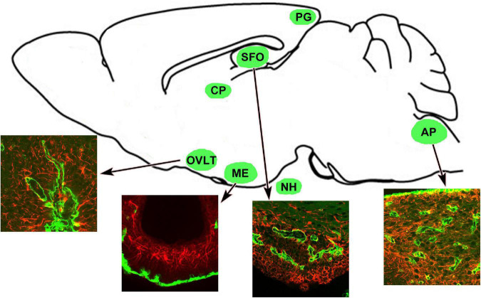
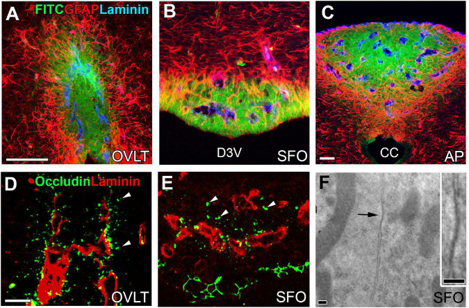
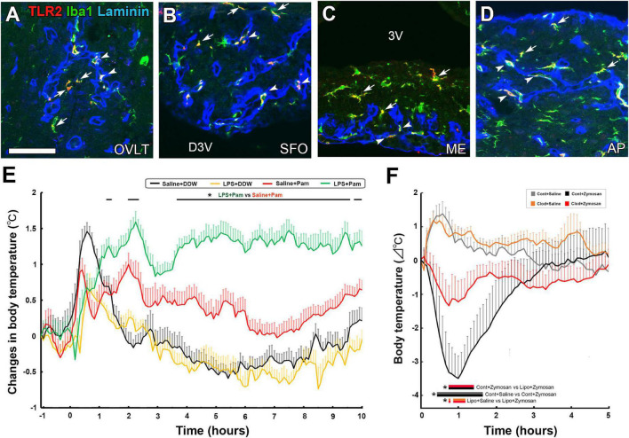
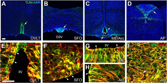
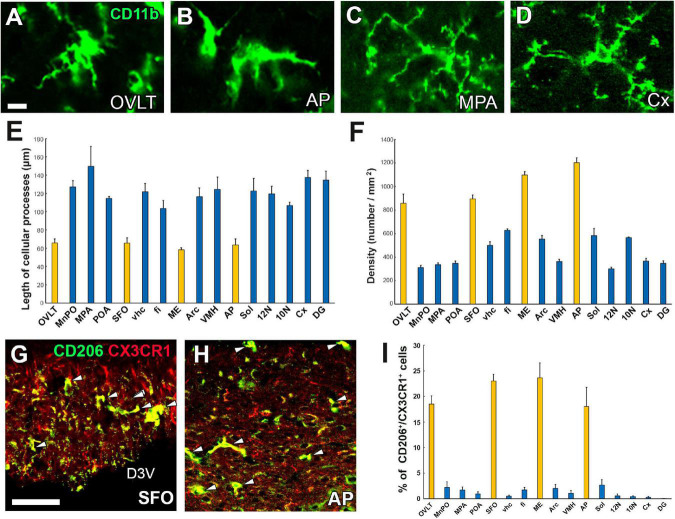
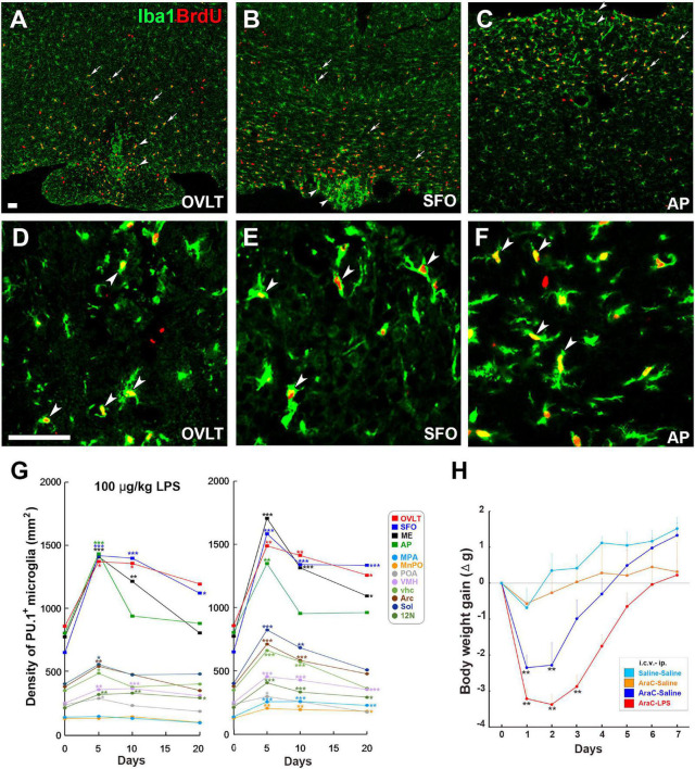
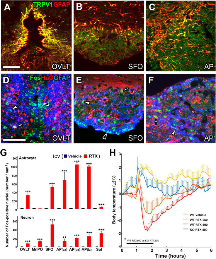
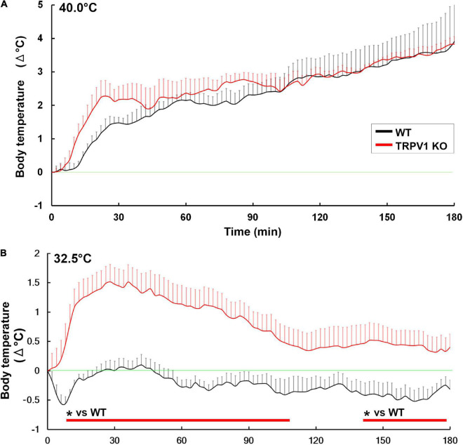
The circumventricular organs (CVOs) are located around the brain ventricles, lack a blood-brain barrier (BBB) and sense blood-derived molecules. This review discusses recent advances in the importance of CVO functions, especially glial cells transferring periphery inflammation signals to the brain. The CVOs show size-limited vascular permeability, allowing the passage of molecules with molecular weight <10,000. This indicates that the lack of an endothelial cell barrier does not mean the free movement of blood-derived molecules into the CVO parenchyma. Astrocytes and tanycytes constitute a dense barrier at the distal CVO subdivision, preventing the free diffusion of blood-derived molecules into neighboring brain regions. Tanycytes in the CVOs mediate communication between cerebrospinal fluid and brain parenchyma via transcytosis. Microglia and macrophages of the CVOs are essential for transmitting peripheral information to other brain regions via toll-like receptor 2 (TLR2). Inhibition of TLR2 signaling or depletion of microglia and macrophages in the brain eliminates TLR2-dependent inflammatory responses. In contrast to TLR2, astrocytes and tanycytes in the CVOs of the brain are crucial for initiating lipopolysaccharide (LPS)-induced inflammatory responses via TLR4. Depletion of microglia and macrophages augments LPS-induced fever and chronic sickness responses. Microglia and macrophages in the CVOs are continuously activated, even under normal physiological conditions, as they exhibit activated morphology and express the M1/M2 marker proteins. Moreover, the microglial proliferation occurs in various regions, such as the hypothalamus, medulla oblongata, and telencephalon, with a marked increase in the CVOs, due to low-dose LPS administration, and after high-dose LPS administration, proliferation is seen in most brain regions, except for the cerebral cortex and hippocampus. A transient increase in the microglial population is beneficial during LPS-induced inflammation for attenuating sickness response. Transient receptor potential receptor vanilloid 1 expressed in astrocytes and tanycytes of the CVOs is responsible for thermoregulation upon exposure to a warm environment less than 37°C. Alternatively, Na x expressed in astrocytes and tanycytes of the CVOs is crucial for maintaining body fluid homeostasis. Thus, recent findings indicate that glial cells in the brain CVOs are essential for initiating neuroinflammatory responses and maintaining body fluid and thermal homeostasis.
Overview
The claims and statistics of this paper haven’t been checked yet.
Run the check — free, no account needed. It reads the open-access text and checks the sources it cites.
Integrity signals
Registry
Cites
Statistics
Figures
Verification
Claims the paper makes
The claims and statistics of this paper haven’t been checked yet.
Run the check — free, no account needed. It reads the open-access text and checks the sources it cites.
Claims other papers make about this one
Each is checked against this paper’s own text: does it say what the citing paper says it does?
The source backs part of the claim, not all of it — or backs it for a different population, condition or comparison.
In the citing paper
“Circumventricular organs (CVOs) are specialized structures located around the third and fourth ventricles that mediate communications between brain, blood and cerebrospinal fluid74,75 (CSF).”
From A high-resolution transcriptomic and spatial atlas of cell types in the whole mouse brain (2023) · cites this paper as [75]
Experimental — accuracy not yet measured
Self-check
Percentages, sums and reported differences re-computed from the paper’s own numbers.
The self-check needs the paper’s full text.
Citations
Cites
The role of pattern-recognition receptors in innate immunity: update on Toll-like receptors
Kawai, T., & Akira, S. (2010) · Nature Immunology · doi:10.1038/ni.1863 · Cited by 9,304
The capsaicin receptor: a heat-activated ion channel in the pain pathway
Caterina, M. J., Schumacher, M. A., Tominaga, M., Rosen, T. A., Levine, J. D., & Julius, D. J. (1997) · Nature · doi:10.1038/39807 · Cited by 9,132
Polazzi, E., Mengoni, I., Caprini, M., Peña‐Altamira, E., Kurtys, E., & Monti, B. (2012) · Neurosignals · doi:10.1159/000337115 · Cited by 9,023
The chemokine system in diverse forms of macrophage activation and polarization
MANTOVANI, A., SICA, A., SOZZANI, S., ALLAVENA, P., VECCHI, A., & LOCATI, M. (2004) · Trends in Immunology · doi:10.1016/j.it.2004.09.015 · Cited by 6,534
Toll-like receptors: critical proteins linking innate and acquired immunity
Akira, S., Takeda, K., & Kaisho, T. (2001) · Nature Immunology · doi:10.1038/90609 · Cited by 4,777
Synaptic Pruning by Microglia Is Necessary for Normal Brain Development
Paolicelli, R. C., Bolasco, G., Pagani, F., Maggi, L., Scianni, M., Panzanelli, P., Giustetto, M., Ferreira, T. A., Guiducci, E., Dumas, L., et al. (2011) · Science · doi:10.1126/science.1202529 · Cited by 4,244
Microglia: Active sensor and versatile effector cells in the normal and pathologic brain
Hanisch, U.-K., & Kettenmann, H. (2007) · Nature Neuroscience · doi:10.1038/nn1997 · Cited by 3,676
Kettenmann, H., Hanisch, U.-K., Noda, M., & Verkhratsky, A. (2011) · Physiological Reviews · doi:10.1152/physrev.00011.2010 · Cited by 3,547
Murray, P. J. (2017) · Annual Review of Physiology · doi:10.1146/annurev-physiol-022516-034339 · Cited by 2,981
Neurovascular pathways to neurodegeneration in Alzheimer’s disease and other disorders
Zlokovic, B. V. (2011) · Nature Reviews Neuroscience · doi:10.1038/nrn3114 · Cited by 2,969
The blood-brain barrier/neurovascular unit in health and disease
Hawkins, B. T., & Davis, T. P. (2005) · Pharmacological Reviews · doi:10.1124/pr.57.2.4 · Cited by 2,661
Multifunctional strands in tight junctions
Tsukita, S., Furuse, M., & Itoh, M. (2001) · Nature reviews. Molecular cell biology · doi:10.1038/35067088 · Cited by 2,578
Development, maintenance and disruption of the blood-brain barrier
Obermeier, B., Daneman, R., & Ransohoff, R. M. (2013) · Nature Medicine · doi:10.1038/nm.3407 · Cited by 2,452
Molecular Mechanisms That Influence the Macrophage M1–M2 Polarization Balance
Wang, N., Liang, H., & Zen, K. (2014) · Frontiers in Immunology · doi:10.3389/fimmu.2014.00614 · Cited by 2,069
Signaling Mechanisms Regulating Endothelial Permeability
Mehta, D., & Malik, A. B. (2005) · Physiological Reviews · doi:10.1152/physrev.00012.2005 · Cited by 1,754
A polarizing question: do M1 and M2 microglia exist?
Ransohoff, R. M. (2016) · Nature Neuroscience · doi:10.1038/nn.4338 · Cited by 1,573
Claudin-based tight junctions are crucial for the mammalian epidermal barrier
Furuse, M., Hata, M., Furuse, K., Yoshida, Y., Haratake, A., Sugitani, Y., Noda, T., Kubo, A., & Tsukita, S. (2002) · The Journal of Cell Biology · doi:10.1083/jcb.200110122 · Cited by 1,512
Astrocytes are active players in cerebral innate immunity
Farina, C., Aloisi, F., & Meinl, E. (2007) · Trends in Immunology · doi:10.1016/j.it.2007.01.005 · Cited by 1,304
Takeuchi, O., Hoshino, K., & Akira, S. (2000) · Journal of Immunology · doi:10.4049/jimmunol.165.10.5392 · Cited by 1,156
Julius, D. J. (2013) · Annual Review of Cell and Developmental Biology · doi:10.1146/annurev-cellbio-101011-155833 · Cited by 1,152
Yang, Y., Estrada, E. Y., Thompson, J. F., Liu, W., & Rosenberg, G. A. (2007) · Journal of Cerebral Blood Flow & Metabolism · doi:10.1038/sj.jcbfm.9600375 · Cited by 1,060
THE CELL BIOLOGY OF THE BLOOD-BRAIN BARRIER
Rubin, L. L., & Staddon, J. M. (1999) · Annual Review of Neuroscience · doi:10.1146/annurev.neuro.22.1.11 · Cited by 1,017
Astrocyte calcium signaling: the third wave
Bazargani, N., & Attwell, D. (2016) · Nature Neuroscience · doi:10.1038/nn.4201 · Cited by 998
New Insights into Neuron-Glia Communication
Fields, R. D., & Stevens, B. (2002) · Science · doi:10.1126/science.298.5593.556 · Cited by 975
Butovsky, O., Ziv, Y., Schwartz, A., Landa, G., Talpalar, A. E., Pluchino, S., Martino, G., & Schwartz, M. (2006) · Molecular and Cellular Neuroscience · doi:10.1016/j.mcn.2005.10.006 · Cited by 923
Heterogeneity of Microglial Activation in the Innate Immune Response in the Brain
Colton, C. A. (2009) · Journal of Neuroimmune Pharmacology · doi:10.1007/s11481-009-9164-4 · Cited by 916
Morita, K., Sasaki, H., Furuse, M., & Tsukita, S. (1999) · The Journal of Cell Biology · doi:10.1083/jcb.147.1.185 · Cited by 867
The Role of TLR2 in Infection and Immunity
Nascimento, L. D. O., Massari, P., & Wetzler, L. M. (2012) · Frontiers in Immunology · doi:10.3389/fimmu.2012.00079 · Cited by 861
Regulation of innate immune responses in the brain
Rivest, S. (2009) · Nature Reviews Immunology · doi:10.1038/nri2565 · Cited by 843
Assembly and localization of Toll-like receptor signalling complexes
Gay, N. J., Symmons, M. F., Gangloff, M., & Bryant, C. E. (2014) · Nature Reviews Immunology · doi:10.1038/nri3713 · Cited by 832
Coupled Proliferation and Apoptosis Maintain the Rapid Turnover of Microglia in the Adult Brain
Askew, K., Li, K., Olmos-Alonso, A., Garcia-Moreno, F., Liang, Y., Richardson, P., Tipton, T., Chapman, M. A., Riecken, K., Beccari, S., et al. (2017) · Cell Reports · doi:10.1016/j.celrep.2016.12.041 · Cited by 726
Furuse, M., Furuse, K., Sasaki, H., & Tsukita, S. (2001) · The Journal of Cell Biology · doi:10.1083/jcb.153.2.263 · Cited by 712
Central mechanisms of osmosensation and systemic osmoregulation
Bourque, C. W. (2008) · Nature reviews. Neuroscience · doi:10.1038/nrn2400 · Cited by 703
Persistent neurologic symptoms and cognitive dysfunction in non-hospitalized COVID-19 “long haulers”
Graham, E. L., Clark, J. R., Orban, Z. S., Lim, P. H., Szymanski, A. L., Taylor, C., DiBiase, R. M., Jia, D. T., Balabanov, R., Ho, S. U., et al. (2021) · Annals of Clinical and Translational Neurology · doi:10.1002/acn3.51350 · Cited by 693
Spinal Cord Injury Reveals Multilineage Differentiation of Ependymal Cells
Meletis, K., Barnabé‐Heider, F., Carlén, M., Evergren, E., Tomilin, N. V., Shupliakov, O., & Frisén, J. (2008) · PLoS Biology · doi:10.1371/journal.pbio.0060182 · Cited by 685
Henry, C. J., Huang, Y., Wynne, A., Hanke, M., Himler, J., Bailey, M. T., Sheridan, J. F., & Godbout, J. P. (2008) · Journal of Neuroinflammation · doi:10.1186/1742-2094-5-15 · Cited by 678
Microglia are an essential component of the neuroprotective scar that forms after spinal cord injury
Bellver‐Landete, V., Bretheau, F., Mailhot, B., Vallières, N., Lessard, M., Janelle, M.-E., Vernoux, N., Tremblay, M., Führmann, T., Shoichet, M. S., et al. (2019) · Nature Communications · doi:10.1038/s41467-019-08446-0 · Cited by 650
Regulation of Endothelial Permeability via Paracellular and Transcellular Transport Pathways
Komarova, Y. A., & Malik, A. B. (2010) · Annual Review of Physiology · doi:10.1146/annurev-physiol-021909-135833 · Cited by 640
Hypothalamic stem cells control ageing speed partly through exosomal miRNAs
Zhang, Y., Kim, M. S., Jia, B., Yan, J., Zuniga-Hertz, J. P., Han, C., & Cai, D. (2017) · Nature · doi:10.1038/nature23282 · Cited by 564
Hypothalamic Tanycytes Are an ERK-Gated Conduit for Leptin into the Brain
Balland, É., Dam, J., Langlet, F., Caron, É., Steculorum, S. M., Messina, A., Rasika, S., Falluel‐Morel, A., Anouar, Y., Dehouck, B., et al. (2014) · Cell Metabolism · doi:10.1016/j.cmet.2013.12.015 · Cited by 535
Olmos-Alonso, A., Schetters, S. T. T., Sri, S., Askew, K., Mancuso, R., Vargas-Caballero, M., Holscher, C., Perry, V. H., & Gomez-Nicola, D. (2016) · Brain · doi:10.1093/brain/awv379 · Cited by 532
Microglia Enhance Neurogenesis and Oligodendrogenesis in the Early Postnatal Subventricular Zone
Shigemoto‐Mogami, Y., Hoshikawa, K., Goldman, J. E., Sekino, Y., & Sato, K. (2014) · Journal of Neuroscience · doi:10.1523/jneurosci.1619-13.2014 · Cited by 524
Buzhdygan, T. P., DeOre, B. J., Baldwin‐Leclair, A., Bullock, T. A., McGary, H. M., Khan, J. A., Razmpour, R., Hale, J. F., Galie, P. A., Potula, R., et al. (2020) · Neurobiology of Disease · doi:10.1016/j.nbd.2020.105131 · Cited by 513
Sensory circumventricular organs and brain homeostatic pathways
Johnson, A. K., & Gross, P. M. (1993) · The FASEB Journal · doi:10.1096/fasebj.7.8.8500693 · Cited by 511
Tanycytes of the hypothalamic median eminence form a diet-responsive neurogenic niche
Lee, D. A., Bedont, J. L., Pak, T., Wang, H., Song, J., Miranda-Angulo, A., Takiar, V., Charubhumi, V., Balordi, F., Takebayashi, H., et al. (2012) · Nature Neuroscience · doi:10.1038/nn.3079 · Cited by 502
LAFLAMME, N., & RIVEST, S. (2001) · The FASEB Journal · doi:10.1096/fj.00-0339com · Cited by 501
Chemical and Pharmacological Aspects of Capsaicin
Reyes-Escogido, M. D. L., González‐Mondragón, E., & Vazquez-Tzompantzi, E. (2011) · Molecules · doi:10.3390/molecules16021253 · Cited by 484
Physiological blood–brain transport is impaired with age by a shift in transcytosis
Yang, A. C., Stevens, M. Y., Chen, M. B., Lee, D. P., Stähli, D., Gate, D., Contrepois, K., Chen, W., Iram, T., Zhang, L., et al. (2020) · Nature · doi:10.1038/s41586-020-2453-z · Cited by 477
Hypothalamic tanycytes: a key component of brain-endocrine interaction
RODRIGUEZ, E., BLAZQUEZ, J., PASTOR, F., PELAEZ, B., PENA, P., PERUZZO, B., & AMAT, P. (2005) · International review of cytology · doi:10.1016/s0074-7696(05)47003-5 · Cited by 475 · cited 2×
“Tanycytes are specialized ependymal cells interfacing with the CSF and have a long basal process that terminates at the capillary (Horstmann, 1954; Rodríguez et al., 2005).”
BackgroundEricsson, A., Liu, C., Hart, R. P., & Sawchenko, P. E. (1995) · Journal of Comparative Neurology · doi:10.1002/cne.903610410 · Cited by 471
Recent advances in the study on capsaicinoids and capsinoids.
Luo, X.-J., Peng, J., & Li, Y. (2011) · European Journal of Pharmacology · doi:10.1016/j.ejphar.2010.09.074 · Cited by 444
Tsai, V. W.-W., Husaini, Y., Sainsbury, A., Brown, D. A., & Breit, S. N. (2018) · Cell Metabolism · doi:10.1016/j.cmet.2018.07.018 · Cited by 424
Zähringer, U., Lindner, B., Inamura, S., Heine, H., & Alexander, C. (2008) · Immunobiology · doi:10.1016/j.imbio.2008.02.005 · Cited by 417
Doobay, M. F., Talman, L. S., Obr, T. D., Tian, X., Davisson, R. L., & Lazartigues, E. D. (2006) · American Journal of Physiology-Regulatory, Integrative and Comparative Physiology · doi:10.1152/ajpregu.00292.2006 · Cited by 405
Chakravarty, S., & Herkenham, M. (2005) · The Journal of Neuroscience · doi:10.1523/jneurosci.4268-04.2005 · Cited by 396
Chai, Z., Gatti, S., Toniatti, C., Poli, V., & Bartfai, T. (1996) · Journal of Experimental Medicine · doi:10.1084/jem.183.1.311 · Cited by 376
The Area Postrema and Vomiting
Miller, A. D., & Leslie, R. A. (1994) · Frontiers in Neuroendocrinology · doi:10.1006/frne.1994.1012 · Cited by 375
Aboudounya, M. M., & Heads, R. J. (2021) · Mediators of Inflammation · doi:10.1155/2021/8874339 · Cited by 359
SARS-CoV-2 spike protein interacts with and activates TLR41
Zhao, Y., Kuang, M., Li, J., Zhu, L., Jia, Z., Guo, X., Hu, Y., Kong, J. B., Yin, H. H., Wang, X., et al. (2021) · Cell Research · doi:10.1038/s41422-021-00495-9 · Cited by 358
Henry, R. J., Ritzel, R. M., Barrett, J. P., Doran, S. J., Jiao, Y., Leach, J. B., Szeto, G. L., Wu, J., Stoica, B. A., Faden, A. I., et al. (2020) · Journal of Neuroscience · doi:10.1523/jneurosci.2402-19.2020 · Cited by 344
Molecular insights on the cerebral innate immune system
Rivest, S. (2003) · Brain, Behavior, and Immunity · doi:10.1016/s0889-1591(02)00055-7 · Cited by 338
The glomerular basement membrane
Miner, J. H. (2012) · Experimental Cell Research · doi:10.1016/j.yexcr.2012.02.031 · Cited by 338
LPS Sensitizes TRPV1 via Activation of TLR4 in Trigeminal Sensory Neurons
Diogenes, A. R., Ferraz, C. C. R., Akopian, A. N., Henry, M. A., & Hargreaves, K. (2011) · Journal of Dental Research · doi:10.1177/0022034511400225 · Cited by 337
Mullier, A., Bouret, S. G., Prevot, V., & Dehouck, B. (2010) · Journal of Comparative Neurology · doi:10.1002/cne.22273 · Cited by 334
Vallières, L., & Rivest, S. (1997) · Journal of Neurochemistry · doi:10.1046/j.1471-4159.1997.69041668.x · Cited by 323
Katsuno, T., Umeda, K., Matsui, T., Hata, M., Tamura, A., Itoh, M., Takeuchi, K., Fujimori, T., Nabeshima, Y.-I., Noda, T., et al. (2008) · Molecular Biology of the Cell · doi:10.1091/mbc.e07-12-1215 · Cited by 309
Rodríguez, E. M., Blázquez, J. L., & Guerra, M. (2010) · Peptides · doi:10.1016/j.peptides.2010.01.003 · Cited by 308
Circumventricular Organs: Definition and Role in the Regulation of Endocrine and Autonomic Function
Ganong, W. F. (2000) · Clinical and Experimental Pharmacology and Physiology · doi:10.1046/j.1440-1681.2000.03259.x · Cited by 307
Robins, S. C., Stewart, I., McNay, D. E., Taylor, V., Giachino, C., Goetz, M., Ninkovic, J., Briancon, N., Maratos-Flier, E., Flier, J. S., et al. (2013) · Nature Communications · doi:10.1038/ncomms3049 · Cited by 306
Age-Dependent Niche Signals from the Choroid Plexus Regulate Adult Neural Stem Cells
Silva-Vargas, V., Maldonado-Soto, A. R., Mizrak, D., Codega, P., & Doetsch, F. (2016) · Cell stem cell · doi:10.1016/j.stem.2016.06.013 · Cited by 306
Hypothalamic tanycytes: potential roles in the control of feeding and energy balance
Bolborea, M., & Dale, N. (2013) · Trends in Neurosciences · doi:10.1016/j.tins.2012.12.008 · Cited by 302
Toll‐like receptor 3 on adult human astrocytes triggers production of neuroprotective mediators
Bsibsi, M., Persoon-Deen, C., Verwer, R. W. H., Meeuwsen, S., Ravid, R., & van Noort, J. M. (2006) · Glia · doi:10.1002/glia.20328 · Cited by 301
Die Faserglia des Selachiergehirns
Horstmann, E. (1954) · Cell and Tissue Research · doi:10.1007/bf00334758 · Cited by 299
Microglial Dysregulation in Psychiatric Disease
Frick, L. R., Williams, K., & Pittenger, C. (2013) · Clinical and Developmental Immunology · doi:10.1155/2013/608654 · Cited by 299
Neurons that regulate mouse torpor
Hrvatin, S., Sun, S., Wilcox, O. F., Yao, H., Lavin-Peter, A. J., Cicconet, M., Assad, E. G., Palmer, M. E., Aronson, S., Banks, A. S., et al. (2020) · Nature · doi:10.1038/s41586-020-2387-5 · Cited by 297
Langlet, F., Mullier, A., Bouret, S. G., Prevot, V., & Dehouck, B. (2013) · Journal of Comparative Neurology · doi:10.1002/cne.23355 · Cited by 295
The blood-brain barrier and glutamate.
Hawkins, R. (2009) · American Journal of Clinical Nutrition · doi:10.3945/ajcn.2009.27462bb · Cited by 294
The Sensory Circumventricular Organs of the Mammalian Brain
McKinley, M. J., McAllen, R. M., Davern, P. J., Giles, M., Penschow, J., Sunn, N., Uschakov, A., & Oldfield, B. J. (2003) · Advances in anatomy, embryology and cell biology · doi:10.1007/978-3-642-55532-9 · Cited by 291
Laflamme, N., Lacroix, S., & Rivest, S. (1999) · Journal of Neuroscience · doi:10.1523/jneurosci.19-24-10923.1999 · Cited by 287
Li, L., Acioglu, C., Heary, R. F., & Elkabes, S. (2021) · Brain, Behavior, and Immunity · doi:10.1016/j.bbi.2020.10.007 · Cited by 283
Ciura, S., & Bourque, C. W. (2006) · Journal of Neuroscience · doi:10.1523/jneurosci.0877-06.2006 · Cited by 277
A discrete neuronal circuit induces a hibernation-like state in rodents
Takahashi, T. M., Sunagawa, G. A., Soya, S., Abe, M., Sakurai, K., Ishikawa, K., Yanagisawa, M., Hama, H., Hasegawa, E., Miyawaki, A., et al. (2020) · Nature · doi:10.1038/s41586-020-2163-6 · Cited by 271
Perivascular macrophages in health and disease
Lapenna, A., Palma, M. D., & Lewis, C. E. (2018) · Nature reviews. Immunology · doi:10.1038/s41577-018-0056-9 · Cited by 268
Area postrema cell types that mediate nausea-associated behaviors
Zhang, C., Kaye, J. A., Cai, Z., Wang, Y., Prescott, S. L., & Liberles, S. D. (2021) · Neuron · doi:10.1016/j.neuron.2020.11.010 · Cited by 263
The ion channel TRPV1 regulates the activation and proinflammatory properties of CD4+ T cells
Bertin, S., Aoki‐Nonaka, Y., de Jong, P. R., Nohara, L. L., Xu, H., Stanwood, S. R., Srikanth, S., Lee, J., To, K., Abramson, L., et al. (2014) · Nature Immunology · doi:10.1038/ni.3009 · Cited by 261
Blood-brain barrier transport machineries and targeted therapy of brain diseases
Barar, J., Rafi, M. A., Pourseif, M. M., & Omidi, Y. (2016) · Bioimpacts · doi:10.15171/bi.2016.30 · Cited by 259
The Area Postrema: A Brain Monitor and Integrator of Systemic Autonomic State
Price, C. J., Hoyda, T. D., & Ferguson, A. V. (2008) · The Neuroscientist · doi:10.1177/1073858407311100 · Cited by 257
Dual Roles for Perivascular Macrophages in Immune-to-Brain Signaling
Serrats, J., Schiltz, J. C., García-Bueno, B., van Rooijen, N., Reyes, T. M., & Sawchenko, P. E. (2010) · Neuron · doi:10.1016/j.neuron.2009.11.032 · Cited by 253
The transient receptor potential vanilloid-1 channel in thermoregulation: a thermosensor it is not
Romanovsky, A. A., Almeida, M. C., Garami, A., Steiner, A. A., Norman, M. H., Morrison, S. F., Nakamura, K., Burmeister, J. J., & Nucci, T. B. (2009) · Pharmacological Reviews · doi:10.1124/pr.109.001263 · Cited by 253
Hashimoto, M., Tawaratsumida, K., Kariya, H., Kiyohara, A., Suda, Y., Krikae, F., Kirikae, T., & Götz, F. (2006) · The Journal of Immunology · doi:10.4049/jimmunol.177.5.3162 · Cited by 239
Konsman, J. P., Kelley, K. W., & Dantzer, R. (1999) · Neuroscience · doi:10.1016/s0306-4522(98)00368-6 · Cited by 232
IL-6-regulated transcription factors
Akira, S. (1997) · The International Journal of Biochemistry & Cell Biology · doi:10.1016/s1357-2725(97)00063-0 · Cited by 231
Brain cell volume regulation in hyponatremia: role of sex, age, vasopressin, and hypoxia
Ayus, J. C., Achinger, S. G., & Arieff, A. I. (2008) · American Journal of Physiology-Renal Physiology · doi:10.1152/ajprenal.00502.2007 · Cited by 231
Lacroix, S., Feinstein, D., & Rivest, S. (1998) · Brain Pathology · doi:10.1111/j.1750-3639.1998.tb00189.x · Cited by 223
The Cellular Code for Mammalian Thermosensation
Pogorzala, L., Mishra, S. K., & Hoon, M. A. (2013) · Journal of Neuroscience · doi:10.1523/jneurosci.5788-12.2013 · Cited by 220
Nadeau, S., & Rivest, S. (2000) · Journal of Neuroscience · doi:10.1523/jneurosci.20-09-03456.2000 · Cited by 217
Suppression of fever after lesions of the anteroventral third ventricle in guinea pigs
Blatteis, C. M., Bealer, S. L., Hunter, W. S., Llanos, J.-Q., Ahokas, R. A., & Mashburn, T. A. (1983) · Brain Research Bulletin · doi:10.1016/0361-9230(83)90124-7 · Cited by 213
Glial Nax Channels Control Lactate Signaling to Neurons for Brain [Na+] Sensing
Shimizu, H., Watanabe, E., Hiyama, T. Y., Nagakura, A., Fujikawa, A., Okado, H., Yanagawa, Y., Obata, K., & Noda, M. (2007) · Neuron · doi:10.1016/j.neuron.2007.03.014 · Cited by 200
Miyata, S. (2015) · Frontiers in Neuroscience · doi:10.3389/fnins.2015.00390 · Cited by 198
Self-citationNeural Mechanisms of Inflammation-Induced Fever
Blomqvist, A. G., & Engblom, D. (2018) · The Neuroscientist · doi:10.1177/1073858418760481 · Cited by 197
The Sensory Coding of Warm Perception
Paricio-Montesinos, R., Schwaller, F., Udhayachandran, A., Rau, F., Walcher, J., Evangelista, R., Vriens, J., Voets, T., Poulet, J. F. A., & Lewin, G. R. (2020) · Neuron · doi:10.1016/j.neuron.2020.02.035 · Cited by 187
Laflamme, N., Soucy, G., & Rivest, S. (2001) · Journal of Neurochemistry · doi:10.1046/j.1471-4159.2001.00603.x · Cited by 185
Vargas-Caraveo, A., Sayd, A., Maus, S. R., Caso, J. R., Madrigal, J. L. M., García‐Bueno, B., & Leza, J. C. (2017) · Scientific Reports · doi:10.1038/s41598-017-13302-6 · Cited by 181
Nax channel involved in CNS sodium-level sensing
Hiyama, T. Y., Watanabe, E., Ono, K., Inenaga, K., Tamkun, M. M., Yoshida, S., & Noda, M. (2002) · Nature Neuroscience · doi:10.1038/nn0602-856 · Cited by 180
Na v 2/NaG Channel Is Involved in Control of Salt-Intake Behavior in the CNS
Watanabe, E., Fujikawa, A., Matsunaga, H., Yasoshima, Y., Sako, N., Yamamoto, T., Saegusa, C., & Noda, M. (2000) · Journal of Neuroscience · doi:10.1523/jneurosci.20-20-07743.2000 · Cited by 170
Rodríguez, E. M., Rodríguez, S. M., & Hein, S. (1998) · Microscopy Research and Technique · doi:10.1002/(sici)1097-0029(19980415)41:2 · Cited by 168
Sensory circumventricular organs in health and disease
Sisó, S., Jeffrey, M., & González, L. (2010) · Acta Neuropathologica · doi:10.1007/s00401-010-0743-5 · Cited by 163
Quan, N., Whiteside, M. B., Kim, L., & Herkenham, M. A. (1997) · Proceedings of the National Academy of Sciences · doi:10.1073/pnas.94.20.10985 · Cited by 159
Brainstem ADCYAP1+ neurons control multiple aspects of sickness behaviour
Ilanges, A., Shiao, R., Shaked, J. T., Luo, J., Yu, X., & Friedman, J. M. (2022) · Nature · doi:10.1038/s41586-022-05161-7 · Cited by 159
Violet-light suppression of thermogenesis by opsin 5 hypothalamic neurons
Zhang, K. X., D’Souza, S., Upton, B. A., Kernodle, S., Vemaraju, S., Nayak, G., Gaitonde, K. D., Holt, A. L., Linne, C. D., Smith, A. N., et al. (2020) · Nature · doi:10.1038/s41586-020-2683-0 · Cited by 154
Stitt, J. T. (1985) · The Journal of Physiology · doi:10.1113/jphysiol.1985.sp015872 · Cited by 153
Hypothalamic tanycytes—masters and servants of metabolic, neuroendocrine, and neurogenic functions
Goodman, T. H., & Hajihosseini, M. K. (2015) · Frontiers in Neuroscience · doi:10.3389/fnins.2015.00387 · Cited by 152
Peering through the Windows of the Brain
Gross, P. M., Weindl, A., & Knigge, K. M. (1987) · Journal of Cerebral Blood Flow & Metabolism · doi:10.1038/jcbfm.1987.120 · Cited by 152
Signaling the brain in systemic inflammation: role of sensory circumventricular organs
Roth, J. (2004) · Frontiers in bioscience · doi:10.2741/1241 · Cited by 152
Yang, Y., Thompson, J. F., Taheri, S., Salayandia, V. M., McAvoy, T. A., Hill, J. W., Yang, Y., Estrada, E. Y., & Rosenberg, G. A. (2013) · Journal of Cerebral Blood Flow & Metabolism · doi:10.1038/jcbfm.2013.56 · Cited by 147
TLR2 and TLR4 in the Brain Injury Caused by Cerebral Ischemia and Reperfusion
Wang, Y., Ge, P., & Zhu, Y. (2013) · Mediators of Inflammation · doi:10.1155/2013/124614 · Cited by 144
Thermoregulatory Phenotype of the Trpv1 Knockout Mouse: Thermoeffector Dysbalance with Hyperkinesis
Garami, A., Pákai, E., de Oliveira, D. L., Steiner, A. A., Wanner, S. P., Almeida, M. C., Lesnikov, V. A., Gavva, N. R., & Romanovsky, A. A. (2011) · Journal of Neuroscience · doi:10.1523/jneurosci.4671-10.2011 · Cited by 140
Nature and Extent of Brain Lesions in Mice Related to Ingestion of Monosodium Glutamate
Lemkey‐Johnston, N., & Reynolds, W. A. (1974) · Journal of Neuropathology & Experimental Neurology · doi:10.1097/00005072-197401000-00006 · Cited by 140
Hiyama, T. Y., Watanabe, E., Okado, H., & Noda, M. (2004) · Journal of Neuroscience · doi:10.1523/jneurosci.2795-04.2004 · Cited by 140
Tanycytes control the hormonal output of the hypothalamic-pituitary-thyroid axis
Müller‐Fielitz, H., Stahr, M., Bernau, M., Richter, M., Abele, S., Krajka, V., Benzin, A. E., Wenzel, J., Kalies, K., Mittag, J., et al. (2017) · Nature Communications · doi:10.1038/s41467-017-00604-6 · Cited by 133
Furube, E., Kawai, S., Inagaki, H., Takagi, S., & Miyata, S. (2018) · Scientific Reports · doi:10.1038/s41598-018-20643-3 · Cited by 126
Self-citationMorita, S., & Miyata, S. (2012) · Cell and Tissue Research · doi:10.1007/s00441-012-1421-9 · Cited by 121
Self-citationVagal control of digestion: Modulation by central neural and peripheral endocrine factors
Rogers, R. C., McTigue, D. M., & Hermann, G. E. (1996) · Neuroscience & Biobehavioral Reviews · doi:10.1016/0149-7634(95)00040-l · Cited by 117
Matsumura, K., Watanabe, Y., Onoe, H., Watanabe, Y., & Hayaishi, O. (1990) · Brain Research · doi:10.1016/0006-8993(90)91808-t · Cited by 117
Ohtaka‐Maruyama, C., & Okado, H. (2015) · Frontiers in Neuroscience · doi:10.3389/fnins.2015.00447 · Cited by 113
Prévot, V., Bellefontaine, N., Baroncini, M., Sharif, A., Hanchate, N. K., Parkash, J., Campagne, C., & Seranno, S. D. (2010) · Journal of Neuroendocrinology · doi:10.1111/j.1365-2826.2010.02033.x · Cited by 111
Attenuated fever response in mice lacking TRPV1
Iida, T., Shimizu, I., Nealen, M. L., Campbell, A., & Caterina, M. J. (2004) · Neuroscience Letters · doi:10.1016/j.neulet.2004.12.007 · Cited by 108
Cellular organization of the lateral and postinfundibular regions of the median eminence in the rat
Rodr�guez, E. M., Gonz�lez, C. B., & Delannoy, L. (1979) · Cell and Tissue Research · doi:10.1007/bf00236998 · Cited by 103
Willis, C. L., Garwood, C. J., & Ray, D. E. (2007) · Neuroscience · doi:10.1016/j.neuroscience.2007.09.023 · Cited by 100
Activity‐related, dynamic neuron‐glial interactions in the hypothalamo‐neurohypophysial system
Miyata, S., & Hatton, G. I. (2002) · Microscopy Research and Technique · doi:10.1002/jemt.10012 · Cited by 98
Self-citationSpecies-Specific Adaptations of Trypanosome Morphology and Motility to the Mammalian Host
Bargul, J. L., Jung, J., McOdimba, F., Omogo, C. O., Adung’a, V. O., Krüger, T., Masiga, D. K., & Engstler, M. (2016) · PLoS Pathogens · doi:10.1371/journal.ppat.1005448 · Cited by 97
Bhattacharya, M., Sharma, A. R., Mallick, B., Sharma, G., Lee, S., & Chakraborty, C. (2020) · Infection Genetics and Evolution · doi:10.1016/j.meegid.2020.104587 · Cited by 96
Fry, M., Hoyda, T. D., & Ferguson, A. V. (2007) · PubMed · Cited by 93
Watanabe, E., Hiyama, T. Y., Shimizu, H., Kodama, R., Hayashi, N., Miyata, S., Yanagawa, Y., Obata, K., & Noda, M. (2005) · American Journal of Physiology-Regulatory, Integrative and Comparative Physiology · doi:10.1152/ajpregu.00618.2005 · Cited by 93
Self-citationDaily body temperature rhythm and heat tolerance in TRPV1 knockout and capsaicin pretreated mice
Szelényi, Z., Hummel, Z., Szolcsányi, J., & Davis, J. B. (2004) · European Journal of Neuroscience · doi:10.1111/j.1460-9568.2004.03221.x · Cited by 92
Nilsberth, C., Elander, L., Hamzic, N., Norell, M., Lönn, J., Engström, L., & Blomqvist, A. G. (2008) · Endocrinology · doi:10.1210/en.2008-0806 · Cited by 92
Rodríguez, E. M., Guerra, M., Peruzzo, B., & Blázquez, J. L. (2019) · Journal of Neuroendocrinology · doi:10.1111/jne.12690 · Cited by 92
Circumventricular organs and fever
Takahashi, Y., Smith, P., Ferguson, A. V., & Pittman, Q. J. (1997) · American Journal of Physiology-Regulatory, Integrative and Comparative Physiology · doi:10.1152/ajpregu.1997.273.5.r1690 · Cited by 91
Yuan, J., Ge, H., Liu, W., Zhu, H., Chen, Y., Zhang, X., Yang, Y., Yin, Y., Chen, W., Wu, W., et al. (2017) · Oncotarget · doi:10.18632/oncotarget.15774 · Cited by 91
Grab, D. J., Garcı́a-Garcı́a, J. C., Nikolskaia, O. V., Kim, Y. V., Brown, A. M., Pardo, C. A., Zhang, Y., Becker, K. G., Wilson, B. A., Lima, A. P. C. A., et al. (2009) · PLoS neglected tropical diseases · doi:10.1371/journal.pntd.0000479 · Cited by 89
Morita, S., Furube, E., Mannari, T., Okuda, H., Tatsumi, K., Wanaka, A., & Miyata, S. (2016) · Cell and Tissue Research · doi:10.1007/s00441-015-2207-7 · Cited by 88
Self-citationProperties of capsaicin and its utility in veterinary and human medicine
Adaszek, Ł., Gadomska, D., MAZUREK, Ł., Łyp, P., Madany, J., & Winiarczyk, S. (2018) · Research in Veterinary Science · doi:10.1016/j.rvsc.2018.12.002 · Cited by 87
Hypothalamic TLR2 triggers sickness behavior via a microglia-neuronal axis
Jin, S., Kim, J. G., Park, J. W., Koch, M., Horváth, T. L., & Lee, B. J. (2016) · Scientific Reports · doi:10.1038/srep29424 · Cited by 84
The choroid plexuses and their impact on developmental neurogenesis
Johansson, P. A. (2014) · Frontiers in Neuroscience · doi:10.3389/fnins.2014.00340 · Cited by 81
Price, M. T., Olney, J. W., Lowry, O. H., & Buchsbaum, S. (1981) · Journal of Neurochemistry · doi:10.1111/j.1471-4159.1981.tb00430.x · Cited by 79
Welser, J. V., Li, L., & Milner, R. (2010) · Journal of Neuroinflammation · doi:10.1186/1742-2094-7-89 · Cited by 79
McKinley, M. J., Denton, D. A., Ryan, P. J., Yao, S. T., Stefanidis, A., & Oldfield, B. J. (2019) · Journal of Neuroendocrinology · doi:10.1111/jne.12689 · Cited by 79
Yoshida, A., Furube, E., Mannari, T., Takayama, Y., Kittaka, H., Tominaga, M., & Miyata, S. (2016) · Scientific Reports · doi:10.1038/srep26088 · Cited by 77
Self-citationThe Subfornical Organ, a Specialized Sodium Channel, and the Sensing of Sodium Levels in the Brain
Noda, M. (2006) · The Neuroscientist · doi:10.1177/1073858405279683 · Cited by 71
Rummel, C. D., Voß, T., Matsumura, K., Korte, S., Gerstberger, R., Roth, J., & Hübschle, T. (2005) · The Journal of Comparative Neurology · doi:10.1002/cne.20653 · Cited by 71
Microglia depletion fails to abrogate inflammation-induced sickness in mice and rats
Vichaya, E. G., Malik, S., Sominsky, L., Ford, B. G., Spencer, S. J., & Dantzer, R. (2020) · Journal of Neuroinflammation · doi:10.1186/s12974-020-01832-2 · Cited by 71
Microglia are continuously activated in the circumventricular organs of mouse brain
Takagi, S., Furube, E., Nakano, Y., Morita, M., & Miyata, S. (2017) · Journal of Neuroimmunology · doi:10.1016/j.jneuroim.2017.10.008 · Cited by 70
Self-citationCircumventricular organs: Targets for integration of circulating fluid and energy balance signals?
Mimee, A., Smith, P., & Ferguson, A. V. (2013) · Physiology & Behavior · doi:10.1016/j.physbeh.2013.02.012 · Cited by 68
Romanovsky, A. A., Sugimoto, N., Simons, C. T., & Hunter, W. S. (2003) · American Journal of Physiology-Regulatory, Integrative and Comparative Physiology · doi:10.1152/ajpregu.00757.2002 · Cited by 68
Nakano, Y., Furube, E., Morita, S., Wanaka, A., Nakashima, T., & Miyata, S. (2014) · Journal of Neuroimmunology · doi:10.1016/j.jneuroim.2014.12.013 · Cited by 67
Self-citationMannari, T., Morita, S., Furube, E., Tominaga, M., & Miyata, S. (2013) · Glia · doi:10.1002/glia.22488 · Cited by 65
Self-citationNeuroinvasion in Prion Diseases: The Roles of Ascending Neural Infection and Blood Dissemination
Sisó, S., González, L., & Jeffrey, M. (2010) · Interdisciplinary Perspectives on Infectious Diseases · doi:10.1155/2010/747892 · Cited by 65
Rummel, C. D., Hübschle, T., Gerstberger, R., & Roth, J. (2004) · The Journal of Physiology · doi:10.1113/jphysiol.2003.058834 · Cited by 63
Vargas-Caraveo, A., Sayd, A., Robledo‐Montaña, J., Caso, J. R., Madrigal, J. L. M., García‐Bueno, B., & Leza, J. C. (2020) · Journal of Neuroinflammation · doi:10.1186/s12974-019-1690-2 · Cited by 60
Noda, M., & Hiyama, T. Y. (2014) · Pflügers Archiv - European Journal of Physiology · doi:10.1007/s00424-014-1662-4 · Cited by 59
Nishihara, E., Hiyama, T. Y., & Noda, M. (2011) · PLoS ONE · doi:10.1371/journal.pone.0022246 · Cited by 59
Circumventricular Organs and Parasite Neurotropism: Neglected Gates to the Brain?
Bentivoglio, M., Kristensson, K., & Rottenberg, M. E. (2018) · Frontiers in Immunology · doi:10.3389/fimmu.2018.02877 · Cited by 59
Taylor, A. C., McCarthy, J. J., & Stocker, S. D. (2008) · American Journal of Physiology-Regulatory, Integrative and Comparative Physiology · doi:10.1152/ajpregu.00003.2008 · Cited by 58
Kaur, C., Singh, J., Lim, M. K., Ng, B. L., & Ling, E. (1997) · Neuroscience Research · doi:10.1016/s0168-0102(97)01164-4 · Cited by 56
Furube, E., Ishii, H., Nambu, Y., Kurganov, E., Nagaoka, S., Morita, M., & Miyata, S. (2020) · Scientific Reports · doi:10.1038/s41598-020-59629-5 · Cited by 55
Self-citationAnatomical organization of the rat organum vasculosum laminae terminalis
Prager‐Khoutorsky, M., & Bourque, C. W. (2015) · American Journal of Physiology-Regulatory, Integrative and Comparative Physiology · doi:10.1152/ajpregu.00134.2015 · Cited by 54
Acute Lesioning and Rapid Repair of Hypothalamic Neurons outside the Blood-Brain Barrier
Yulyaningsih, E., Rudenko, I. A., Valdearcos, M., Dahlén, E., Vagena, E., Chan, A. Y., Álvarez-Buylla, A., Vaisse, C., Koliwad, S. K., & Xu, A. W. (2017) · Cell Reports · doi:10.1016/j.celrep.2017.05.060 · Cited by 53
Neurogenesis in the circumventricular organs of adult mouse brains
Hourai, A., & Miyata, S. (2013) · Journal of Neuroscience Research · doi:10.1002/jnr.23206 · Cited by 52
Self-citationMolecular Aspects of Fever and Hyperthermia
Roth, J., Rummel, C. D., Barth, S. W., Gerstberger, R., & Hübschle, T. (2009) · Immunology and Allergy Clinics of North America · doi:10.1016/j.iac.2009.02.005 · Cited by 48
Michels, M., Ávila, P. R. M., Pescador, B., Vieira, A. A. D. S., Abatti, M. R., Cucker, L., Borges, H. D. M., Goulart, A. I., Carvalho, C. C., Barichello, T., et al. (2019) · Molecular Neurobiology · doi:10.1007/s12035-019-1606-2 · Cited by 48
Furube, E., Morita, M., & Miyata, S. (2015) · Cell and Tissue Research · doi:10.1007/s00441-015-2201-0 · Cited by 47
Self-citationHiyama, T. Y., Yoshida, M., Matsumoto, M., Suzuki, R., Matsuda, T., Watanabe, E., & Noda, M. (2013) · Cell Metabolism · doi:10.1016/j.cmet.2013.02.018 · Cited by 47
Furube, E., Mannari, T., Morita, S., Nishikawa, K., Yoshida, A., Itoh, M., & Miyata, S. (2014) · Journal of Endocrinology · doi:10.1530/joe-14-0075 · Cited by 46
Self-citationRiediger, T. (2012) · Proceedings of The Nutrition Society · doi:10.1017/s0029665112000778 · Cited by 46
Morita, S., Furube, E., Mannari, T., Okuda, H., Tatsumi, K., Wanaka, A., & Miyata, S. (2015) · Cell and Tissue Research · doi:10.1007/s00441-014-2080-9 · Cited by 46
Self-citationGlomerular basement membrane and related glomerular disease
Chen, Y. M., & Miner, J. H. (2012) · Translational research · doi:10.1016/j.trsl.2012.03.004 · Cited by 46
The crosstalk between brain and periphery: Implications for brain health and disease
Gonçalves, R. A., & Felice, F. G. D. (2021) · Neuropharmacology · doi:10.1016/j.neuropharm.2021.108728 · Cited by 45
Integration of thermal and osmotic regulation of water homeostasis: the role of TRPV channels
Sladek, C. D., & Johnson, A. K. (2013) · American Journal of Physiology-Regulatory, Integrative and Comparative Physiology · doi:10.1152/ajpregu.00270.2013 · Cited by 44
Advances in Understanding of Structural Reorganization in the Hypothalamic Neurosecretory System
Miyata, S. (2017) · Frontiers in Endocrinology · doi:10.3389/fendo.2017.00275 · Cited by 44
Self-citationKorim, W. S., Elsaafien, K., Basser, J. R., Setiadi, A., May, C. N., & Yao, S. T. (2018) · Cardiovascular Research · doi:10.1093/cvr/cvy268 · Cited by 44
Miyata, S., & Morita, S. (2011) · Journal of Neuroscience Methods · doi:10.1016/j.jneumeth.2011.08.002 · Cited by 42
Self-citationHydromineral neuroendocrinology: mechanism of sensing sodium levels in the mammalian brain
Noda, M. (2007) · Experimental Physiology · doi:10.1113/expphysiol.2006.035659 · Cited by 42
Laminin-Dependent Interaction between Astrocytes and Microglia
Biswas, S., Bachay, G., Chu, J., Hunter, D. D., & Brunken, W. J. (2017) · American Journal Of Pathology · doi:10.1016/j.ajpath.2017.05.016 · Cited by 41
Tesoriero, C., Gallo, F. D., & Bentivoglio, M. (2018) · Brain Research Bulletin · doi:10.1016/j.brainresbull.2018.07.002 · Cited by 41
Morita, S., & Miyata, S. (2013) · Cell Biochemistry and Function · doi:10.1002/cbf.2953 · Cited by 39
Self-citationKinsman, B. J., Cowles, J., Lay, J., Simmonds, S., Browning, K. N., & Stocker, S. D. (2014) · American Journal of Physiology-Regulatory, Integrative and Comparative Physiology · doi:10.1152/ajpregu.00102.2014 · Cited by 39
Mechanisms of CNS invasion and damage by parasites
Kristensson, K., Masocha, W., & Bentivoglio, M. (2013) · Handbook of clinical neurology · doi:10.1016/b978-0-444-53490-3.00002-9 · Cited by 37
VEGF ‐dependent continuous angiogenesis in the median eminence of adult mice
Morita, S., Ukai, S., & Miyata, S. (2012) · European Journal of Neuroscience · doi:10.1111/ejn.12047 · Cited by 37
Self-citationFukushima, S., Furube, E., Itoh, M., Nakashima, T., & Miyata, S. (2015) · Journal of Neuroimmunology · doi:10.1016/j.jneuroim.2015.05.014 · Cited by 35
Self-citationNOX2 Activation in COVID-19: Possible Implications for Neurodegenerative Diseases
Sindona, C., Schepici, G., Contestabile, V., Bramanti, P., & Mazzon, E. (2021) · Medicina · doi:10.3390/medicina57060604 · Cited by 35
Yonghak, P., Miyata, S., & Kurganov, E. (2020) · Scientific Reports · doi:10.1038/s41598-020-65703-9 · Cited by 32
Self-citationStegink, L. D., Filer, L. J., & Baker, G. L. (1982) · Journal of Nutrition · doi:10.1093/jn/112.10.1953 · Cited by 31
Knoll, J. G., Krasnow, S. M., & Marks, D. L. (2017) · Journal of Neuroinflammation · doi:10.1186/s12974-017-0990-7 · Cited by 31
Hou, B., Jiang, C. S., Wang, D., Wang, G., Wang, Z.-N., Zhu, M., Kang, Y., Su, J., Wei, P., Ren, H., et al. (2020) · Frontiers in Cellular Neuroscience · doi:10.3389/fncel.2020.00267 · Cited by 28
ACE2, Circumventricular Organs and the Hypothalamus, and COVID-19
Ong, W., Satish, R. L., & Herr, D. R. (2022) · NeuroMolecular Medicine · doi:10.1007/s12017-022-08706-1 · Cited by 27
Distribution of microsomal prostaglandin E synthase‐1 in the mouse brain
Eskilsson, A., Tachikawa, M., Hosoya, K., & Blomqvist, A. G. (2014) · The Journal of Comparative Neurology · doi:10.1002/cne.23593 · Cited by 25
Characterization of different proteolytic activities in Trypanosoma brucei brucei
Huet, G., Richet, C., Demeyer, D., Bisiau, H., Soudan, B., Tétaert, D., KAIKIHAN, & Degand, P. (1992) · Biochimica et Biophysica Acta (BBA) - Molecular Basis of Disease · doi:10.1016/0925-4439(92)90040-t · Cited by 25
Knorr, C. C., Hübschle, T., Murgott, J., Mühlradt, P. F., Gerstberger, R., & Roth, J. (2008) · Brain Research · doi:10.1016/j.brainres.2008.02.021 · Cited by 24
The Perivascular Phagocyte of the Mouse Pineal Gland: an Antigen‐Presenting Cell
Møller, M., Rath, M. F., & Klein, D. C. (2006) · Chronobiology International · doi:10.1080/07420520500521855 · Cited by 24
Hypernatremia-induced vasopressin secretion is not altered in TRPV1−/− rats
Tucker, A. B., & Stocker, S. D. (2016) · American Journal of Physiology-Regulatory, Integrative and Comparative Physiology · doi:10.1152/ajpregu.00483.2015 · Cited by 23
Research works printed but not linked: 1
- Ehrlich, 1885, Das Sauerstoff-Bedurfinis dews Oraganisms. Eine Farenanalytisch Studie, 69
Cited by
Sorted by citations
A high-resolution transcriptomic and spatial atlas of cell types in the whole mouse brain
Yao, Z., van Velthoven, C. T. J., Kunst, M., Zhang, M., McMillen, D., Lee, C., Jung, W., Goldy, J., Abdelhak, A., Aitken, M., et al. (2023) · Nature · doi:10.1038/s41586-023-06812-z · Cited by 1,203
Breaking the Barrier: The Role of Proinflammatory Cytokines in BBB Dysfunction
Gryka-Marton, M., Grabowska, A. D., & Szukiewicz, D. (2025) · International Journal of Molecular Sciences · doi:10.3390/ijms26083532 · Cited by 144
Border-associated macrophages as gatekeepers of brain homeostasis and immunity
Vara‐Pérez, M., & Movahedi, K. (2025) · Immunity · doi:10.1016/j.immuni.2025.04.005 · Cited by 84
Astroglia support, regulate and reinforce brain barriers
Verkhratsky, A., & Pivoriūnas, A. (2023) · Neurobiology of Disease · doi:10.1016/j.nbd.2023.106054 · Cited by 59
Central Nervous System Macrophages in Health and Disease
Hove, H. V., Feo, D. D., Greter, M., & Becher, B. (2025) · Annual Review of Immunology · doi:10.1146/annurev-immunol-082423-041334 · Cited by 53
Metadata
- Authors
- Miyata, S.
- Affiliations
- Kyoto Institute of Technology (JP)
- Corresponding author
- Miyata, Seiji
- ORCID
- Miyata, Seiji
- Venue
- Frontiers in Neuroscience
- Publisher
- Frontiers Media SA
- Published
- 2022-10-06
- Volume
- 16
- Pages
- 991779-991779
- ISSN
- 1662-453X (ISSN-L)
- PMC
- PMC9583022
- OpenAlex
- W4302305006
- Cited by
- 66
- References
- 224
- Citations by year
- 2026: 24 · 2025: 22 · 2024: 13 · 2023: 7
- FWCI
- 1.08
- Type
- journal article
- Language
- en
- Open access
- gold
- doi.org
- Licence
- Locations
- Frontiers in Neuroscience (publishedVersion, cc-by)
- PubMed Central (publishedVersion, cc-by)
- PubMed (publishedVersion)
- DOAJ (DOAJ: Directory of Open Access Journals) (submittedVersion, cc-by-sa)
- Funders
- Japan Society for the Promotion of Science (22K06456, 19K06921)
- Topics
- Life Sciences › Neuroscience › Neurology › Neuroinflammation and Neurodegeneration Mechanisms
- Life Sciences › Neuroscience › Neurology › Barrier Structure and Function Studies
- Health Sciences › Medicine › Critical Care and Intensive Care Medicine › Thermal Regulation in Medicine
- Keywords
- circumventricular organs, blood-brain barrier, glial cells, tanycytes, astrocytes, microglia, macrophages, TLR2, TLR4, lipopolysaccharide, neuroinflammation, sickness behavior, fever, microglial proliferation, TRPV1, Na+/Ca2+ exchanger, thermoregulation, fluid homeostasis, transcytosis, cerebrospinal fluid, hypothalamus, medulla oblongata
About these checks
Registry, retraction, Cites and statistics results are computed, not judged, with one exception: a printed reference that no index lists is read once by a model before it counts as a citation error. Claims are extracted by a model and kept only when their quote appears verbatim in the text; each cited source is then checked, with a model’s help, for whether it says what the claim relies on. Those verdicts can be wrong — read the claim’s quote and the cited source before relying on one.
Statistics and arithmetic are recomputed, not judged.
Confidence bands are shown only for fields calibrated on our annotated test set. None is calibrated yet, so no band is shown.
What counts as a claim ›How we measure confidence ›How we check this ›
Are you an author, or do you see a problem with this page? Request a review
