

The DOI matches its Crossref record; 2 of the 2 references it cites were found in a registry or an index.
MR imaging and outcome in neonatal HIBD models are correlated with sex: the value of diffusion tensor MR imaging and diffusion kurtosis MR imaging
Abstract
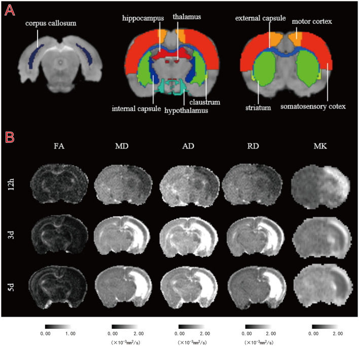
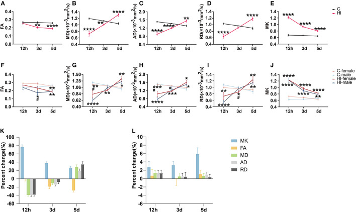
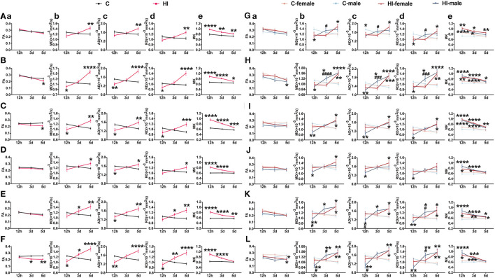
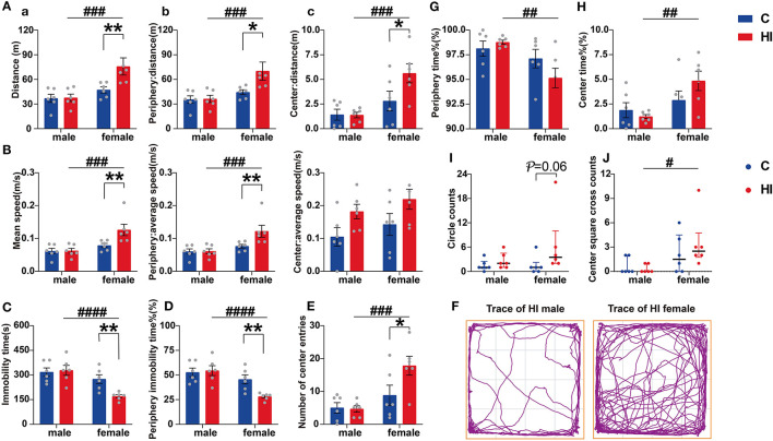
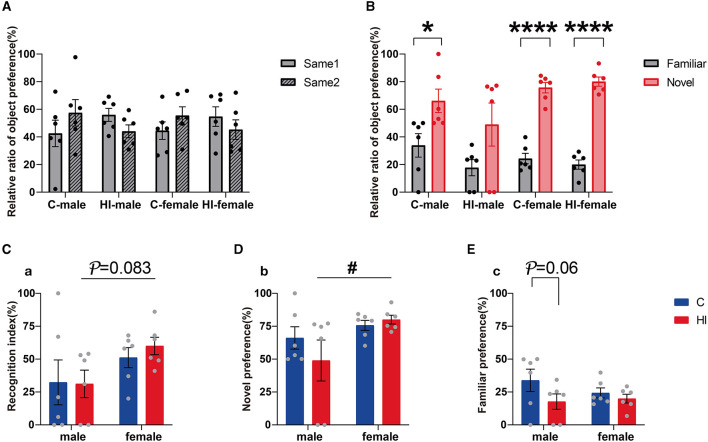
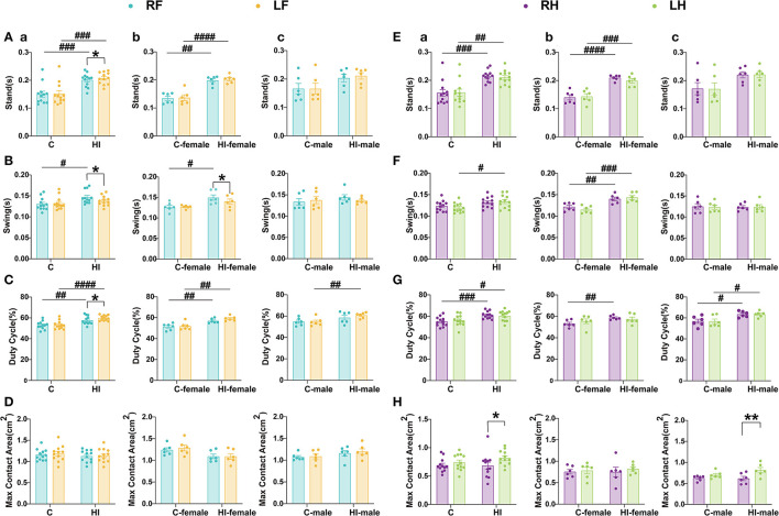
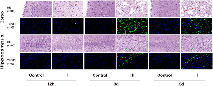
Objective: Hypoxic-ischemic encephalopathy can lead to lifelong morbidity and premature death in full-term newborns. Here, we aimed to determine the efficacy of diffusion kurtosis (DK) [mean kurtosis (MK)] and diffusion tensor (DT) [fractional anisotropy (FA), mean diffusion (MD), axial diffusion (AD), and radial diffusion (RD)] parameters for the early diagnosis of early brain histopathological changes and the prediction of neurodegenerative events in a full-term neonatal hypoxic-ischemic brain injury (HIBD) rat model. Methods: The HIBD model was generated in postnatal day 7 Sprague-Dawley rats to assess the changes in DK and DT parameters in 10 specific brain structural regions involving the gray matter, white matter, and limbic system during acute (12 h) and subacute (3 d and 5 d) phases after hypoxic ischemia (HI), which were validated against histology. Sensory and cognitive parameters were assessed by the open field, novel object recognition, elevated plus maze, and CatWalk tests. Results: Repeated-measures ANOVA revealed that specific brain structures showed similar trends to the lesion, and the temporal pattern of MK was substantially more varied than DT parameters, particularly in the deep gray matter. The change rate of MK in the acute phase (12 h) was significantly higher than that of DT parameters. We noted a delayed pseudo-normalization for MK. Additionally, MD, AD, and RD showed more pronounced differences between males and females after HI compared to MK, which was confirmed in behavioral tests. HI females exhibited anxiolytic hyperactivity-like baseline behavior, while the memory ability of HI males was affected in the novel object recognition test. CatWalk assessments revealed chronic deficits in limb gait parameters, particularly the left front paw and right hind paw, as well as poorer performance in HI males than HI females. Conclusions: Our results suggested that DK and DT parameters were complementary in the immature brain and provided great value in assessing early tissue microstructural changes and predicting long-term neurobehavioral deficits, highlighting their ability to detect both acute and long-term changes. Thus, the various diffusion coefficient parameters estimated by the DKI model are powerful tools for early HIBD diagnosis and prognosis assessment, thus providing an experimental and theoretical basis for clinical treatment.
Overview
The claims and statistics of this paper haven’t been checked yet.
Run the check — free, no account needed. It reads the open-access text and checks the sources it cites.
Integrity signals
Registry
Cites
Statistics
Figures
Verification
Claims the paper makes
The claims and statistics of this paper haven’t been checked yet.
Run the check — free, no account needed. It reads the open-access text and checks the sources it cites.
Claims other papers make about this one
Each is checked against this paper’s own text: does it say what the citing paper says it does?
In the citing paper
“The numbers of crossings and total distance traveled by HI-insult mice increased as previously reported [21–23], while miR-100-5p antagomir administration significantly decreased the crossing number and total distance traveled compared with the HI + NC group (Fig. 2A, B).”
From Engineered Extracellular Vesicles Loaded with MiR-100-5p Antagonist Selectively Target the Lesioned Region to Promote Recovery from Brain Damage (2025) · cites this paper as [22]
Self-check
Percentages, sums and reported differences re-computed from the paper’s own numbers.
The self-check needs the paper’s full text.
Citations
Cites
Estradiol and the Developing Brain
McCARTHY, M. M. (2008) · Physiological Reviews · doi:10.1152/physrev.00010.2007 · Cited by 885
Gliomas: diffusion kurtosis MR imaging in grading
Van Cauter, S., Veraart, J., Sijbers, J., Peeters, R. R., Himmelreich, U., De Keyzer, F., Van Gool, S. W., Van Calenbergh, F., De Vleeschouwer, S., Van Hecke, W., et al. (2012) · Radiology · doi:10.1148/radiol.12110927 · Cited by 340
Research works printed but not linked: 119
- Akamatsu, 2019, A pilot study of soluble form of LOX-1 as a novel biomarker for neonatal hypoxic-ischemic encephalopathy, J. Pediatr., 206, 49, 10.1016/j.jpeds.2018.10.036 · DOI
- Al Mamun, 2018, Inflammatory responses are sex specific in chronic hypoxic-ischemic encephalopathy, Cell Transplant., 27, 1328, 10.1177/0963689718766362 · DOI
- Almli, 2000, BDNF protects against spatial memory deficits following neonatal hypoxia-ischemia, Exp. Neurol., 166, 99, 10.1006/exnr.2000.7492 · DOI
- Aravamuthan, 2020, Sex may influence motor phenotype in a novel rodent model of cerebral palsy, Neurobiol. Dis., 134, 104711, 10.1016/j.nbd.2019.104711 · DOI
- Arteni, 2010, Lateralized and sex-dependent behavioral and morphological effects of unilateral neonatal cerebral hypoxia-ischemia in the rat, Behav. Brain Res., 210, 92, 10.1016/j.bbr.2010.02.015 · DOI
- Askalan, 2015, Mechanisms of neurodegeneration after severe hypoxic-ischemic injury in the neonatal rat brain, Brain Res., 1629, 94, 10.1016/j.brainres.2015.10.020 · DOI
- Awal, 2016, EEG background features that predict outcome in term neonates with hypoxic ischaemic encephalopathy: a structured review, Clin. Neurophysiol., 127, 285, 10.1016/j.clinph.2015.05.018 · DOI
- Azzopardi, 2009, Moderate hypothermia to treat perinatal asphyxial encephalopathy, N. Engl. J. Med., 361, 1349, 10.1056/NEJMoa0900854 · DOI
- Badawi, 2006, Autism following a history of newborn encephalopathy: more than a coincidence?, Dev. Med. Child Neurol., 48, 85, 10.1017/S001216220600020X · DOI
- Bai, 2021, Progressive microstructural alterations in subcortical nuclei in Parkinson's disease: a diffusion magnetic resonance imaging study, Parkinson. Relat. Disord., 88, 82, 10.1016/j.parkreldis.2021.06.003 · DOI
- Balduini, 2000, Long-lasting behavioral alterations following a hypoxic/ischemic brain injury in neonatal rats, Brain Res., 859, 318, 10.1016/S0006-8993(00)01997-1 · DOI
- Barkhuizen, 2019, Systemic multipotent adult progenitor cells improve long-term neurodevelopmental outcomes after preterm hypoxic-ischemic encephalopathy, Behav. Brain Res., 362, 77, 10.1016/j.bbr.2019.01.016 · DOI
- Barkovich, 1990, Brain damage from perinatal asphyxia: correlation of MR findings with gestational age, Am. J. Neuroradiol., 11, 1087
- Bazarian, 2007, Diffusion tensor imaging detects clinically important axonal damage after mild traumatic brain injury: a pilot study, J. Neurotrauma, 24, 1447, 10.1089/neu.2007.0241 · DOI · Paper page
- Black, 2010, Global, regional, and national causes of child mortality in 2008: a systematic analysis, Lancet, 375, 1969, 10.1016/S0140-6736(10)60549-1 · DOI
- Blockx, 2012, Identification and characterization of Huntington related pathology: an in vivo DKI imaging study, Neuroimage, 63, 653, 10.1016/j.neuroimage.2012.06.032 · DOI
- Bonnin, 2020, Cerebral vasodilator property of poly(ADP-ribose) polymerase inhibitor (PJ34) in the neonatal and adult mouse is mediated by the nitric oxide pathway, Int. J. Mol. Sci., 21, 10.3390/ijms21186569 · DOI
- Bonnin, 2021, Poly(ADP-Ribose) polymerase inhibitor PJ34 reduces brain damage after stroke in the neonatal mouse brain, Curr. Issues Mol. Biol., 43, 301, 10.3390/cimb43010025 · DOI
- Borjini, 2019, Potential biomarkers for neuroinflammation and neurodegeneration at short and long term after neonatal hypoxic-ischemic insult in rat, J. Neuroinflamm., 16, 194, 10.1186/s12974-019-1595-0 · DOI
- Bosemani, 2014, Susceptibility-weighted imaging in pediatric neuroimaging, J. Magn. Reson. Imaging, 40, 530, 10.1002/jmri.24410 · DOI
- Carwile, 2009, Estrogen and stroke: a review of the current literature, J. Neurosci. Nurs., 41, 18, 10.1097/JNN.0b013e31819345f8 · DOI
- Cercignani, 2001, Magnetisation transfer ratio and mean diffusivity of normal appearing white and grey matter from patients with multiple sclerosis, J. Neurol. Neurosurg. Psychiatr., 70, 311, 10.1136/jnnp.70.3.311 · DOI
- Charriaut-Marlangue, 2018, Sex differences in the effects of PARP inhibition on microglial phenotypes following neonatal stroke, Brain Behav. Immun., 73, 375, 10.1016/j.bbi.2018.05.022 · DOI
- Cheung, 2009, Does diffusion kurtosis imaging lead to better neural tissue characterization? A rodent brain maturation study, Neuroimage, 45, 386, 10.1016/j.neuroimage.2008.12.018 · DOI
- Chu, 2022, Comparison of brain microstructure alterations on diffusion kurtosis imaging among Alzheimer's disease, mild cognitive impairment, and cognitively normal individuals, Front. Aging Neurosci., 14, 919143, 10.3389/fnagi.2022.919143 · DOI
- Cserjesi, 2012, Functioning of 7-year-old children born at 32 to 35 weeks' gestational age, Pediatrics, 130, e838, 10.1542/peds.2011-2079 · DOI
- De Haan, 2006, Brain and cognitive-behavioural development after asphyxia at term birth, Dev. Sci., 9, 350, 10.1111/j.1467-7687.2006.00499.x · DOI
- De Vis, 2015, Arterial spin-labelling perfusion MRI and outcome in neonates with hypoxic-ischemic encephalopathy, Eur. Radiol., 25, 113, 10.1007/s00330-014-3352-1 · DOI
- DeLong, 1992, Autism, amnesia, hippocampus, and learning, Neurosci. Biobehav. Rev., 16, 63, 10.1016/S0149-7634(05)80052-1 · DOI
- DiBella, 2022, Beyond diffusion tensor MRI methods for improved characterization of the brain after ischemic stroke: a review, Am. J. Neuroradiol., 43, 661, 10.3174/ajnr.A7414 · DOI
- Dilenge, 2001, Long-term developmental outcome of asphyxiated term neonates, J. Child Neurol., 16, 781, 10.1177/08830738010160110201 · DOI
- Donders, 2002, Gender differences in learning and memory after pediatric traumatic brain injury, Neuropsychology, 16, 491, 10.1037/0894-4105.16.4.491 · DOI
- Douglas-Escobar, 2015, Hypoxic-ischemic encephalopathy: a review for the clinician, JAMA Pediatr., 169, 397, 10.1001/jamapediatrics.2014.3269 · DOI · Paper page
- East, 2010, Intrapartum fetal scalp lactate sampling for fetal assessment in the presence of a non-reassuring fetal heart rate trace, Cochrane Database Syst. Rev., 10.1002/14651858.CD006174.pub2 · DOI
- Falangola, 2008, Age-related non-Gaussian diffusion patterns in the prefrontal brain, J. Magn. Reson. Imaging, 28, 1345, 10.1002/jmri.21604 · DOI
- Fatemi, 2009, Hypoxic-ischemic encephalopathy in the term infant, Clin. Perinatol., 36, 835, 10.1016/j.clp.2009.07.011 · DOI
- Gadian, 2000, Developmental amnesia associated with early hypoxic-ischaemic injury, Brain, 123, 499, 10.1093/brain/123.3.499 · DOI
- Grinberg, 2017, Diffusion kurtosis metrics as biomarkers of microstructural development: a comparative study of a group of children and a group of adults, Neuroimage, 144, 12, 10.1016/j.neuroimage.2016.08.033 · DOI
- Guo, 2022, Diffusion abnormality in temporal lobe epilepsy patients with sleep disorders: a diffusion kurtosis imaging study, Front. Psychiatry, 13, 885477, 10.3389/fpsyt.2022.885477 · DOI
- Hagberg, 2015, The role of inflammation in perinatal brain injury, Nat. Rev. Neurol., 11, 192, 10.1038/nrneurol.2015.13 · DOI
- He, 2020, Diffusion kurtosis imaging of leptin intervention in early hypoxic-ischemic brain edema, Neuroscience, 431, 176, 10.1016/j.neuroscience.2020.02.009 · DOI
- Herrera, 2018, Outcomes of preterm infants treated with hypothermia for hypoxic-ischemic encephalopathy, Early Hum. Dev., 125, 1, 10.1016/j.earlhumdev.2018.08.003 · DOI
- Hill, 2011, Early testosterone modulated sex differences in behavioral outcome following neonatal hypoxia ischemia in rats, Int. J. Dev. Neurosci., 29, 381, 10.1016/j.ijdevneu.2011.03.005 · DOI
- Hindmarsh, 2000, Gender differences in cognitive abilities at 2 years in ELBW infants. Extremely low birth weight, Early Hum. Dev., 60, 115, 10.1016/S0378-3782(00)00105-5 · DOI
- Hu, 2008, “CSF partial volume effect for diffusion kurtosis imaging,”
- Huang, 2016, Sex differences in brain MRI abnormalities and neurodevelopmental outcomes in a rat model of neonatal hypoxia-ischemia, Int. J. Neurosci., 126, 647, 10.3109/00207454.2015.1047016 · DOI
- Huang, 2020, Impact of hypoxic-ischemic injury on brain development in neonatal rats of different sexes, Zhonghua Er Ke Za Zhi, 58, 30, 10.3760/cma.j.issn.0578-1310.2020.01.008 · DOI
- Hui, 2008, Towards better MR characterization of neural tissues using directional diffusion kurtosis analysis, Neuroimage, 42, 122, 10.1016/j.neuroimage.2008.04.237 · DOI
- Hui, Spatiotemporal dynamics of diffusional kurtosis, mean diffusivity and perfusion changes in experimental stroke, Brain Res., 1451, 100, 10.1016/j.brainres.2012.02.044 · DOI
- Hui, Stroke assessment with diffusional kurtosis imaging, Stroke, 43, 2968, 10.1161/STROKEAHA.112.657742 · DOI
- Immonen, 2009, Distinct MRI pattern in lesional and perilesional area after traumatic brain injury in rat−11 months follow-up, Exp. Neurol., 215, 29, 10.1016/j.expneurol.2008.09.009 · DOI
- Jensen, 2002, The role of glutamate receptor maturation in perinatal seizures and brain injury, Int. J. Dev. Neurosci., 20, 339, 10.1016/S0736-5748(02)00012-6 · DOI
- Jensen, 2010, MRI quantification of non-Gaussian water diffusion by kurtosis analysis, NMR Biomed., 23, 698, 10.1002/nbm.1518 · DOI · Paper page
- Jensen, 2005, Diffusional kurtosis imaging: the quantification of non-gaussian water diffusion by means of magnetic resonance imaging, Magn. Reson. Med., 53, 1432, 10.1002/mrm.20508 · DOI · Paper page
- Jeong, 2022, Neonatal encephalopathy prediction of poor outcome with diffusion-weighted imaging connectome and fixel-based analysis, Pediatr. Res., 91, 1505, 10.1038/s41390-021-01550-2 · DOI
- Johnston, 2007, Sex and the pathogenesis of cerebral palsy, Dev. Med. Child Neurol., 49, 74, 10.1017/S0012162207000199.x · DOI
- Kent, 2012, Mortality and adverse neurologic outcomes are greater in preterm male infants, Pediatrics, 129, 124, 10.1542/peds.2011-1578 · DOI
- Kitamura, 2011, Hypoxic-ischemic injury: utility of susceptibility-weighted imaging, Pediatr. Neurol., 45, 220, 10.1016/j.pediatrneurol.2011.06.009 · DOI
- Kurinczuk, 2010, Epidemiology of neonatal encephalopathy and hypoxic-ischaemic encephalopathy, Early Hum. Dev., 86, 329, 10.1016/j.earlhumdev.2010.05.010 · DOI
- Lally, 2019, Magnetic resonance spectroscopy assessment of brain injury after moderate hypothermia in neonatal encephalopathy: a prospective multicentre cohort study, Lancet Neurol., 18, 35, 10.1016/S1474-4422(18)30325-9 · DOI
- Lampinen, 2021, Time dependence in diffusion MRI predicts tissue outcome in ischemic stroke patients, Magn. Reson. Med., 86, 754, 10.1002/mrm.28743 · DOI
- Lawn, 2010, 3.6 million neonatal deaths–what is progressing and what is not?, Semin. Perinatol., 34, 371, 10.1053/j.semperi.2010.09.011 · DOI
- Leversen, 2011, Prediction of neurodevelopmental and sensory outcome at 5 years in Norwegian children born extremely preterm, Pediatrics, 127, e630, 10.1542/peds.2010-1001 · DOI
- Li, 2017, Brain-immune interactions in perinatal hypoxic-ischemic brain injury, Prog. Neurobiol., 159, 50, 10.1016/j.pneurobio.2017.10.006 · DOI
- Li, 2019, Evaluation of diffusion kurtosis imaging in stratification of nonalcoholic fatty liver disease and early diagnosis of nonalcoholic steatohepatitis in a rabbit model, Magn. Reson. Imaging, 63, 267, 10.1016/j.mri.2019.08.032 · DOI
- Lou, 1996, Etiology and pathogenesis of attention-deficit hyperactivity disorder (ADHD): significance of prematurity and perinatal hypoxic-haemodynamic encephalopathy, Acta Paediatr., 85, 1266, 10.1111/j.1651-2227.1996.tb13909.x · DOI
- Lubics, 2005, Neurological reflexes and early motor behavior in rats subjected to neonatal hypoxic-ischemic injury, Behav. Brain Res., 157, 157, 10.1016/j.bbr.2004.06.019 · DOI
- Ma, 2022, Evaluation of crossed cerebellar diaschisis after cerebral infarction in MCAO rats based on DKI, Eur. J. Clin. Invest., 52, e13716, 10.1111/eci.13716 · DOI
- Mac Donald, 2007, Diffusion tensor imaging reliably detects experimental traumatic axonal injury and indicates approximate time of injury, J. Neurosci., 27, 11869, 10.1523/JNEUROSCI.3647-07.2007 · DOI
- Mañeru, 2003, Residual hippocampal atrophy in asphyxiated term neonates, J. Neuroimaging, 13, 68, 10.1111/j.1552-6569.2003.tb00159.x · DOI
- Marlow, 2005, Neuropsychological and educational problems at school age associated with neonatal encephalopathy, Arch. Dis. Child. Fetal Neonatal Ed., 90, F380, 10.1136/adc.2004.067520 · DOI
- McAuliffe, 2006, Adult neurological function following neonatal hypoxia-ischemia in a mouse model of the term neonate: water maze performance is dependent on separable cognitive and motor components, Brain Res., 1118, 208, 10.1016/j.brainres.2006.08.030 · DOI
- McKenna, 2020, Diffusion kurtosis imaging of gray matter in young adults with autism spectrum disorder, Sci. Rep., 10, 21465, 10.1038/s41598-020-78486-w · DOI
- McKenna, 2019, Diffusion kurtosis imaging of gray matter in schizophrenia, Cortex, 121, 201, 10.1016/j.cortex.2019.08.013 · DOI
- Moster, 2002, Joint association of Apgar scores and early neonatal symptoms with minor disabilities at school age, Arch. Dis. Child. Fetal Neonatal Ed., 86, F16, 10.1136/fn.86.1.F16 · DOI
- Muntsant, 2019, Severe perinatal hypoxic-ischemic brain injury induces long-term sensorimotor deficits, anxiety-like behaviors and cognitive impairment in a sex-, age- and task-selective manner in C57BL/6 mice but can be modulated by neonatal handling, Front. Behav. Neurosci., 13, 7, 10.3389/fnbeh.2019.00007 · DOI · Paper page
- Nematipour, 2020, Estrogen and progesterone attenuate glutamate neurotoxicity via regulation of EAAT3 and GLT-1 in a rat model of ischemic stroke, Iran. J. Basic Med. Sci., 23, 1346, 10.22038/ijbms.2020.48090.11039 · DOI
- Netto, 2017, Sex-dependent consequences of neonatal brain hypoxia-ischemia in the rat, J. Neurosci. Res., 95, 409, 10.1002/jnr.23828 · DOI
- Niogi, 2008, Extent of microstructural white matter injury in postconcussive syndrome correlates with impaired cognitive reaction time: a 3T diffusion tensor imaging study of mild traumatic brain injury, Am. J. Neuroradiol., 29, 967, 10.3174/ajnr.A0970 · DOI
- Pereira, 2008, Early enriched housing results in partial recovery of memory deficits in female, but not in male, rats after neonatal hypoxia-ischemia, Brain Res., 1218, 257, 10.1016/j.brainres.2008.04.010 · DOI
- Raab, 2010, Cerebral gliomas: diffusional kurtosis imaging analysis of microstructural differences, Radiology, 254, 876, 10.1148/radiol.09090819 · DOI · Paper page
- Rademakers, 1995, Central cortico-subcortical involvement: a distinct pattern of brain damage caused by perinatal and postnatal asphyxia in term infants, J. Comput. Assist. Tomogr., 19, 256, 10.1097/00004728-199503000-00017 · DOI
- Raz, 2013, A better characterization of spinal cord damage in multiple sclerosis: a diffusional kurtosis imaging study, Am. J. Neuroradiol., 34, 1846, 10.3174/ajnr.A3512 · DOI
- Renolleau, 2008, Gender-related differences in apoptotic pathways after neonatal cerebral ischemia, Neuroscientist, 14, 46, 10.1177/1073858407308889 · DOI
- Renolleau, 2007, Specific caspase inhibitor Q-VD-OPh prevents neonatal stroke in P7 rat: a role for gender, J. Neurochem., 100, 1062, 10.1111/j.1471-4159.2006.04269.x · DOI
- Rice III, 1981, The influence of immaturity on hypoxic-ischemic brain damage in the rat, Ann. Neurol., 9, 131, 10.1002/ana.410090206 · DOI
- Robertson, 1999, MR line-scan diffusion-weighted imaging of term neonates with perinatal brain ischemia, Am. J. Neuroradiol., 20, 1658
- Sanches, 2015, Sexual dimorphism and brain lateralization impact behavioral and histological outcomes following hypoxia-ischemia in P3 and P7 rats, Neuroscience, 290, 581, 10.1016/j.neuroscience.2014.12.074 · DOI
- Sanches, 2019, Mild neonatal brain hypoxia-ischemia in very immature rats causes long-term behavioral and cerebellar abnormalities at adulthood, Front. Physiol., 10, 634, 10.3389/fphys.2019.00634 · DOI
- Shankaran, 2009, Neonatal encephalopathy: treatment with hypothermia, J. Neurotrauma, 26, 437, 10.1089/neu.2008.0678 · DOI
- Shen, 1991, The behavioral and anatomical effects of prenatal umbilical cord clamping in the rat and their alteration by the prior maternal administration of nimodipine, Restor. Neurol. Neurosci., 3, 11, 10.3233/RNN-1991-3102 · DOI
- Shi, 2019, Detecting normal pediatric brain development with diffusional kurtosis imaging, Eur. J. Radiol., 120, 108690, 10.1016/j.ejrad.2019.108690 · DOI
- Shibasaki, 2021, Comparison of predictive values of magnetic resonance biomarkers based on scan timing in neonatal encephalopathy following therapeutic hypothermia, J. Pediatr., 239, 101, 10.1016/j.jpeds.2021.08.011 · DOI
- Simon, 2023, “APGAR score,”
- Simon, 1999, Long-term neurodevelopmental outcome of asphyxiated newborns, Clin. Perinatol., 26, 767, 10.1016/S0095-5108(18)30050-2 · DOI
- Spampinato, 2017, Diffusional kurtosis imaging and motor outcome in acute ischemic stroke, Am. J. Neuroradiol., 38, 1328, 10.3174/ajnr.A5180 · DOI
- Tabacaru, 2023, Diffusion tensor imaging to predict neurodevelopmental impairment in infants after hypoxic-ischemic injury, Am. J. Perinatol, 10.1055/a-2071-3057 · DOI
- Toft, 1999, Prenatal and perinatal striatal injury: a hypothetical cause of attention-deficit-hyperactivity disorder?, Pediatr. Neurol., 21, 602, 10.1016/S0887-8994(99)00046-6 · DOI
- Tsuji, 2010, Sex differences in the benefits of rehabilitative training during adolescence following neonatal hypoxia-ischemia in rats, Exp. Neurol., 226, 285, 10.1016/j.expneurol.2010.09.002 · DOI
- Tusor, 2012, Prediction of neurodevelopmental outcome after hypoxic-ischemic encephalopathy treated with hypothermia by diffusion tensor imaging analyzed using tract-based spatial statistics, Pediatr. Res., 72, 63, 10.1038/pr.2012.40 · DOI
- Umesh Rudrapatna, 2014, Can diffusion kurtosis imaging improve the sensitivity and specificity of detecting microstructural alterations in brain tissue chronically after experimental stroke? Comparisons with diffusion tensor imaging and histology, Neuroimage, 97, 363, 10.1016/j.neuroimage.2014.04.013 · DOI
- van de Looij, 2011, Longitudinal MR assessment of hypoxic ischemic injury in the immature rat brain, Magn. Reson. Med., 65, 305, 10.1002/mrm.22617 · DOI
- Veraart, 2011, More accurate estimation of diffusion tensor parameters using diffusion Kurtosis imaging, Magn. Reson. Med., 65, 138, 10.1002/mrm.22603 · DOI
- Vexler, 2001, Molecular and biochemical mechanisms of perinatal brain injury, Semin. Neonatol., 6, 99, 10.1053/siny.2001.0041 · DOI
- Villa, 2016, Estrogens, neuroinflammation, and neurodegeneration, Endocr. Rev., 37, 372, 10.1210/er.2016-1007 · DOI · Paper page
- Volpe, 2009, Brain injury in premature infants: a complex amalgam of destructive and developmental disturbances, Lancet Neurol., 8, 110, 10.1016/S1474-4422(08)70294-1 · DOI
- Waddell, 2016, Sex differences in cell genesis, hippocampal volume and behavioral outcomes in a rat model of neonatal HI, Exp. Neurol., 275, 285, 10.1016/j.expneurol.2015.09.003 · DOI
- Wagner, 2002, Delayed postischemic hypothermia improves long-term behavioral outcome after cerebral hypoxia-ischemia in neonatal rats, Pediatr. Res., 51, 354, 10.1203/00006450-200203000-00015 · DOI
- Wang, 2018, Longitudinal microstructural changes in traumatic brain injury in rats: a diffusional kurtosis imaging, histology, and behavior study, Am. J. Neuroradiol., 39, 1650, 10.3174/ajnr.A5737 · DOI
- Wiberg-Itzel, 2008, Determination of pH or lactate in fetal scalp blood in management of intrapartum fetal distress: randomised controlled multicentre trial, BMJ, 336, 1284, 10.1136/bmj.39553.406991.25 · DOI
- Wieshmann, 1999, Anisotropy of water diffusion in corona radiata and cerebral peduncle in patients with hemiparesis, Neuroimage, 10, 225, 10.1006/nimg.1999.0456 · DOI
- Wintermark, 2011, Brain perfusion in asphyxiated newborns treated with therapeutic hypothermia, Am. J. Neuroradiol., 32, 2023, 10.3174/ajnr.A2708 · DOI
- Xiao, 2020, Diffusion kurtosis imaging and pathological comparison of early hypoxic-ischemic brain damage in newborn piglets, Sci. Rep., 10, 17242, 10.1038/s41598-020-74387-0 · DOI
- Yang, 2013, Effect of cerebral spinal fluid suppression for diffusional kurtosis imaging, J. Magn. Reson. Imaging, 37, 365, 10.1002/jmri.23840 · DOI
- Yin, 2018, Diffusion kurtosis imaging of acute infarction: comparison with routine diffusion and follow-up MR imaging, Radiology, 287, 651, 10.1148/radiol.2017170553 · DOI
- Zheng, 2015, 17β-Estradiol attenuates hematoma expansion through estrogen receptor α/silent information regulator 1/nuclear factor-kappa b pathway in hyperglycemic intracerebral hemorrhage mice, Stroke, 46, 485, 10.1161/STROKEAHA.114.006372 · DOI
- Zhu, 2006, Different apoptotic mechanisms are activated in male and female brains after neonatal hypoxia-ischaemia, J. Neurochem., 96, 1016, 10.1111/j.1471-4159.2005.03639.x · DOI
- Zhu, 2019, Diffusion kurtosis imaging of microstructural changes in brain tissue affected by acute ischemic stroke in different locations, Neural Regen. Res., 14, 272, 10.4103/1673-5374.244791 · DOI
- Zhu, 2008, Proton magnetic resonance spectroscopy in neonates with hypoxic-ischemic injury and its prognostic value, Transl. Res., 152, 225, 10.1016/j.trsl.2008.09.004 · DOI
Cited by
Sorted by citations
Cheng, Y., Gai, C., Zhao, Y., Li, T., Song, Y., Luo, Q., Xin, D., Jiang, Z., Chen, W., Liu, D., et al. (2025) · Neuroscience Bulletin · doi:10.1007/s12264-025-01376-6 · Cited by 5
Metadata
- Authors
- Bao, J., Zhang, X., & Zhao, X.
- Venue
- Frontiers in Neuroscience
- Publisher
- Frontiers Media SA
- Published
- 2023-09-15
- Volume
- 17
- ISSN
- 1662-453X
- PMC
- PMC10543095
- Cited by
- 6
- References
- 2
About these checks
Registry, retraction, Cites and statistics results are computed, not judged, with one exception: a printed reference that no index lists is read once by a model before it counts as a citation error. Claims are extracted by a model and kept only when their quote appears verbatim in the text; each cited source is then checked, with a model’s help, for whether it says what the claim relies on. Those verdicts can be wrong — read the claim’s quote and the cited source before relying on one.
Statistics and arithmetic are recomputed, not judged.
Confidence bands are shown only for fields calibrated on our annotated test set. None is calibrated yet, so no band is shown.
What counts as a claim ›How we measure confidence ›How we check this ›
Are you an author, or do you see a problem with this page? Request a review
How to cite
Cite CoChat for the checks on this page, and the paper for its own findings.
- This page
- CoChat. (2026). Citation check: MR imaging and outcome in neonatal HIBD models are correlated with sex: the value of diffusion tensor MR imaging and diffusion kurtosis MR imaging. https://app.cochat.ai/papers/10.3389/fnins.2023.1234049
- The paper
- Bao, J., Zhang, X., & Zhao, X. (2023). MR imaging and outcome in neonatal HIBD models are correlated with sex: the value of diffusion tensor MR imaging and diffusion kurtosis MR imaging. Frontiers in Neuroscience. https://doi.org/10.3389/fnins.2023.1234049
