

The DOI matches its Crossref record; 15 of the 15 references it cites were found in a registry or an index.
The effects of enhancing angiotensin converting enzyme in myelomonocytes on ameliorating Alzheimer’s-related disease and preserving cognition
Abstract
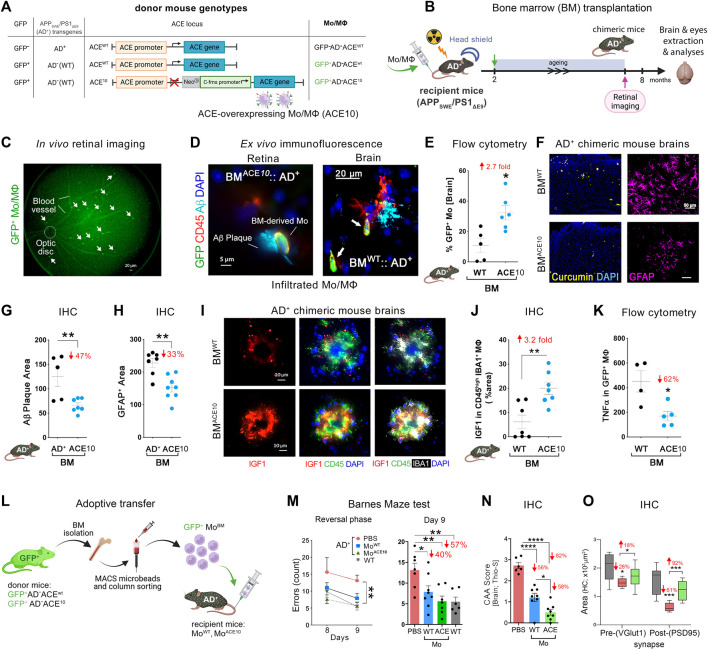
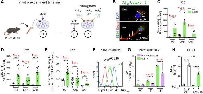
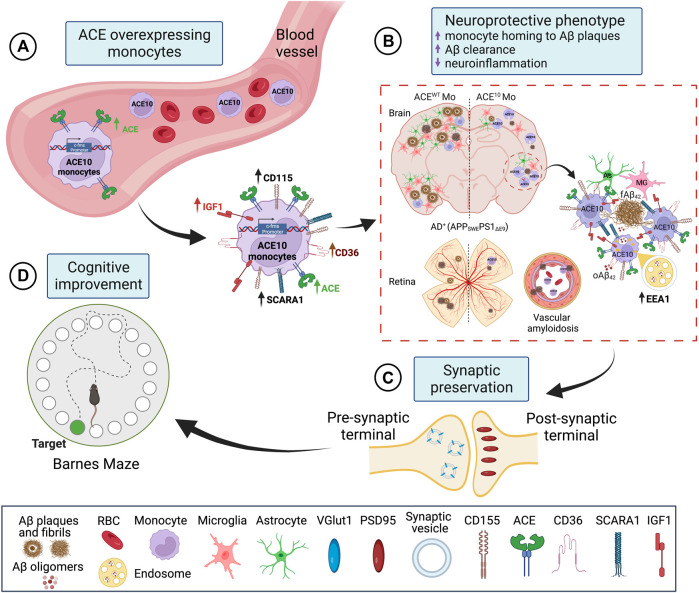
This review examines the role of angiotensin-converting enzyme (ACE) in the context of Alzheimer’s disease (AD) and its potential therapeutic value. ACE is known to degrade the neurotoxic 42-residue long alloform of amyloid β-protein (Aβ 42), a peptide strongly associated with AD. Previous studies in mice, demonstrated that targeted overexpression of ACE in CD115+ myelomonocytic cells (ACE10 models) improved their immune responses to effectively reduce viral and bacterial infection, tumor growth, and atherosclerotic plaque. We further demonstrated that introducing ACE10 myelomonocytes (microglia and peripheral monocytes) into the double transgenic APP SWE/PS1 ΔE9 murine model of AD (AD+ mice), diminished neuropathology and enhanced the cognitive functions. These beneficial effects were dependent on ACE catalytic activity and vanished when ACE was pharmacologically blocked. Moreover, we revealed that the therapeutic effects in AD+ mice can be achieved by enhancing ACE expression in bone marrow (BM)-derived CD115+ monocytes alone, without targeting central nervous system (CNS) resident microglia. Following blood enrichment with CD115+ ACE10-monocytes versus wild-type (WT) monocytes, AD+ mice had reduced cerebral vascular and parenchymal Aβ burden, limited microgliosis and astrogliosis, as well as improved synaptic and cognitive preservation. CD115+ ACE10-versus WT-monocyte-derived macrophages (Mo/MΦ) were recruited in higher numbers to the brains of AD+ mice, homing to Aβ plaque lesions and exhibiting a highly Aβ-phagocytic and anti-inflammatory phenotype (reduced TNFα/iNOS and increased MMP-9/IGF-1). Moreover, BM-derived ACE10-Mo/MΦ cultures had enhanced capability to phagocytose Aβ 42 fibrils, prion-rod-like, and soluble oligomeric forms that was associated with elongated cell morphology and expression of surface scavenger receptors (i.e., CD36, Scara-1). This review explores the emerging evidence behind the role of ACE in AD, the neuroprotective properties of monocytes overexpressing ACE and the therapeutic potential for exploiting this natural mechanism for ameliorating AD pathogenesis.
Overview
The claims and statistics of this paper haven’t been checked yet.
Run the check — free, no account needed. It reads the open-access text and checks the sources it cites.
Integrity signals
Registry
Cites
Statistics
Figures
Verification
Claims the paper makes
The claims and statistics of this paper haven’t been checked yet.
Run the check — free, no account needed. It reads the open-access text and checks the sources it cites.
Claims other papers make about this one
Each is checked against this paper’s own text: does it say what the citing paper says it does?
The cited source says what the claim attributes to it.
In the citing paper
“Analysis of the risk genes with the highest expression in our iMGs showed that the majority of the genes significantly downregulated by miR-132 (MS4A6A, INPP5D, ACE, CR1) are either increased in the human AD brain or their high expression levels were associated with increased risk of AD [158–161].”
From microRNA-132 attenuates inflammation in induced pluripotent stem cell-derived microglia from Alzheimer’s disease patients (2026) · cites this paper as [160]
Self-check
Percentages, sums and reported differences re-computed from the paper’s own numbers.
The self-check needs the paper’s full text.
Citations
Cites
Neuropathological stageing of Alzheimer-related changes.
Braak, H., & Braak, E. (1991) · Acta Neuropathologica · doi:10.1007/bf00308809 · Cited by 16,123
Neurotoxic reactive astrocytes are induced by activated microglia
Liddelow, S. A., Guttenplan, K. A., Clarke, L. E., Bennett, F. C., Bohlen, C. J., Schirmer, L., Bennett, M. L., Münch, A. E., Chung, W.-S., Peterson, T. C., et al. (2017) · Nature · doi:10.1038/nature21029 · Cited by 7,971
Donanemab in Early Alzheimer’s Disease
Mintun, M. A., Lo, A. C., Duggan Evans, C., Wessels, A. M., Ardayfio, P. A., Andersen, S. W., Shcherbinin, S., Sparks, J., Sims, J. R., Brys, M., et al. (2021) · New England Journal of Medicine · doi:10.1056/nejmoa2100708 · Cited by 1,728 · cited 1×
“Given the injurious role that soluble Aβ42 oligomers play in AD pathophysiology, there has been considerable effort to target and remove them as a strategy to prevent disease progression (Viola and Klein, 2015; Honig et al., 2018; Alexander et al., 2021; Mintun et al., 2021).”
BackgroundMicroglial Dysfunction and Defective β-Amyloid Clearance Pathways in Aging Alzheimer's Disease Mice
Hickman, S. E., Allison, E. K., & El Khoury, J. (2008) · The Journal of Neuroscience · doi:10.1523/jneurosci.0616-08.2008 · Cited by 1,329 · cited 1×
“In previous studies, these receptors have been associated with increased Aβ uptake (Hickman et al., 2008; Frenkel et al., 2013; Kim et al., 2017).”
BackgroundInflammation in Neurodegenerative Disease—A Double-Edged Sword
Wyss-Coray, T., & Mucke, L. (2002) · Neuron · doi:10.1016/s0896-6273(02)00794-8 · Cited by 1,235
Trial of Solanezumab for Mild Dementia Due to Alzheimer’s Disease
Honig, L. S., Vellas, B., Woodward, M., Boada, M., Bullock, R., Borrie, M., Hager, K., Andreasen, N., Scarpini, E., Liu-Seifert, H., et al. (2018) · New England Journal of Medicine · doi:10.1056/nejmoa1705971 · Cited by 1,049
Amyloid β oligomers in Alzheimer’s disease pathogenesis, treatment, and diagnosis
Viola, K. L., & Klein, W. L. (2015) · Acta Neuropathologica · doi:10.1007/s00401-015-1386-3 · Cited by 628
Coraci, I. S., Husemann, J., Berman, J. W., Hulette, C., Dufour, J. H., Campanella, G. K., Luster, A. D., Silverstein, S. C., & El Khoury, J. B. (2002) · The American Journal of Pathology · doi:10.1016/s0002-9440(10)64354-4 · Cited by 398
Real-Time In Vivo Imaging Reveals the Ability of Monocytes to Clear Vascular Amyloid Beta
Michaud, J.-P., Bellavance, M.-A., Préfontaine, P., & Rivest, S. (2013) · Cell Reports · doi:10.1016/j.celrep.2013.10.010 · Cited by 257
Frenkel, D., Wilkinson, K., Zhao, L., Hickman, S. E., Means, T. K., Puckett, L., Farfara, D., Kingery, N. D., Weiner, H. L., & El Khoury, J. (2013) · Nature Communications · doi:10.1038/ncomms3030 · Cited by 214
Research works printed but not linked: 78
- Arjunan, 2023, Identification of the molecular mechanism of insulin-like growth factor-1 (IGF-1): A promising therapeutic target for neurodegenerative diseases associated with metabolic syndrome, Cell. Biosci., 13, 16, 10.1186/s13578-023-00966-z · DOI
- Baudin, 2002, New aspects on angiotensin-converting enzyme: From gene to disease, Clin. Chem. Lab. Med., 40, 256, 10.1515/CCLM.2002.042 · DOI
- Bernstein, 2018, Angiotensin-converting enzyme in innate and adaptive immunity, Nat. Rev. Nephrol., 14, 325, 10.1038/nrneph.2018.15 · DOI
- Bernstein, 2014, Angiotensin-converting enzyme overexpression in myelomonocytes prevents Alzheimer's-like cognitive decline, J. Clin. Invest., 124, 1000, 10.1172/JCI66541 · DOI
- Bernstein, 2013, A modern understanding of the traditional and nontraditional biological functions of angiotensin-converting enzyme, Pharmacol. Rev., 65, 1, 10.1124/pr.112.006809 · DOI
- Bertram, 2007, Systematic meta-analyses of alzheimer disease genetic association studies: The AlzGene database, Nat. Genet., 39, 17, 10.1038/ng1934 · DOI · Paper page
- Brice, 1995, Serum angiotensin-converting enzyme activity, concentration, and specific activity in granulomatous interstitial lung disease, tuberculosis, and COPD, Chest, 107, 706, 10.1378/chest.107.3.706 · DOI
- Brody, 2018, Synaptotoxic signaling by amyloid beta oligomers in Alzheimer’s disease through prion protein and mGluR5, Adv Pharmacol., 82, 293, 10.1016/bs.apha.2017.09.007 · DOI
- Butovsky, 2006, Glatiramer acetate fights against Alzheimer's disease by inducing dendritic-like microglia expressing insulin-like growth factor 1, Proc. Natl. Acad. Sci. U. S. A., 103, 11784, 10.1073/pnas.0604681103 · DOI · Paper page
- Butovsky, 2007, Selective ablation of bone marrow-derived dendritic cells increases amyloid plaques in a mouse Alzheimer's disease model, Eur. J. Neurosci., 26, 413, 10.1111/j.1460-9568.2007.05652.x · DOI
- Cao, 2023, Macrophage angiotensin-converting enzyme (ACE) reduces atherosclerosis by increasing PPARα and fundamentally changing lipid metabolism, Cardiovasc Res, 10.1093/cvr/cvad082 · DOI
- Cao, Role of angiotensin-converting enzyme in myeloid cell immune responses, Cell. Mol. Biol. Lett., 25, 31, 10.1186/s11658-020-00225-w · DOI
- Cao, ACE overexpression in myeloid cells increases oxidative metabolism and cellular ATP, J. Biol. Chem., 295, 1369, 10.1074/jbc.RA119.011244 · DOI
- Cenini, 2010, Generation of reactive oxygen species by beta amyloid fibrils and oligomers involves different intra/extracellular pathways, Amino Acids, 38, 1101, 10.1007/s00726-009-0319-7 · DOI
- Chang, 2017, Tumor necrosis factor alpha Inhibition for Alzheimer's Disease, J. Cent. Nerv. Syst. Dis., 9, 9278, 10.1177/1179573517709278 · DOI · Paper page
- Charidimou, 2017, Emerging concepts in sporadic cerebral amyloid angiopathy, Brain, 140, 1829, 10.1093/brain/awx047 · DOI · Paper page
- Cunningham, 2013, Microglia and neurodegeneration: The role of systemic inflammation, Glia, 61, 71, 10.1002/glia.22350 · DOI · Paper page
- Deane, 2009, Clearance of amyloid-beta peptide across the blood-brain barrier: Implication for therapies in Alzheimer's disease, CNS Neurol. Disord. Drug Targets, 8, 16, 10.2174/187152709787601867 · DOI
- Diet, 1996, Increased accumulation of tissue ACE in human atherosclerotic coronary artery disease, Circulation, 94, 2756, 10.1161/01.cir.94.11.2756 · DOI
- Doustar, 2020, Parallels between retinal and brain pathology and response to immunotherapy in old, late-stage Alzheimer's disease mouse models, Aging Cell., 19, 13246, 10.1111/acel.13246 · DOI
- Dvir-Szternfeld, 2022, Alzheimer's disease modification mediated by bone marrow-derived macrophages via a TREM2-independent pathway in mouse model of amyloidosis, Nat. Aging, 2, 60, 10.1038/s43587-021-00149-w · DOI
- George, 2017, The Alzheimer's disease transcriptome mimics the neuroprotective signature of IGF-1 receptor-deficient neurons, Brain, 140, 2012, 10.1093/brain/awx132 · DOI
- Grober, 1999, Memory and mental status correlates of modified Braak staging, Neurobiol. Aging, 20, 573, 10.1016/s0197-4580(99)00063-9 · DOI
- Han, 2017, Amyloid beta-42 induces neuronal apoptosis by targeting mitochondria, Mol. Med. Rep., 16, 4521, 10.3892/mmr.2017.7203 · DOI
- Hardy, 2002, The amyloid hypothesis of Alzheimer's disease: Progress and problems on the road to therapeutics, Science, 297, 353, 10.1126/science.1072994 · DOI · Paper page
- He, 2006, ACE activity in CSF of patients with mild cognitive impairment and Alzheimer disease, Neurology, 67, 1309, 10.1212/01.wnl.0000238102.04582.ec · DOI
- Hemming, 2005, Amyloid beta-protein is degraded by cellular angiotensin-converting enzyme (ACE) and elevated by an ACE inhibitor, J. Biol. Chem., 280, 37644, 10.1074/jbc.M508460200 · DOI
- Jochemsen, 2014, The association of angiotensin-converting enzyme with biomarkers for Alzheimer's disease, Alzheimers Res. Ther., 6, 27, 10.1186/alzrt257 · DOI · Paper page
- Kanemitsu, 2003, Human neprilysin is capable of degrading amyloid beta peptide not only in the monomeric form but also the pathological oligomeric form, Neurosci. Lett., 350, 113, 10.1016/s0304-3940(03)00898-x · DOI
- Kehoe, 2019, Cerebrospinal fluid changes in the renin-angiotensin system in Alzheimer's disease, J. Alzheimers Dis., 72, 525, 10.3233/JAD-190721 · DOI
- Kehoe, 2009, Angiotensins in Alzheimer's disease - friend or foe?, Trends Neurosci., 32, 619, 10.1016/j.tins.2009.07.006 · DOI
- Keren-Shaul, 2017, A unique microglia type associated with restricting development of Alzheimer's disease, Cell., 169, 1276, 10.1016/j.cell.2017.05.018 · DOI · Paper page
- Koronyo, 2017, Retinal amyloid pathology and proof-of-concept imaging trial in Alzheimer's disease, JCI Insight, 2, 93621, 10.1172/jci.insight.93621 · DOI
- Koronyo, 2023, Retinal pathological features and proteome signatures of Alzheimer's disease, Acta Neuropathol., 145, 409, 10.1007/s00401-023-02548-2 · DOI
- Koronyo-Hamaoui, 2009, Attenuation of AD-like neuropathology by harnessing peripheral immune cells: Local elevation of IL-10 and MMP-9, J. Neurochem., 111, 1409, 10.1111/j.1471-4159.2009.06402.x · DOI
- Koronyo-Hamaoui, 2011, Identification of amyloid plaques in retinas from Alzheimer's patients and noninvasive in vivo optical imaging of retinal plaques in a mouse model, Neuroimage, 54, S204, 10.1016/j.neuroimage.2010.06.020 · DOI · Paper page
- Koronyo-Hamaoui, 2014, ACE overexpression in myelomonocytic cells: effect on a mouse model of Alzheimer’s disease, Curr. Hypertens. Rep., 16, 444, 10.1007/s11906-014-0444-x · DOI
- Koronyo-Hamaoui, 2020, Peripherally derived angiotensin converting enzyme-enhanced macrophages alleviate Alzheimer-related disease, Brain, 143, 336, 10.1093/brain/awz364 · DOI
- Krasemann, 2017, The TREM2-APOE pathway drives the transcriptional phenotype of dysfunctional microglia in neurodegenerative diseases, Immunity, 47, 566, 10.1016/j.immuni.2017.08.008 · DOI · Paper page
- La Morgia, 2016, Melanopsin retinal ganglion cell loss in Alzheimer disease, Ann. Neurol., 79, 90, 10.1002/ana.24548 · DOI
- Lebson, 2010, Trafficking CD11b-positive blood cells deliver therapeutic genes to the brain of amyloid-depositing transgenic mice, J. Neurosci., 30, 9651, 10.1523/JNEUROSCI.0329-10.2010 · DOI
- Li, 2020, Activated bone marrow-derived macrophages eradicate alzheimer's-related Aβ42 oligomers and protect synapses, Front. Immunol., 11, 49, 10.3389/fimmu.2020.00049 · DOI
- Lieberman, 1975, Elevation of serum angiotensin-converting-enzyme (ACE) level in sarcoidosis, Am. J. Med., 59, 365, 10.1016/0002-9343(75)90395-2 · DOI
- Marioni, 2019, Correction: GWAS on family history of Alzheimer's disease, Transl. Psychiatry, 9, 161, 10.1038/s41398-019-0498-2 · DOI
- Marques, 2009, Peripheral amyloid-beta levels regulate amyloid-beta clearance from the central nervous system, J. Alzheimers Dis., 16, 325, 10.3233/JAD-2009-0964 · DOI
- Mateos, 2011, Upregulation of brain renin angiotensin system by 27-hydroxycholesterol in Alzheimer's disease, J. Alzheimers Dis., 24, 669, 10.3233/JAD-2011-101512 · DOI
- Meng, 2006, Association of polymorphisms in the Angiotensin-converting enzyme gene with Alzheimer disease in an Israeli Arab community, Am. J. Hum. Genet., 78, 871, 10.1086/503687 · DOI · Paper page
- Miners, 2008, Angiotensin-converting enzyme (ACE) levels and activity in Alzheimer's disease, and relationship of perivascular ACE-1 to cerebral amyloid angiopathy, Neuropathol. Appl. Neurobiol., 34, 181, 10.1111/j.1365-2990.2007.00885.x · DOI
- Miners, 2009, Angiotensin-converting enzyme levels and activity in Alzheimer's disease: Differences in brain and CSF ACE and association with ACE1 genotypes, Am. J. Transl. Res., 1, 163
- Morgan, 2005, Dynamic complexity of the microglial activation response in transgenic models of amyloid deposition: Implications for alzheimer therapeutics, J. Neuropathol. Exp. Neurol., 64, 743, 10.1097/01.jnen.0000178444.33972.e0 · DOI
- Mukherjee, 2000, Insulysin hydrolyzes amyloid beta peptides to products that are neither neurotoxic nor deposit on amyloid plaques, J. Neurosci., 20, 8745, 10.1523/JNEUROSCI.20-23-08745.2000 · DOI
- Ohishi, 1997, Enhanced expression of angiotensin-converting enzyme is associated with progression of coronary atherosclerosis in humans, J. Hypertens., 15, 1295, 10.1097/00004872-199715110-00014 · DOI
- Okwan-Duodu, 2010, Angiotensin-converting enzyme overexpression in mouse myelomonocytic cells augments resistance to Listeria and methicillin-resistant Staphylococcus aureus, J. Biol. Chem., 285, 39051, 10.1074/jbc.M110.163782 · DOI
- Pimentel-Coelho, 2012, The early contribution of cerebrovascular factors to the pathogenesis of Alzheimer's disease, Eur. J. Neurosci., 35, 1917, 10.1111/j.1460-9568.2012.08126.x · DOI
- Prinz, 2017, The role of peripheral immune cells in the CNS in steady state and disease, Nat. Neurosci., 20, 136, 10.1038/nn.4475 · DOI · Paper page
- Rentsendorj, 2018, A novel role for osteopontin in macrophage-mediated amyloid-beta clearance in Alzheimer's models, Brain Behav. Immun., 67, 163, 10.1016/j.bbi.2017.08.019 · DOI
- Rossi, 2021, Common peripheral immunity mechanisms in multiple sclerosis and Alzheimer's disease, Front. Immunol., 12, 639369, 10.3389/fimmu.2021.639369 · DOI
- Selkoe, 2001, Alzheimer's disease results from the cerebral accumulation and cytotoxicity of amyloid beta-protein, J. Alzheimers Dis., 3, 75, 10.3233/jad-2001-3111 · DOI · Paper page
- Selkoe, 2008, Soluble oligomers of the amyloid beta-protein impair synaptic plasticity and behavior, Behav. Brain Res., 192, 106, 10.1016/j.bbr.2008.02.016 · DOI · Paper page
- Shankar, 2007, Natural oligomers of the Alzheimer amyloid-beta protein induce reversible synapse loss by modulating an NMDA-type glutamate receptor-dependent signaling pathway, J. Neurosci., 27, 2866, 10.1523/JNEUROSCI.4970-06.2007 · DOI · Paper page
- Shankar, 2008, Amyloid-beta protein dimers isolated directly from Alzheimer's brains impair synaptic plasticity and memory, Nat. Med., 14, 837, 10.1038/nm1782 · DOI · Paper page
- Shankar, 2009, Alzheimer's disease: Synaptic dysfunction and abeta, Mol. Neurodegener., 4, 48, 10.1186/1750-1326-4-48 · DOI · Paper page
- Shen, 2007, Mice with enhanced macrophage angiotensin-converting enzyme are resistant to melanoma, Am. J. Pathol., 170, 2122, 10.2353/ajpath.2007.061205 · DOI
- Shi, 2020, Identification of early pericyte loss and vascular amyloidosis in Alzheimer's disease retina, Acta Neuropathol., 139, 813, 10.1007/s00401-020-02134-w · DOI · Paper page
- Simard, 2006, Bone marrow-derived microglia play a critical role in restricting senile plaque formation in Alzheimer's disease, Neuron, 49, 489, 10.1016/j.neuron.2006.01.022 · DOI · Paper page
- Tu, 2014, Oligomeric Aβ-induced synaptic dysfunction in Alzheimer's disease, Mol. Neurodegener., 9, 48, 10.1186/1750-1326-9-48 · DOI
- Umeda, 2015, Intracellular amyloid beta oligomers impair organelle transport and induce dendritic spine loss in primary neurons, Acta Neuropathol. Commun., 3, 51, 10.1186/s40478-015-0230-2 · DOI
- Walsh, 2002, Naturally secreted oligomers of amyloid beta protein potently inhibit hippocampal long-term potentiation in vivo, Nature, 416, 535, 10.1038/416535a · DOI · Paper page
- Wei, 1991, The two homologous domains of human angiotensin I-converting enzyme are both catalytically active, J. Biol. Chem., 266, 9002, 10.1016/s0021-9258(18)31543-6 · DOI
- Williams, 1983, Granulomatous inflammation-a review, J. Clin. Pathol., 36, 723, 10.1136/jcp.36.7.723 · DOI
- Xin, 2021, Angiotensin-converting enzyme polymorphisms AND Alzheimer's disease susceptibility: An updated meta-analysis, PLoS One, 16, 0260498, 10.1371/journal.pone.0260498 · DOI
- Xu, 2016, Microglia-mediated inflammation and neurodegenerative disease, Mol. Neurobiol., 53, 6709, 10.1007/s12035-015-9593-4 · DOI
- Yan, 2022, Peripheral monocyte-derived cells counter amyloid plaque pathogenesis in a mouse model of Alzheimer's disease, J. Clin. Invest., 132, 152565, 10.1172/JCI152565 · DOI · Paper page
- Yin, 2006, Matrix metalloproteinases expressed by astrocytes mediate extracellular amyloid-beta peptide catabolism, J. Neurosci., 26, 10939, 10.1523/JNEUROSCI.2085-06.2006 · DOI · Paper page
- Yu, 2019, Association of cortical beta-amyloid protein in the absence of insoluble deposits with alzheimer disease, JAMA Neurol., 76, 818, 10.1001/jamaneurol.2019.0834 · DOI
- Zhang, 2013, Systemic immune system alterations in early stages of Alzheimer's disease, J. Neuroimmunol., 256, 38, 10.1016/j.jneuroim.2013.01.002 · DOI
- Zou, 2007, Angiotensin-converting enzyme converts amyloid beta-protein 1-42 (Abeta(1-42)) to Abeta(1-40), and its inhibition enhances brain Abeta deposition, J. Neurosci., 27, 8628, 10.1523/JNEUROSCI.1549-07.2007 · DOI
- Zuroff, 2017, Clearance of cerebral Aβ in Alzheimer's disease: Reassessing the role of microglia and monocytes, Cell. Mol. Life Sci., 74, 2167, 10.1007/s00018-017-2463-7 · DOI
Cited by
Sorted by citations
Penning, A., Snoeck, S., Ormaechea, O. R., Ayyildiz, D., Polzer, O., Buitrago-Arango, M., Capobianco, R., de Winter, F., Balusu, S., Verhaagen, J., et al. (2026) · Acta Neuropathologica Communications · doi:10.1186/s40478-026-02228-8 · Cited by 6
Metadata
- Authors
- Danziger, R., Fuchs, D.-T., Koronyo, Y., Rentsendorj, A., Sheyn, J., Hayden, E. Y., Teplow, D. B., Black, K. L., Fuchs, S., Bernstein, K. E., & Koronyo-Hamaoui, M.
- Venue
- Frontiers in Physiology
- Publisher
- Frontiers Media SA
- Published
- 2023-06-23
- Volume
- 14
- ISSN
- 1664-042X
- PMC
- PMC10326285
- Cited by
- 16
- References
- 15
- Funders
- National Institute on Aging (R01 AG042195 R01 AG056478 R01 AG075998 R01 AG055865)
- BrightFocus Foundation (A2013328S00)
- Coins for Alzheimer’s Research Trust (CART fund)
- Clinical and Translational Science Institute, University of California, Los Angeles (UL1TR000124)
About these checks
Registry, retraction, Cites and statistics results are computed, not judged, with one exception: a printed reference that no index lists is read once by a model before it counts as a citation error. Claims are extracted by a model and kept only when their quote appears verbatim in the text; each cited source is then checked, with a model’s help, for whether it says what the claim relies on. Those verdicts can be wrong — read the claim’s quote and the cited source before relying on one.
Statistics and arithmetic are recomputed, not judged.
Confidence bands are shown only for fields calibrated on our annotated test set. None is calibrated yet, so no band is shown.
What counts as a claim ›How we measure confidence ›How we check this ›
Are you an author, or do you see a problem with this page? Request a review
