

The DOI matches its Crossref record; 104 of the 113 references it cites were found in a registry or an index.
Lipid Metabolism and Ferroptosis
Open accessAbstract
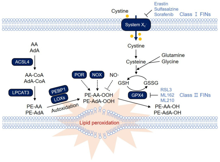
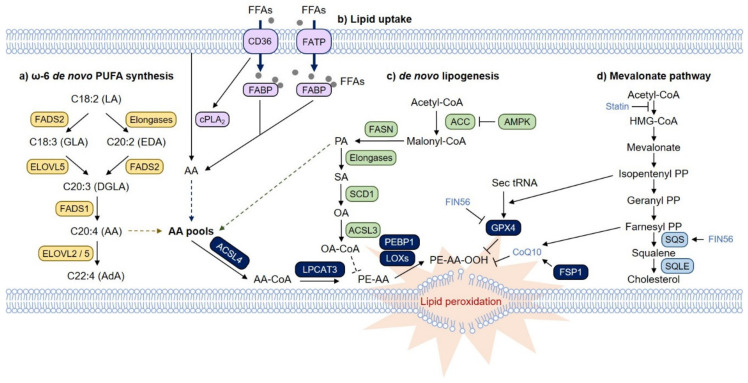
Ferroptosis is a type of iron-dependent regulated necrosis induced by lipid peroxidation that occurs in cellular membranes. Among the various lipids, polyunsaturated fatty acids (PUFAs) associated with several phospholipids, such as phosphatidylethanolamine (PE) and phosphatidylcholine (PC), are responsible for ferroptosis-inducing lipid peroxidation. Since the de novo synthesis of PUFAs is strongly restricted in mammals, cells take up essential fatty acids from the blood and lymph to produce a variety of PUFAs via PUFA biosynthesis pathways. Free PUFAs can be incorporated into the cellular membrane by several enzymes, such as ACLS4 and LPCAT3, and undergo lipid peroxidation through enzymatic and non-enzymatic mechanisms. These pathways are tightly regulated by various metabolic and signaling pathways. In this review, we summarize our current knowledge of how various lipid metabolic pathways are associated with lipid peroxidation and ferroptosis. Our review will provide insight into treatment strategies for ferroptosis-related diseases.
Overview
The claims and statistics of this paper haven’t been checked yet.
Run the check — free, no account needed. It reads the open-access text and checks the sources it cites.
Integrity signals
Registry
Cites
Statistics
Figures
Verification
Claims the paper makes
The claims and statistics of this paper haven’t been checked yet.
Run the check — free, no account needed. It reads the open-access text and checks the sources it cites.
Claims other papers make about this one
Each is checked against this paper’s own text: does it say what the citing paper says it does?
The cited source says what the claim attributes to it.
In the citing paper
“Growing evidence indicates that this type of regulated cell death (RCD) represents a cancer cell vulnerability caused by incorporating polyunsaturated fatty acids (PUFAs) into cellular membrane lipids (8).”
From Secreted Apoe rewires melanoma cell state vulnerability to ferroptosis (2024) · cites this paper as [8]
Self-check
Percentages, sums and reported differences re-computed from the paper’s own numbers.
The self-check needs the paper’s full text.
Citations
Cites
Ferroptosis: An Iron-Dependent Form of Nonapoptotic Cell Death
Dixon, S. J., Lemberg, K. M., Lamprecht, M. R., Skouta, R., Zaitsev, E. M., Gleason, C. E., Patel, D. N., Bauer, A. J., Cantley, A. M., Yang, W. S., et al. (2012) · Cell · doi:10.1016/j.cell.2012.03.042 · Cited by 18,691
Regulation of Ferroptotic Cancer Cell Death by GPX4
Yang, W. S., SriRamaratnam, R., Welsch, M. E., Shimada, K., Skouta, R., Viswanathan, V. S., Cheah, J. H., Clemons, P. A., Shamji, A. F., Clish, C. B., et al. (2014) · Cell · doi:10.1016/j.cell.2013.12.010 · Cited by 7,713
Ayala, A., Muñoz, M., & Argüelles, S. (2014) · Oxidative Medicine and Cellular Longevity · doi:10.1155/2014/360438 · Cited by 6,837
Mitochondrial Dysfunction and Oxidative Stress in Neurodegenerative Diseases
Lin, M. T., & Beal, M. F. (2006) · Nature · doi:10.1038/nature05292 · Cited by 6,455
AMPK: a nutrient and energy sensor that maintains energy homeostasis
Hardie, D. G., Ross, F. A., & Hawley, S. A. (2012) · Nature Reviews Molecular Cell Biology · doi:10.1038/nrm3311 · Cited by 4,602
Ferroptosis: molecular mechanisms and health implications
Tang, D., Chen, X., Kang, R., & Kroemer, G. (2020) · Cell Research · doi:10.1038/s41422-020-00441-1 · Cited by 4,495
Inactivation of the ferroptosis regulator Gpx4 triggers acute renal failure in mice
Friedmann Angeli, J. P., Schneider, M., Proneth, B., Tyurina, Y. Y., Tyurin, V. A., Hammond, V. J., Herbach, N., Aichler, M., Walch, A., Eggenhofer, E., et al. (2014) · Nature Cell Biology · doi:10.1038/ncb3064 · Cited by 3,732
The CoQ oxidoreductase FSP1 acts parallel to GPX4 to inhibit ferroptosis
Bersuker, K., Hendricks, J. M., Li, Z., Magtanong, L., Ford, B., Tang, P. H., Roberts, M. A., Tong, B., Maimone, T. J., Zoncu, R., et al. (2019) · Nature · doi:10.1038/s41586-019-1705-2 · Cited by 3,703
ROS as signalling molecules: Mechanisms that generate specificity in ROS homeostasis
D'Autréaux, B., & Toledano, M. B. (2007) · Nature Reviews Molecular Cell Biology · doi:10.1038/nrm2256 · Cited by 3,642
Ferroptosis as a p53-mediated activity during tumour suppression
Jiang, L., Kon, N., Li, T., Wang, S.-J., Su, T., Hibshoosh, H., Baer, R., & Gu, W. (2015) · Nature · doi:10.1038/nature14344 · Cited by 3,398
Research works printed but not linked: 1
- Xu, S., Chaudhary, O., Rodríguez-Morales, P., Sun, X., Zappasodi, R., Xu, Z., Pinto, A.F.M., Williams, A., Chen, D., and Low, J.S. (2020). Oxidized Lipids and CD36-Mediated Lipid Peroxidation in CD8 T Cells Suppress Anti-Tumor Immune Responses. bioRxiv. · DOI
Cited by
Sorted by citations
Items 201–300 of 375
Unraveling the Molecular Regulation of Ferroptosis in Respiratory Diseases
Zhu, L., Zhou, J., Yu, C., Gu, L., Wang, Q., Xu, H., Zhu, Y., Guo, M., Hu, M., Peng, W., et al. (2024) · Journal of Inflammation Research · doi:10.2147/jir.s457092 · Cited by 9
Epigenetic regulation of targeted ferroptosis: A new strategy for drug development
Ouyang, S., Zeng, Z., He, J., & Luo, L. (2024) · Journal of Pharmaceutical Analysis · doi:10.1016/j.jpha.2024.101012 · Cited by 10
Role of Ferroptosis in ALD: Focusing on Hepcidin and Besides Hepcidin
Chen, L., Zhou, H., Cheng, Z., Liu, D., & Tan, Y. (2024) · Current Medicinal Chemistry · doi:10.2174/0109298673317526240924050651 · Cited by 9
Hou, Y., Zhong, B., Zhao, L., Wang, H., Zhu, Y., Wang, X., Zheng, H., Yu, J. Z., Liu, G., Wang, X., et al. (2024) · Acta Pharmaceutica Sinica B · doi:10.1016/j.apsb.2024.12.005 · Cited by 9
Biltekin, E., Kahraman, N., Gul, O. A., Akay, Y. M., Akay, M., & Ozpolat, B. (2025) · International Journal of Molecular Sciences · doi:10.3390/ijms26146792 · Cited by 12
Shi, M., Yang, X., Ding, Y., Sun, L., Zhang, P., Liu, M., Han, X., Huang, Z., & Li, R. (2022) · Biology · doi:10.3390/biology11070950 · Cited by 9
A promising anti-tumor targeting on ERMMDs mediated abnormal lipid metabolism in tumor cells
Pang, M., Yu, L., Li, X., Lu, C., Xiao, C., & Liu, Y. (2024) · Cell Death and Disease · doi:10.1038/s41419-024-06956-4 · Cited by 8
Subramanian, C., McNamara, K., Croslow, S. W., Tan, Y., Hess, D. L., Kiseljak‐Vassiliades, K., Wierman, M. E., Sweedler, J. V., & Cohen, M. S. (2024) · Surgery · doi:10.1016/j.surg.2024.07.075 · Cited by 8
Ding, R., Zhong, S., Deng, L., & Luo, L. (2024) · The American Journal of Chinese Medicine · doi:10.1142/s0192415x24500691 · Cited by 8
Simvastatin-Induced Ferroptosis in Orbital Fibroblasts in Graves’ Ophthalmopathy
Wang, L., Li, Y., Niu, T., Deng, J., Shi, Y.-X., Liu, Y.-T., Tong, B., Qi, X., Cao, D., Tao, Y.-G., et al. (2025) · Investigative Ophthalmology and Visual Science · doi:10.1167/iovs.66.1.56 · Cited by 7
Wu, X., He, S., Lu, J., Chen, Y., Jiang, X., Wang, X., Zhu, Y., Zhu, Y., Chen, R., Xu, L., et al. (2025) · Free Radical Biology and Medicine · doi:10.1016/j.freeradbiomed.2025.07.029 · Cited by 9
Ruan, W., Peng, Z., Huang, M., Zhang, H., Li, X., Ma, T., Deng, J., Pan, M., Mai, Z., Meng, X., et al. (2025) · Chemico-Biological Interactions · doi:10.1016/j.cbi.2025.111697 · Cited by 9
Xia, C., Sun, X., Wang, Y., Min, J., Zhang, W., Wei, C., Gao, L., Zhao, F., Aleem, A. R., Peng, W., et al. (2025) · Cancer Letters · doi:10.1016/j.canlet.2025.217992 · Cited by 8
Lin, W., Gu, S., Zhang, X., Li, K., Zhao, D., Ma, B., Pan, C., Xu, Z., Liu, T., Wang, H.-H., et al. (2025) · Nature Communications · doi:10.1038/s41467-025-63376-4 · Cited by 9
Ferroptosis in Myocardial Fibrosis: Mechanisms and Therapeutic Insights
Lin, X., Li, W., Ye, J., & Li, L. (2026) · Antioxidants · doi:10.3390/antiox15010070 · Cited by 8
Kordi, N., Bagherzadeh-Rahmani, B., Kheirandish, R., Rezaali, R., & Stockwell, B. R. (2026) · npj Metabolic Health and Disease · doi:10.1038/s44324-025-00092-z · Cited by 8
Non-canonical cell death in neurodegeneration: emerging mechanisms and therapeutic frontiers
Ercin, N., Beşli, N., Beker, M., & Kılıç, Ü. (2026) · APOPTOSIS · doi:10.1007/s10495-026-02260-y · Cited by 10
Gigliotti, C. L., Boggio, E. M., Favero, F., Incarnato, D., Santoro, C., Oliviero, S., Rojo, J. M., Zucchelli, S., Persichetti, F., Baldanzi, G., et al. (2022) · Frontiers in Immunology · doi:10.3389/fimmu.2022.915963 · Cited by 8
Hacking the Lipidome: New Ferroptosis Strategies in Cancer Therapy
Варинський, Б. О., & Schick, J. (2024) · Biomedicines · doi:10.3390/biomedicines12030541 · Cited by 9
The Emerging Roles of Hydrogen Sulfide in Ferroptosis
Zhu, Y., Liu, Z.-T., Tang, A.-Q., Liang, X., Wang, Y., Liu, Y., Jin, Y., Gao, W., Yuan, H., Wang, D., et al. (2024) · Antioxidants and Redox Signaling · doi:10.1089/ars.2023.0535 · Cited by 8
Broadening horizons: research on ferroptosis in lung cancer and its potential therapeutic targets
Gao, G., & Zhang, X. (2025) · Frontiers in Immunology · doi:10.3389/fimmu.2025.1542844 · Cited by 8
Liu, L., Xue, Y., Wang, J., Shen, C., Li, Y., & Huang, Y. (2024) · Skin Research and Technology · doi:10.1111/srt.13608 · Cited by 8
New curcumin derivative induces ferroptosis in MCF-7 cells through activating SLC7A11/GPX4 axis
Wu, Z., Zhang, G., Shang, Y., Huang, J., Liu, Y., Zhou, H., & Wang, T. (2025) · Bioorganic & Medicinal Chemistry · doi:10.1016/j.bmc.2025.118078 · Cited by 7
Safari, M. H., Rahimzadeh, P., Alaei, E., Alimohammadi, M., Esfandiari, N., Daneshi, S., Malgard, N., Farahani, N., Taheriazam, A., & Hashemi, M. (2025) · Molecular and Cellular Probes · doi:10.1016/j.mcp.2025.102013 · Cited by 8
Ferroptosis as a potential therapeutic target for obesity-related metabolic diseases
Huang, C., Luo, T., Zhang, H., Cheng, L.-H., Peng, S., Zhou, X., Cao, L., Chen, F.-Y., He, J., Chen, C., et al. (2025) · Frontiers in Pharmacology · doi:10.3389/fphar.2025.1581632 · Cited by 7
Mbah, N. E., Sutton, D., Hong, H. S., Singhal, R., Menjivar, R. E., Perricone, M. D., Giza, H., Sajjakulnukit, P., Myers, A. L., Nwosu, Z. C., et al. (2026) · Cancer & Metabolism · doi:10.1186/s40170-026-00428-3 · Cited by 7
Zheng, Y., Wang, W., Chen, J., Peng, K., Chen, X., Shen, Q., Liang, B., Mao, Z., & Tan, C. (2024) · Advanced Science · doi:10.1002/advs.202411629 · Cited by 7
Novel impact of metal ion-induced cell death on diabetic cardiomyopathy pathogenesis and therapy
Jiang, J., Hu, S., Hu, K., Xiao, L., Lin, J., Chen, Y., Zhang, D., Ou, Y.-L., Zhang, J., Yuan, L., et al. (2025) · APOPTOSIS · doi:10.1007/s10495-025-02090-4 · Cited by 8
Feng, B., Guo, H., Ning, Y., Zhao, Y., Wang, X., & Cui, R. (2025) · Discover Oncology · doi:10.1007/s12672-025-02283-y · Cited by 9
Yang, X., Wu, H., Liu, D., Zhou, G., Zhang, D., Yang, Q., Liu, Y., & Li, Y. (2025) · Journal of Cardiovascular Translational Research · doi:10.1007/s12265-025-10590-6 · Cited by 8
DHODH Blockade Induces Ferroptosis in Neuroblastoma by Modulating the Mevalonate Pathway
Shir, J.-C., Chen, P.-Y., Kuo, C.-H., Hsieh, C.-H., Chang, H., Lee, H.-C., Huang, C.-H., Hsu, C., Hsu, W., Huang, H., et al. (2025) · Molecular & Cellular Proteomics · doi:10.1016/j.mcpro.2025.101014 · Cited by 6
Li, S., Rao, P., Yu, W., Tang, Y., Jiang, X., & Liu, J. (2025) · Biomolecules & Therapeutics · doi:10.4062/biomolther.2025.037 · Cited by 8
NFS1 Plays a Critical Role in Regulating Ferroptosis Homeostasis
Sun, S., Cao, H., Li, X., & Liao, H. (2025) · Biomolecules · doi:10.3390/biom16010032 · Cited by 7
Xue, J., Wang, C., & Fan, H. (2026) · Frontiers in Immunology · doi:10.3389/fimmu.2026.1715665 · Cited by 6
Liu, X., Sun, Y., Lv, X., Song, M., Sun, J., & Sun, Y. (2024) · International Immunopharmacology · doi:10.1016/j.intimp.2024.112518 · Cited by 6
Rahmati‐Dehkordi, F., Khanifar, H., Zare-Hoseinabadi, A., Dadgostar, E., Jafarpour, H., Aschner, M., Mirzaei, H., Tamtaji, O. R., & Nabavizadeh, F. (2024) · Molecular Biology Reports · doi:10.1007/s11033-024-09952-1 · Cited by 6
Pan, R., Luo, Z., You, Y.-S., Wang, J.-D., Chen, Y.-Q., Zhou, R.-R., Chen, S.-Z.-Q., Wang, L.-M., Zhao, J.-X., Su, H., et al. (2024) · Biological Trace Element Research · doi:10.1007/s12011-024-04459-z · Cited by 6
Qiu, T.-X., Zhang, X., Hu, Y., Liu, L., Shan, L., & Chen, J. (2024) · Fish & Shellfish Immunology · doi:10.1016/j.fsi.2024.109977 · Cited by 6
NINJ1 in Cell Death and Ferroptosis: Implications for Tumor Invasion and Metastasis
Chen, S.-Y., Shyu, I., & Chi, J. (2025) · Cancers · doi:10.3390/cancers17050800 · Cited by 7
Song, Z.-M., Wang, X., Zeng, J., Ren, F., Wang, Y., Li, M., Lin, Q., Li, W., Liao, X., & Wang, D. (2025) · Journal of Lipid Research · doi:10.1016/j.jlr.2025.100876 · Cited by 5
Therapeutic Materials for Ferroptosis Inhibition: from Design to Applications
Zhao, Z., Li, H., Wang, X., Wan, Q., Wang, J., Chen, J., Zhao, W., Cheng, B., & Pei, X. (2025) · Advanced Healthcare Materials · doi:10.1002/adhm.202503407 · Cited by 5
Fan, R., Liu, F., Gong, Q., Liu, D., Tang, S., & Shen, D. (2024) · Frontiers in Immunology · doi:10.3389/fimmu.2024.1393801 · Cited by 5
Gedaly, R., Orozco, G., Lewis, L. J., Valvi, D., Chapelin, F., Khurana, A., Hidalgo, G. E., Shmookler, A. D., Tripathi, A., Zhang, C., et al. (2024) · American Journal of Transplantation · doi:10.1016/j.ajt.2024.10.024 · Cited by 5
Autophagy is involved in Salmonella Typhimurium-induced ferroptosis in macrophages
Liu, W., Fu, D. C., Di, C., Sun, J. F., & Liu, P. (2025) · Veterinary Microbiology · doi:10.1016/j.vetmic.2025.110538 · Cited by 5
The Molecular Interplay Between p53-Mediated Ferroptosis and Non-Coding RNAs in Cancer
Punziano, C., Trombetti, S., Grosso, M., Tornesello, M. L., & Faraonio, R. (2025) · International Journal of Molecular Sciences · doi:10.3390/ijms26146588 · Cited by 5
Enlightening the Mechanism of Ferroptosis in Epileptic Heart
Akyüz, E., Saleem, Q. H., Sarı, Ç., Auzmendi, J., & Lazarowski, A. (2023) · Current Medicinal Chemistry · doi:10.2174/0929867330666230223103524 · Cited by 5
Unique lipid composition maintained by extracellular blockade leads to prooncogenicity
Kudo, K., Yanagiya, R., Hasegawa, M., Carreras, J., Miki, Y., Nakayama, S., Nagashima, E., Miyatake, Y., Torii, K., Ando, K., et al. (2024) · Cell Death Discovery · doi:10.1038/s41420-024-01971-y · Cited by 5
Computational pipeline predicting cell death suppressors as targets for cancer therapy
Vinik, Y., Maimon, A., Raj, H., Dubey, V., Geist, F., Wienke, D., & Lev, S. (2024) · iScience · doi:10.1016/j.isci.2024.110859 · Cited by 5
Sridhar, S., Bhalla, P., Kullu, J., Veerapaneni, S., Sahoo, S., Bhatt, N., & Suraishkumar, G. K. (2023) · Metabolic Engineering · doi:10.1016/j.ymben.2023.08.006 · Cited by 8
Ferroptosis by nanomaterials: targeting cancer stem cells and overcoming drug resistance
Wang, X., Luo, J., Zeng, X., & Yan, S. (2025) · Acta Materia Medica · doi:10.15212/amm-2024-0083 · Cited by 4
Ferroptosis and epilepsy: bidirectional pathogenic links and therapeutic implications
Xu, J., Dong, X., Yuan, M., Chen, X., Shu, H., & Yu, S. (2025) · Frontiers in Neurology · doi:10.3389/fneur.2025.1635441 · Cited by 5
Zhang, X., Zhou, Y., Li, H., Chen, M., Peng, F., & Li, N. (2025) · Biomedicines · doi:10.3390/biomedicines13040942 · Cited by 4
Shahid, M. M., Hohman, G., & Eldeeb, M. A. (2025) · ChemBioChem · doi:10.1002/cbic.202401052 · Cited by 4
Wang, K., Wang, Q., Chen, Y., Cao, Y., Zhao, R., Yu, S., Sun, X., Zhang, Y., & Li, J.-T. (2025) · Fishes · doi:10.3390/fishes10050234 · Cited by 4
Liu, J., Yan, G., Gui, H., Luo, K., Liang, H., Guan, A., Liu, W., Zhou, H., Lin, Z., & Yan, B. (2025) · Journal of Hazardous Materials · doi:10.1016/j.jhazmat.2025.140749 · Cited by 4
Dou, T., Zhu, X., Li, H., Wang, R., He, J., Geng, H., Zhang, W., Yu, Q., Zhao, H., & Yang, H. (2025) · Genes and Immunity · doi:10.1038/s41435-025-00370-2 · Cited by 4
Ferroptosis: A Key Mechanism in Alcohol-Induced Mitochondrial Dysfunction and Oxidative Stress
Kuruvalli, G., Nagajyothi, P. C., Pannuru, P., Maity, S., Santra, A., Shim, J., Syed, K., & Reddy, V. D. (2026) · ACS Pharmacology & Translational Science · doi:10.1021/acsptsci.5c00759 · Cited by 4
Screening of Natural Compounds for CYP11A1 Stimulation Against Cell Renal Cell Carcinoma
Ong, H. T. M., Ates, E., Kwon, O., & Kang, M. (2023) · Biological Procedures Online · doi:10.1186/s12575-023-00225-y · Cited by 4
The role of ferroptosis resistance in lymph-associated tumour metastasis
Li, X., Tian, M., Yu, L., Qian, J., Yang, J., Wang, X., Lu, C., Xiao, C., & Liu, Y. (2024) · Biochimica et Biophysica Acta (BBA) - Reviews on Cancer · doi:10.1016/j.bbcan.2024.189200 · Cited by 4
Metal-Dependent Cell Death in Renal Fibrosis: Now and in the Future
Li, T., & Yu, C. (2024) · International Journal of Molecular Sciences · doi:10.3390/ijms252413279 · Cited by 4
Enzymatic Oxidants, Antioxidants, and Inflammatory Bowel Disease
Esworthy, R. S. (2025) · Applied Biosciences · doi:10.3390/applbiosci4020019 · Cited by 5
Ferroptosis: Opportunities and Challenges in Cancer
Jia, Y., Zhang, Y., Ma, X., Wang, Y., Tian, Y.-Q., He, P., & Xu, Y. (2023) · Journal of Exploratory Research in Pharmacology · doi:10.14218/jerp.2023.00003 · Cited by 6
Pharmacologic Inhibition of Ferroptosis Attenuates Experimental Abdominal Aortic Aneurysm FormationPreprint
Krebs, J. R., Bellotti, P., Valisno, J. A. C., Su, G., Sharma, S., Kollareth, D. J. M., Hartman, J. B., Aravinthan, A., Spinosa, M. D., Kamat, M. N., et al. (2024) · bioRxiv (Cold Spring Harbor Laboratory) · doi:10.1101/2024.06.18.599427 · Cited by 4
Liu, K., Yu, M., He, Y., Wang, T., Li, G., Wang, L., & Zhong, X. (2025) · Frontiers in Pharmacology · doi:10.3389/fphar.2025.1610573 · Cited by 3
Ferroptosis: a new dawn in the treatment of acute lymphoblastic leukemia
Han, J., Yang, Y., Wei, Y., Jin, J., & Feng, W. (2026) · Frontiers in Oncology · doi:10.3389/fonc.2025.1669432 · Cited by 3
Functional lipid analysis via index-based lipidomics profile: a new computational module in LipidOne
Alabed, H. B. R., Mancini, D. F., Pergola, M., Romani, L., Martino, S., Koulman, A., & Pellegrino, R. M. (2026) · Bioinformatics · doi:10.1093/bioinformatics/btag090 · Cited by 3
FSP1, a key ferroptosis suppressor with therapeutic potential
Bai, X., Zhu, J., Xiong, B., Chen, Y., Xu, C., Zhao, Q., Wu, Y., Tang, Z., Shi, R., Liu, Z., et al. (2026) · Biochemical Pharmacology · doi:10.1016/j.bcp.2026.118151 · Cited by 3
Live and let die: analyzing ultrastructural features in cell death
Perrotta, I. D. (2024) · Ultrastructural Pathology · doi:10.1080/01913123.2024.2428703 · Cited by 9
Pharmacological Targeting of Ferroptosis in Cancer Treatment
Khan, M. A., Satapathy, T., Sen, K., Sahu, S., Pradhan, B., Gupta, A., Satapathy, A., & Satapathy, A. (2024) · Journal of Drug Delivery and Therapeutics · doi:10.22270/jddt.v14i2.6371 · Cited by 3
Zhang, X., Xiong, W. G., Gao, F., Yu, Z., Ren, F., & Lei, X. G. (2025) · Free Radical Biology and Medicine · doi:10.1016/j.freeradbiomed.2025.05.387 · Cited by 3
Toczyłowska, B., Skowrońska, M., Kurkowska‐Jastrzębska, I., Ruszczyńska, A., & Ziemińska, E. (2025) · Scientific Reports · doi:10.1038/s41598-025-94838-w · Cited by 2
Liu, Y., Zhang, Y., Zhang, H., Xu, D., Zhou, X., Xie, L., Xi, H., Zhu, Y., Yang, H., Zhu, X., et al. (2025) · Scientific Reports · doi:10.1038/s41598-025-07201-4 · Cited by 2
Ferroptosis as a potential therapeutic target for post-traumatic stress disorder
Zhang, Q., Mao, J.-D., Chen, H., Wang, M., Wu, Y., & Wang, C. (2025) · Frontiers in Molecular Neuroscience · doi:10.3389/fnmol.2025.1648047 · Cited by 2
Du, L., Liu, W., Cui, Y., Chen, W., & He, Z. (2025) · Research · doi:10.34133/research.1005 · Cited by 2
Wang, Z., Liu, Z., Yang, Y., Wang, W., Chen, Q., Liang, S., Guo, L., & Li, M. (2025) · Cell & Bioscience · doi:10.1186/s13578-025-01496-6 · Cited by 2
Wu, N., Wang, G., Wang, J., Wu, M., Wang, X., Ma, J., & He, W. (2025) · European Journal of Medicinal Chemistry · doi:10.1016/j.ejmech.2025.118448 · Cited by 2
Hormetic Effects of 7‐Ketocholesterol in Preventing Ferroptosis in Hepatocytes
Anavi, S., Giltman, N., Zeigerman, H., Madar, Z., & Tirosh, O. (2026) · Oxidative Medicine and Cellular Longevity · doi:10.1155/omcl/7958511 · Cited by 2
Proteolysis-targeting Chimeras induce ferroptosis in cancer: From Mechanism to clinical application
Nx, Z., Zhong, X.-Y., Zhang, S.-H., Liu, M.-C., Xia, Q. D., Yu, X., & Wang, S.-G. (2026) · Pharmacological Research · doi:10.1016/j.phrs.2026.108141 · Cited by 2
Ferroptosis: A Double-Edged Sword in Cisplatin-Based Cancer Therapy and Acute Kidney Injury
Zhou, S., Xiao, Y., & Fang, L. (2026) · Cancer Management and Research · doi:10.2147/cmar.s589114 · Cited by 2
Beyond Feature Selection: Interpretable Machine Learning for Mechanistic Insights in Metabolomics
Bai, H., Ren, Y., Wang, J., Wang, Y., & Yang, Y. (2026) · Biology · doi:10.3390/biology15070572 · Cited by 2
He, S., Wu, Z., & Li, S. (2026) · Frontiers in Pharmacology · doi:10.3389/fphar.2026.1808167 · Cited by 2
Tyurina, Y. Y., Mikulska‐Ruminska, K., Tyurin, V. A., Kleiboeker, B. A., Kapralov, A. A., Hashimoto, A., Sparvero, L. J., Dar, H. H., Akdogan, M., Yamada, K., et al. (2026) · Nature Communications · doi:10.1038/s41467-026-71869-z · Cited by 4
Lysosomes in Ferroptosis: Regulatory Mechanisms and Molecular Targets
Luo, T., Wang, C., Zhou, N., ZHANG, Y., & Mou, X. (2026) · Molecules · doi:10.3390/molecules31132373 · Cited by 2
Enfeksiyon Hastalıklarında Ferroptozun Rolü
Işkın, A. E., & Budak, F. (2023) · Uludağ Üniversitesi Tıp Fakültesi Dergisi · doi:10.32708/uutfd.1337979 · Cited by 2
Tunçer, S., Elibol, B., Severcan, F., Gursoy, E. B., Tiftikçioğlu, B. İ., Güngördü, Z., Celik, C., Dai, A. S., & Karaçam, S. (2024) · International Immunopharmacology · doi:10.1016/j.intimp.2024.112787 · Cited by 2
Hao, J., Ren, J., Chang, B., Xu, H., Wang, H., & Ji, L. (2024) · Heliyon · doi:10.1016/j.heliyon.2024.e36664 · Cited by 2
Intracellular biliverdin dynamics during ferroptosis
Nakajima, K., Nishizawa, H., Chen, G., Tsuge, S., Yamanaka, M., Kiyohara, M., Irikura, R., Matsumoto, M., Tanaka, K., Narikawa, R., et al. (2024) · The Journal of Biochemistry · doi:10.1093/jb/mvae067 · Cited by 3
A Systems Biology Approach Towards a Comprehensive Understanding of Ferroptosis
Arbatskiy, M. S., Balandin, D. E., Akberdin, I. R., & Churov, A. V. (2024) · International Journal of Molecular Sciences · doi:10.3390/ijms252111782 · Cited by 4
Liu, C., Shang, M., Sun, Y., Tang, C., & Cheng, Y. (2025) · Comparative Biochemistry and Physiology Part D Genomics and Proteomics · doi:10.1016/j.cbd.2025.101608 · Cited by 2
Chappell, M. K., Shelestak, J., Irfan, M., Shelestak, E., Nemes, A. D., Mey, G. M., Venkat, L., Surabhi, A., & DeSilva, T. M. (2025) · iScience · doi:10.1016/j.isci.2025.113458 · Cited by 2
Fan, L. (2025) · American Journal of Cancer Research · doi:10.62347/jtqo8098 · Cited by 2
Das, J., Adhikari, N., Pal, A., Chongder, T., Dule, S., Sarma, A., Dutta, S., Gupta, P., & Shome, A. (2025) · Molecular Biology Reports · doi:10.1007/s11033-025-10986-2 · Cited by 2
Chen, S., Zhao, M., Chen, K., Xu, J., & Li, H. (2024) · Genes · doi:10.3390/genes15060812 · Cited by 2
Peng, T., & Li, M. (2024) · International Urology and Nephrology · doi:10.1007/s11255-024-04302-3 · Cited by 2
Oh, M., Jang, S. Y., Lee, J.-Y., Kim, J. W., Jung, Y., Seo, J., Han, T., Jang, E., Son, H. Y., Kim, D. R., et al. (2023) · bioRxiv (Cold Spring Harbor Laboratory) · doi:10.1101/2023.04.08.536136 · Cited by 2
Self-citationYe, K., Zhao, X. L., Liu, L., Ge, F., Zheng, F., Liu, Z., Tian, M., Han, X., Gao, X., Xia, Q. H., et al. (2024) · Molecular Neurobiology · doi:10.1007/s12035-024-04642-2 · Cited by 1
He, H., & Zhao, D. (2025) · Gene Reports · doi:10.1016/j.genrep.2025.102185 · Cited by 1
Shadmani, Y., Pourshojaei, Y., Karami‐Mohajeri, S., Amirheidari, B., & Sadeghzadeh, M. (2025) · Research in Pharmaceutical Sciences · doi:10.4103/rps.rps_120_24 · Cited by 1
Guo, M., Zhao, G., Cai, Z., Zhang, Z., & Jie, Z. H. O. U. (2025) · Journal of Zhejiang University (Medical Sciences) · doi:10.3724/zdxbyxb-2025-0329 · Cited by 2
Exploring the chemotherapeutic potential of five plant species through in vitro screening
Neves, V. F., Soto‐Blanco, B., Oliveira, M. F. C., & Oloris, S. C. S. (2025) · Arquivo Brasileiro de Medicina Veterinária e Zootecnia · doi:10.1590/1678-4162-13419 · Cited by 1
Metadata
- Authors
- Lee, J.-Y., Kim, W. K., Bae, K.-H., Lee, S. C., & Lee, E.-W.
- Affiliations
- Korea Research Institute of Bioscience and Biotechnology (KR)
- Corresponding author
- Lee, Sang Chul
- Lee, Eun-Woo
- Venue
- Biology
- Publisher
- MDPI AG
- Published
- 2021-03-02
- Volume
- 10
- Issue
- 3
- Pages
- 184
- ISSN
- 2079-7737 (ISSN-L)
- PMC
- PMC8000263
- OpenAlex
- W3134191433
- Cited by
- 374
- References
- 113
- Citations by year
- 2026: 58 · 2025: 103 · 2024: 96 · 2023: 69 · 2022: 38 · 2021: 10
- FWCI
- 8.60
- Type
- journal article
- Language
- en
- Open access
- gold
- doi.org
- Licence
- Locations
- Biology (publishedVersion, cc-by)
- PubMed Central (publishedVersion, cc-by)
- PubMed (publishedVersion)
- DOAJ (DOAJ: Directory of Open Access Journals) (submittedVersion, cc-by-sa)
- Funders
- Korea Research Institute of Bioscience and Biotechnology (Research Initiative Progam)
- National Research Foundation of Korea (NRF-2017M3A9G5083321, NRF-2019R1C1C1002831, NRF-2020R1A2C1006841)
- Topics
- Life Sciences › Biochemistry, Genetics and Molecular Biology › Cancer Research › Ferroptosis and cancer prognosis
- Health Sciences › Medicine › Hematology › Iron Metabolism and Disorders
- Life Sciences › Biochemistry, Genetics and Molecular Biology › Cancer Research › Cancer, Lipids, and Metabolism
- Keywords
- ferroptosis, lipid metabolism, lipid peroxidation, polyunsaturated fatty acids, phosphatidylethanolamine, phosphatidylcholine, LPCAT3
About these checks
Registry, retraction, Cites and statistics results are computed, not judged, with one exception: a printed reference that no index lists is read once by a model before it counts as a citation error. Claims are extracted by a model and kept only when their quote appears verbatim in the text; each cited source is then checked, with a model’s help, for whether it says what the claim relies on. Those verdicts can be wrong — read the claim’s quote and the cited source before relying on one.
Statistics and arithmetic are recomputed, not judged.
Confidence bands are shown only for fields calibrated on our annotated test set. None is calibrated yet, so no band is shown.
What counts as a claim ›How we measure confidence ›How we check this ›
Are you an author, or do you see a problem with this page? Request a review
