

The DOI matches its Crossref record; 57 of the 57 references it cites were found in a registry or an index.
Redefining Radiation Metrics: Evaluating Actual Doses in Computed Tomography Scans
Open accessAbstract
Background: Computed tomography (CT) contributes significantly to the collective dose from medical sources, raising concerns about potential health risks. However, existing radiation dose estimation tools, such as volume computed tomography dose index (CTDIvol), dose-length product (DLP), effective dose (ED), and size-specific dose estimate (SSDE), have limitations in accurately reflecting patient exposure. This study introduces a new parameter, size-specific dose-length product (DLPss), aiming to enhance the precision of radiation dose estimation in real-life scenarios. Methods: A retrospective analysis of 134 chest CT studies was conducted. Relationships between CTDIvol and anthropometric parameters were examined, and SSDE was calculated based on effective diameter. Additionally, the novel parameter, DLPss, was introduced, considering scan length and cross-sectional dimensions. Results: Analysis reveals variations in scan length, effective diameter, and CTDIvol between genders. Strong correlations were observed between CTDIvol and effective diameter, particularly in men. The average CTDIvol for the entire group was 7.83 ± 2.92 mGy, with statistically significant differences between women (7.38 ± 3.23 mGy) and men (8.30 ± 2.49 mGy). SSDE values showed significant gender differences, with men exhibiting higher values. The average SSDE values for women and men were 9.15 ± 2.5 mGy and 9.6 ± 2.09 mGy, respectively, with a statistically significant difference (p = 0.03). The newly introduced DLPss values ranged around 343.90 ± 81.66 mGy·cm for the entire group, with statistically significant differences between women (323.53 ± 78.69 mGy·cm) and men (364.89 ± 79.87 mGy·cm) (p < 0.05), providing a comprehensive assessment of total radiation dose. Conclusion: The study highlights the need for accurate radiation dose estimation, emphasizing the impact of CT examination parameters on dose variability. The proposed DLPss parameter offers a promising approach to enhancing precision in assessing radiation risk during CT scans. Further research is warranted to explore additional parameters for a comprehensive understanding of radiation exposure and to optimize imaging protocols for patient safety.
Overview
The claims and statistics of this paper haven’t been checked yet.
Run the check — free, no account needed. It reads the open-access text and checks the sources it cites.
Integrity signals
Registry
Cites
Statistics
Figures
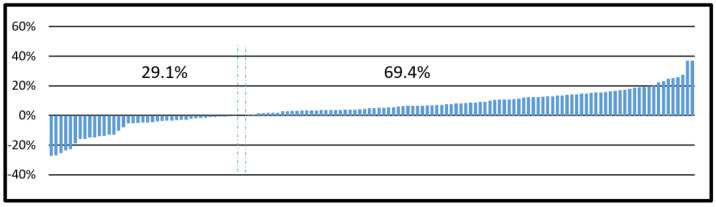
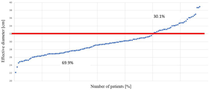
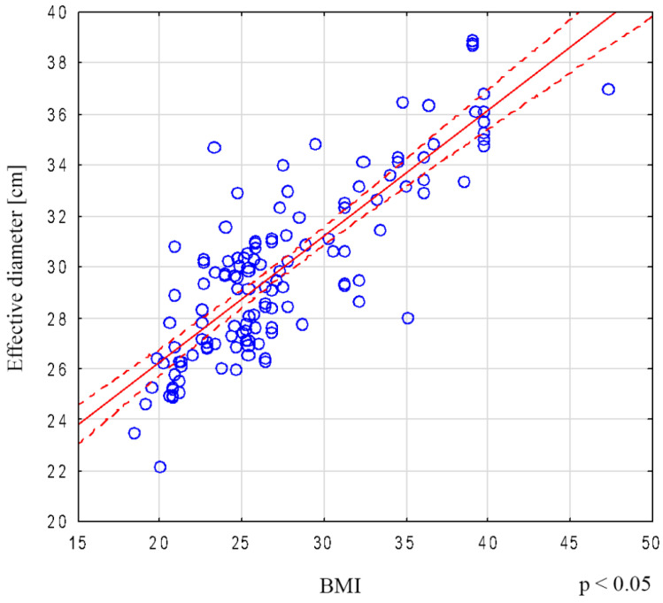
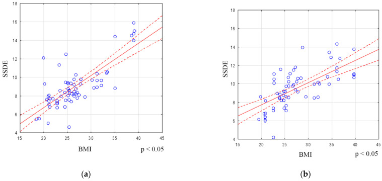
Verification
Claims the paper makes
The claims and statistics of this paper haven’t been checked yet.
Run the check — free, no account needed. It reads the open-access text and checks the sources it cites.
Claims other papers make about this one
Each is checked against this paper’s own text: does it say what the citing paper says it does?
The cited source says what the claim attributes to it.
In the citing paper
“While CTDIvol is not a direct indicator of the radiation received by the patient or tailored to specific anatomy, it is nonetheless a crucial comparison parameter among scanners and procedures.89”
From Computed Tomography Diagnostic Reference Levels Worldwide and in Jordan: Implications for Patient Dose Reduction (2026) · cites this paper as [89]
Self-check
Percentages, sums and reported differences re-computed from the paper’s own numbers.
The self-check needs the paper’s full text.
Citations
Cites
Computed Tomography — An Increasing Source of Radiation Exposure
Brenner, D. J., & Hall, E. J. (2007) · New England Journal of Medicine · doi:10.1056/nejmra072149 · Cited by 8,738
Sources and effects of ionizing radiation
United Nations Scientific Committee on the Effects of Atomic Radiation (2018) · Report of the United Nations Scientific Committee on the Effects of Atomic Radiation · doi:10.18356/f48d655d-en · Cited by 3,981
Health Risks from Exposure to Low Levels of Ionizing Radiation:: BEIR VII Phase 2
Earth, D. O. (2006) · CERN Document Server (European Organization for Nuclear Research) · Cited by 2,190
Preface, Executive Summary and Glossary
(2007) · Annals of the ICRP · doi:10.1016/j.icrp.2007.10.003 · Cited by 2,116
Size-Specific Dose Estimates (SSDE) in Pediatric and Adult Body CT Examinations
Boone, J. M., Strauss, K. J., CODY, D. D., et al. (2011) · doi:10.37206/143 · Cited by 554
McCollough, C. H., Bakalyar, D., Bostani, M., Brady, S. L., Boedeker, K., Boone, J. M., Chen‐Mayer, H. H., Christianson, O. I., Leng, S., Li, B., et al. (2014) · PubMed · Cited by 269
Radiation Exposure From Medical Imaging
Brenner, D. J., & Hricak, H. (2010) · JAMA · doi:10.1001/jama.2010.973 · Cited by 209
CT scanning: A major source of radiation exposure
Wiest, P. W., Locken, J. A., Heintz, P. H., & Mettler, F. A. (2002) · Seminars in Ultrasound CT and MRI · doi:10.1016/s0887-2171(02)90011-9 · Cited by 173
McCollough, C. H., Bakalyar, D., Bostani, M., et al. (2014) · doi:10.37206/146 · Cited by 171
Verdun, F. R., Gutiérrez, D., Vader, J. P., Aroua, A., Alamo, L., Bochud, F. O., & Gudinchet, F. (2008) · European Radiology · doi:10.1007/s00330-008-0963-4 · Cited by 169
Tsapaki, V., Aldrich, J. E., Sharma, R., et al. (2006) · Radiology · doi:10.1148/radiol.2403050993 · Cited by 141
Should We be Concerned About the Rapid Increase in CT Usage?
Brenner, D. J. (2010) · Reviews on Environmental Health · doi:10.1515/reveh.2010.25.1.63 · Cited by 137
Jarry, G., DeMarco, J. J., BEIFUSS, U., et al. (2003) · Physics in Medicine and Biology · doi:10.1088/0031-9155/48/16/306 · Cited by 134
Slowing the Increase in the Population Dose Resulting from CT Scans
Brenner, D. J. (2010) · Radiation Research · doi:10.1667/rr1859.1 · Cited by 134
Radiation Doses in Consecutive CT Examinations from Five University of California Medical Centers
Smith‐Bindman, R., Moghadassi, M., Wilson, N., et al. (2015) · Radiology · doi:10.1148/radiol.2015142728 · Cited by 127
Israel, G. M., Cicchiello, L. A., Brink, J. A., & Huda, W. (2010) · American Journal of Roentgenology · doi:10.2214/ajr.09.3331 · Cited by 114
Diagnostic Reference Ranges for Pediatric Abdominal CT
Goske, M. J., Strauss, K. J., Coombs, L. P., et al. (2013) · Radiology · doi:10.1148/radiol.13120730 · Cited by 113
Ding, A., Mille, M. M., Liu, T., Caracappa, P. F., & Xu, X. G. (2012) · Physics in Medicine and Biology · doi:10.1088/0031-9155/57/9/2441 · Cited by 96
Patient Dose in Cardiac Computed Tomography
McCollough, C. H. (2003) · Herz · doi:10.1007/s00059-003-2447-2 · Cited by 89
Wang, J., Christner, J. A., Duan, X., et al. (2012) · Medical Physics · doi:10.1118/1.4757586 · Cited by 85
Campbell, J., Kalra, M. K., Rizzo, S., et al. (2005) · American Journal of Roentgenology · doi:10.2214/ajr.04.1512 · Cited by 81
McLaughlin, D. J., & Mooney, R. B. (2004) · Clinical Radiology · doi:10.1016/j.crad.2003.10.016 · Cited by 79
Individually Adapted Examination Protocols for Reduction of Radiation Exposure in Chest CT
Wildberger, J. E., Mahnken, A. H., Schmitz‐Rode, T., Flohr, T. G., Stargardt, A., Haage, P., SCHALLER, S., & Günther, R. W. (2001) · Investigative Radiology · doi:10.1097/00004424-200110000-00006 · Cited by 72
Artificial Intelligence for Radiation Dose Optimization in Pediatric Radiology: A Systematic Review
Ng, C. K. C. (2022) · Children · doi:10.3390/children9071044 · Cited by 70
Frija, G., Damilakis, J. E., Paulo, G., et al. (2021) · European Radiology · doi:10.1007/s00330-021-07696-1 · Cited by 69
Patient Radiation Exposure in a Modern, Large-Volume, Pediatric Cardiac Catheterization Laboratory
Glatz, A. C., Patel, A., Zhu, X., Dori, Y., Hanna, B. D., Gillespie, M. J., & Rome, J. J. (2014) · Pediatric Cardiology · doi:10.1007/s00246-014-0869-7 · Cited by 63
Simplifying Size-Specific Radiation Dose Estimates in Pediatric CT
Khawaja, R. D. A., Singh, S., Vettiyil, B., Lim, R., Gee, M. S., Westra, S. J., & Kalra, M. K. (2014) · American Journal of Roentgenology · doi:10.2214/ajr.13.12191 · Cited by 54
Zanca, F., Demeter, M., Oyen, R., & Bosmans, H. (2011) · European Radiology · doi:10.1007/s00330-011-2332-y · Cited by 53
Brady, S. L., Mirro, A. E., Moore, B. M., & Kaufman, R. A. (2015) · American Journal of Roentgenology · doi:10.2214/ajr.14.13317 · Cited by 53
Radiation doses from head, neck, chest and abdominal CT examinations: an institutional dose report
Atlı, E., Uyanık, S. A., Öğüşlü, U., et al. (2020) · Diagnostic and Interventional Radiology · doi:10.5152/dir.2020.19560 · Cited by 52
Boos, J., Lanzman, R. S., Heusch, P., Aissa, J., Schleich, C., Thomas, C. K., Sawicki, L. M., Antoch, G., & Kröpil, P. (2015) · British Journal of Radiology · doi:10.1259/bjr.20150734 · Cited by 49
Das, M., Mahnken, A. H., Mühlenbruch, G., et al. (2005) · American Journal of Roentgenology · doi:10.2214/ajr.184.5.01841437 · Cited by 45
U.S. National Diagnostic Reference Levels: Closing the Gap
Brink, J. A., & Miller, D. L. (2015) · Radiology · doi:10.1148/radiol.2015150971 · Cited by 42
Subtle excess in lifetime cancer risk related to CT scanning in Spanish young people
de Basea, M. B., Moriña, D., Figuerola, J., Barber, I., Muchart, J., Lee, C., & Cardis, E. (2018) · Environment International · doi:10.1016/j.envint.2018.07.020 · Cited by 31
ACCURACY OF SIZE-SPECIFIC DOSE ESTIMATE CALCULATION FROM CENTER SLICE IN COMPUTED TOMOGRAPHY
Boos, J., Kröpil, P., Bethge, O., et al. (2017) · Radiation Protection Dosimetry · doi:10.1093/rpd/ncx069 · Cited by 31
Nyman, U., Ahl, T. L., Kristiansson, M., et al. (2005) · Acta Radiologica · doi:10.1080/02841850510021193 · Cited by 30
CT is still not a low‐dose imaging modality
Rehani, M. M., Szczykutowicz, T. P., & Zaidi, H. (2019) · Medical Physics · doi:10.1002/mp.14000 · Cited by 30
Automated Estimation of Abdominal Effective Diameter for Body Size Normalization of CT Dose
Cheng, P. M. (2012) · Journal of Imaging Informatics in Medicine · doi:10.1007/s10278-012-9525-z · Cited by 28
Extra Z-Axis Coverage at CT Imaging Resulting in Excess Radiation Dose
Liao, E. A., Quint, L. E., Goodsitt, M. M., et al. (2011) · Journal of Computer Assisted Tomography · doi:10.1097/rct.0b013e3181f5a652 · Cited by 27
Anam, C., Haryanto, F., Widita, R., Arif, I., & Dougherty, G. (2017) · Atom Indonesia · doi:10.17146/aij.2017.617 · Cited by 27
Justifying referrals for paediatric CT
Brady, Z., Cain, T. M., & Johnston, P. N. (2012) · The Medical Journal of Australia · doi:10.5694/mja11.11124 · Cited by 25
Cohen, S. L., Ward, T. J., Makhnevich, A., et al. (2020) · Clinical Imaging · doi:10.1016/j.clinimag.2019.11.020 · Cited by 25
Artificial intelligence and radiation protection. A game changer or an update?
Andresz, S., Zéphir, A., Bez, J., Karst, M., & Danieli, J. (2022) · Radioprotection · doi:10.1051/radiopro/2022004 · Cited by 25
Garg, M. K., Karami, V., Moazen, J., Kwee, T. C., Bhalla, A. S., Shahbazi‐Gahrouei, D., & Shao, Y. J. (2022) · Diagnostics · doi:10.3390/diagnostics12123043 · Cited by 25
Meeson, S., Alvey, C., & Golding, S. J. (2010) · Journal of Radiological Protection · doi:10.1088/0952-4746/30/2/003 · Cited by 21
Accuracy of Size-Specific Dose Estimate Calculation from Center Slice in Computed Tomography
Boos, J., Kröpil, P., Bethge, O., et al. (2017) · Radiation Protection Dosimetry · doi:10.1093/rpd/ncx119 · Cited by 21
The use of the effective dose equivalent,HE, as a risk parameter in computed tomography
Huda, W., & Sandison, G. A. (1986) · British Journal of Radiology · doi:10.1259/0007-1285-59-708-1236 · Cited by 18
Optimisedz-axis coverage at multidetector-row CT in adults suspected of acute appendicitis
Brassart, N., Winant, C., Tack, D., et al. (2013) · British Journal of Radiology · doi:10.1259/bjr.20130115 · Cited by 17
Jansen, J. T. M., & Shrimpton, P. C. (2011) · Progress in Nuclear Science and Technology · doi:10.15669/pnst.2.165 · Cited by 14
Li, Z., Zhang, J., Xia, C., Zhao, F., Zhang, K., Li, Y., Li, L., Pu, J., Peng, W., Liu, K., et al. (2020) · Annals of Translational Medicine · doi:10.21037/atm-20-5443 · Cited by 14
IONIZING RADIATION EXPOSURE IN PATIENTS WITH COVID-19: MORE THAN NEEDED
Yurdaışık, I., Nurili, F., Aksoy, S. H., Agirman, A. G., & Aktan, A. (2021) · Radiation Protection Dosimetry · doi:10.1093/rpd/ncab092 · Cited by 11
Haba, T., Koyama, S., Kinomura, Y., et al. (2017) · Medical Physics · doi:10.1002/mp.12601 · Cited by 9
Fiagbedzi, E. W., Gorleku, P. N., Nyarko, S., Atuwo-Ampoh, V. D., Fiagan, Y. A. C., & Asare, A. (2022) · Open Journal of Medical Imaging · doi:10.4236/ojmi.2022.121004 · Cited by 9
(2015) · Radiology · doi:10.1148/radiol.2771201510 · Cited by 8
A new Monte Carlo tool for organ dose estimation in computed tomography
Adrien, C., Loirec, C. L., Dreuil, S., et al. (2020) · Radioprotection · doi:10.1051/radiopro/2020006 · Cited by 7
Socol, Y., Richardson, A., Garali, I., Grison, S., Varès, G., & Klokov, D. Y. (2024) · Frontiers in Artificial Intelligence · doi:10.3389/frai.2023.1291136 · Cited by 6
Lamoureux, R. H., Sinclair, L., Mench, A. M., et al. (2015) · Medical Physics · doi:10.1118/1.4924025 · Cited by 2
Research works printed but not linked: 3
- Andersson, J., Pavlicek, W., Al-Senan, R., Bolch, W., Bosmans, H., Cody, D., Dixon, R., Colombo, P., Dong, F., and Edyvean, S. (2023, September 27). Estimating Patient Organ Dose with Computed Tomography: A Review of Present Methodology and Required DICOM Information a Joint Report of AAPM Task Group 246 and the European Federation of Organizations for Medical Physics (EFOMP). AAPM Raport. Available online: https://www.aapm.org/pubs/reports/RPT_246.pdf.
- Damilakis, J., Frija, G., Jaschke, W., Paulo, G., Repussard, J., Schegerer, A., Tsapaki, V., Clark, J., and Hierath, M. (2023, September 18). European Study on Clinical Diagnostic Reference Levels for X-ray Medical Imaging—EUCLID, Publications Office of the European Union. Available online: https://www.eurosafeimaging.org/wp/wp-content/uploads/2021/03/20210325_D5.3-EUCLID-Workshop-Report-FINAL_with-updates_for-website.pdf.
- The 2007 Recommendations of the International Commission on Radiological Protection (2007). ICRP Publication 103. Ann. ICRP, 37, 1–332. Available online: https://journals.sagepub.com/doi/pdf/10.1177/ANIB_37_2-4. · DOI
Cited by
Sorted by citations
Oglat, A., Shdaifat, S., Sayah, M., Abukonna, A., & Khogali, W. (2026) · Drug, Healthcare and Patient Safety · doi:10.2147/dhps.s595721
“89 The DLP, expressed in mGy·cm, is determined by multiplying CTDIvol by the scan duration.”
Metadata
- Authors
- Sabiniewicz-Ziajka, D., Szarmach, A., Grzywińska, M., Gać, P., & Piskunowicz, M.
- Affiliations
- Gdańsk Medical University (PL)
- Wroclaw Medical University (PL)
- Corresponding author
- Szarmach, Arkadiusz
- ORCID
- Venue
- Biomedicines
- Publisher
- MDPI AG
- Published
- 2024-03-07
- Volume
- 12
- Issue
- 3
- Pages
- 600
- ISSN
- 2227-9059 (ISSN-L)
- PMC
- PMC10967789
- OpenAlex
- W4392562802
- Cited by
- 8
- References
- 57
- Citations by year
- 2026: 5 · 2025: 3
- FWCI
- 2.03
- Type
- journal article
- Language
- en
- Open access
- gold
- doi.org
- Licence
- Locations
- Biomedicines (publishedVersion, cc-by)
- PubMed Central (publishedVersion, cc-by)
- PubMed (publishedVersion)
- DOAJ (DOAJ: Directory of Open Access Journals) (submittedVersion)
- Topics
- Health Sciences › Medicine › Radiology, Nuclear Medicine and Imaging › Radiation Dose and Imaging
- Physical Sciences › Physics and Astronomy › Radiation › Advanced Radiotherapy Techniques
- Health Sciences › Medicine › Radiology, Nuclear Medicine and Imaging › Digital Radiography and Breast Imaging
- Keywords
- computed tomography radiation dose, CTDIvol, effective dose, size-specific dose estimate, dose-length product, chest CT, anthropometric factors, effective diameter, sex differences in radiation dose, radiation risk, patient safety
About these checks
Registry, retraction, Cites and statistics results are computed, not judged, with one exception: a printed reference that no index lists is read once by a model before it counts as a citation error. Claims are extracted by a model and kept only when their quote appears verbatim in the text; each cited source is then checked, with a model’s help, for whether it says what the claim relies on. Those verdicts can be wrong — read the claim’s quote and the cited source before relying on one.
Statistics and arithmetic are recomputed, not judged.
Confidence bands are shown only for fields calibrated on our annotated test set. None is calibrated yet, so no band is shown.
What counts as a claim ›How we measure confidence ›How we check this ›
Are you an author, or do you see a problem with this page? Request a review
