

The DOI matches its Crossref record; 266 of the 267 references it cites were found in a registry or an index.
The Urokinase Receptor (uPAR) as a “Trojan Horse” in Targeted Cancer Therapy: Challenges and Opportunities
Open accessAbstract
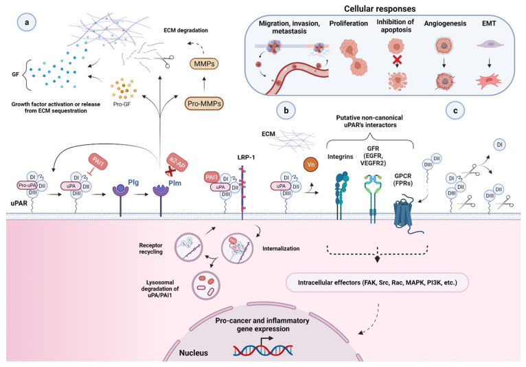
One of the largest challenges to the implementation of precision oncology is identifying and validating selective tumor-driving targets to enhance the therapeutic efficacy while limiting off-target toxicity. In this context, the urokinase-type plasminogen activator receptor (uPAR) has progressively emerged as a promising therapeutic target in the management of aggressive malignancies. By focalizing the plasminogen activation cascade and subsequent extracellular proteolysis on the cell surface of migrating cells, uPAR endows malignant cells with a high proteolytic and migratory potential to dissolve the restraining extracellular matrix (ECM) barriers and metastasize to distant sites. uPAR is also assumed to choreograph multiple other neoplastic stages via a complex molecular interplay with distinct cancer-associated signaling pathways. Accordingly, high uPAR expression is observed in virtually all human cancers and is frequently associated with poor patient prognosis and survival. The promising therapeutic potential unveiled by the pleiotropic nature of this receptor has prompted the development of distinct targeted intervention strategies. The present review will focus on recently emerged cytotoxic approaches emphasizing the novel technologies and related limits hindering their application in the clinical setting. Finally, future research directions and emerging opportunities in the field of uPAR targeting are also discussed.
Overview
The claims and statistics of this paper haven’t been checked yet.
Run the check — free, no account needed. It reads the open-access text and checks the sources it cites.
Integrity signals
Registry
Cites
Statistics
Figures
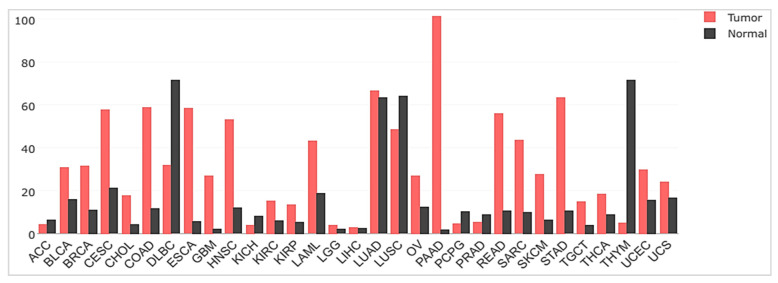
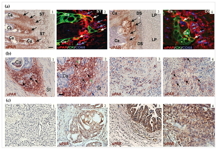
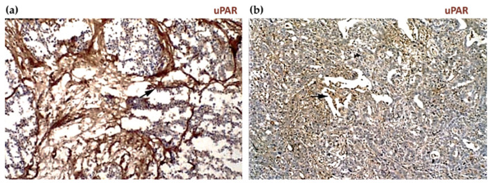
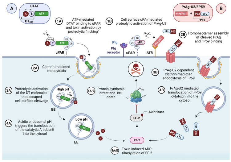
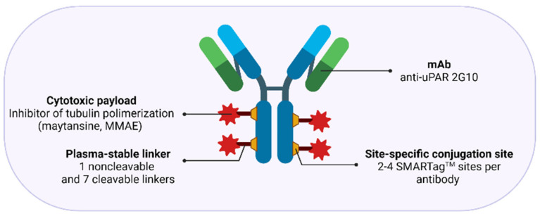
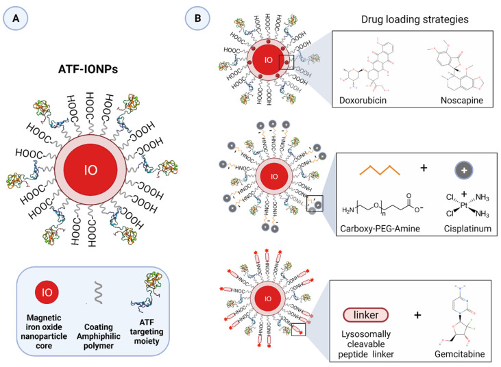
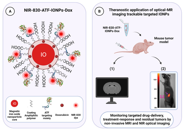
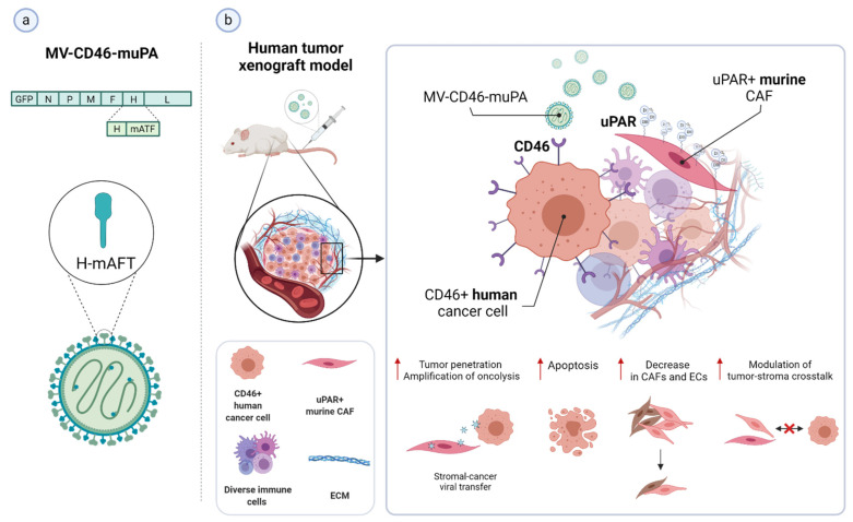
Verification
Claims the paper makes
The claims and statistics of this paper haven’t been checked yet.
Run the check — free, no account needed. It reads the open-access text and checks the sources it cites.
Claims other papers make about this one
Each is checked against this paper’s own text: does it say what the citing paper says it does?
The source backs part of the claim, not all of it — or backs it for a different population, condition or comparison.
In the citing paper
“Radiopharmaceutical case studies vividly illustrate instances where this approach resulted in tumor regression and prolonged survival, underscoring the promise of such tailored strategies [97].”
From Radiopharmaceuticals: navigating the frontier of precision medicine and therapeutic innovation (2024) · cites this paper as [97]
Experimental — accuracy not yet measured
Self-check
Percentages, sums and reported differences re-computed from the paper’s own numbers.
The self-check needs the paper’s full text.
Citations
Cites
Items 201–266 of 266
Schafer, J., Peters, D. E., Morley, T., Liu, S., Molinolo, A., Leppla, S. H., & Bugge, T. H. (2011) · PLoS ONE · doi:10.1371/journal.pone.0020532 · Cited by 36
Jing, Y., Zaias, J., DUNCAN, R. C., Russell, S. J., & Merchan, J. R. (2014) · Gene Therapy · doi:10.1038/gt.2013.84 · Cited by 36
Molecular Effects of Stromal-Selective Targeting by uPAR-Retargeted Oncolytic Virus in Breast Cancer
Jing, Y., Chavez, V. A., Ban, Y. J., Acquavella, N., El-Ashry, D., Pronin, A. N., Chen, X., & Merchan, J. R. (2017) · Molecular Cancer Research · doi:10.1158/1541-7786.mcr-17-0016 · Cited by 36
Molecular imaging of the urokinase plasminogen activator receptor: opportunities beyond cancer
Baart, V. M., Houvast, R. D., de Geus‐Oei, L., Quax, P. H. A., Kuppen, P. J. K., Vahrmeijer, A. L., & Sier, C. F. M. (2020) · EJNMMI Research · doi:10.1186/s13550-020-00673-7 · Cited by 36
Oh, S., Tsai, A. K., Ohlfest, J. R., Panoskaltsis‐Mortari, A., & Vallera, D. A. (2011) · Journal of neurosurgery · doi:10.3171/2010.11.jns101214 · Cited by 35
Yamashita, K., Tanaka, Y., Mimori, K., Inoue, H., & Mori, M. (2004) · International Journal of Cancer · doi:10.1002/ijc.20067 · Cited by 35
Tsai, A. K., Oh, S., Chen, H., Shu, Y., Ohlfest, J. R., & Vallera, D. A. (2010) · Journal of Neuro-Oncology · doi:10.1007/s11060-010-0392-5 · Cited by 35
Hildenbrand, R., Glienke, W., Magdolen, V., Graeff, H., Stutte, H.-J., & Schmitt, M. (1998) · Histochemistry and Cell Biology · doi:10.1007/s004180050261 · Cited by 35
Giannopoulou, I., Mylona, E., Kapranou, A., Mavrommatis, J., Markaki, S., Zoumbouli, C., Keramopoulos, A., & Nakopoulou, L. (2006) · Cancer Letters · doi:10.1016/j.canlet.2006.03.003 · Cited by 35
Li, Z., Wang, C., Chen, J., Lian, X., Xiong, C., Tian, R., Hu, L., Xiong, X., & Tian, J. (2020) · Materials & Design · doi:10.1016/j.matdes.2020.109386 · Cited by 35
Abi‐Habib, R. J., Liu, S., Bugge, T. H., Leppla, S. H., & Frankel, A. E. (2004) · Blood · doi:10.1182/blood-2004-01-0339 · Cited by 34
Ippoliti, R. (2000) · The FASEB Journal · doi:10.1096/fj.14.10.1335 · Cited by 34
Murad, J., Graber, D. J., & Sentman, C. L. (2018) · Best Practice & Research Clinical Haematology · doi:10.1016/j.beha.2018.03.003 · Cited by 33
Structural Basis for Therapeutic Intervention of uPA/uPAR System
Ngo, J. C. K., Jiang, L., Lin, Z., Yuan, C., Chen, Z., Zhang, X., Yu, H., Wang, J., Lin, L., & Huang, M. (2011) · Current Drug Targets · doi:10.2174/138945011797635911 · Cited by 32
Ploug, M. (2013) · Theranostics · doi:10.7150/thno.3791 · Cited by 32
Self-citationUrokinase Plasminogen Activator Receptor (uPAR) Targeted Nuclear Imaging and Radionuclide Therapy
Li, D., Liu, S., Shan, H., Conti, P., & Li, Z. (2013) · Theranostics · doi:10.7150/thno.5557 · Cited by 32
Reprogramming Urokinase into an Antibody-Recruiting Anticancer Agent
Jakobsche, C. E., McEnaney, P. J., Zhang, A. X., & Spiegel, D. A. (2011) · ACS Chemical Biology · doi:10.1021/cb200374e · Cited by 32
Hansen, L., Larsen, E., Nielsen, E. N., Iversen, F., Liu, Z., Thomsen, K. L., Pedersen, M., Skrydstrup, T., Nielsen, N. C., Ploug, M., et al. (2013) · Nanoscale · doi:10.1039/c3nr32922d · Cited by 31
Self-citationImmunotoxin Therapy for CNS Tumor
Rustamzadeh, E., Low, W. C., Vallera, D. A., & Hall, W. A. (2003) · Journal of Neuro-Oncology · doi:10.1007/bf02700025 · Cited by 31
Development of inhibitors for uPAR: blocking the interaction of uPAR with its partners
Yuan, C., Guo, Z., Yu, S., Jiang, L., & Huang, M. (2021) · Drug Discovery Today · doi:10.1016/j.drudis.2021.01.016 · Cited by 31 · cited 1×
“These include monoclonal antibodies, small molecules- and peptide-derived antagonists (recently reviewed in [62]), recombinant uPA-derived fusion proteins, and various gene therapy approaches.”
BackgroundRustamzadeh, E., Vallera, D. A., Todhunter, D. A., Low, W. C., Panoskaltsis‐Mortari, A., & Hall, W. A. (2005) · Journal of Neuro-Oncology · doi:10.1007/s11060-005-9051-7 · Cited by 30
Gårdsvoll, H., Kjærgaard, M., Jacobsen, B., Kriegbaum, M. C., Huang, M., & Ploug, M. (2011) · Journal of Biological Chemistry · doi:10.1074/jbc.m111.300020 · Cited by 30
Self-citationSu, Y., Ortiz, J. M., Liu, S., Bugge, T. H., Singh, R., Leppla, S. H., & Frankel, A. E. (2007) · Cancer Research · doi:10.1158/0008-5472.can-06-4642 · Cited by 30
Wang, L., Yang, R., Zhao, L., Zhang, X., Xu, T., & Cui, M. (2019) · Biomedicine & Pharmacotherapy · doi:10.1016/j.biopha.2019.109173 · Cited by 30
Harel, E., Drake, P. M., Barfield, R. M., Lui, I., Farr-Jones, S., Veer, L. V., Gartner, Z. J., Green, E. M., Lourenço, A. L., Cheng, Y., et al. (2019) · Antibodies · doi:10.3390/antib8040054 · Cited by 30
Differential Roles of uPAR in Peritoneal Ovarian Carcinomatosis
Al-Hassan, N., Behzadian, A., Caldwell, R. B., Ivanova, V. S., Syed, V., Motamed, K., & Said, N. A. (2012) · Neoplasia · doi:10.1593/neo.12442 · Cited by 29
Dohn, L. H., Illemann, M., Høyer‐Hansen, G., Christensen, I. J., Hostmark, J. G., Litlekalsøy, J., von der Maase, H., Pappot, H., & Lærum, O. D. (2015) · Urologic Oncology Seminars and Original Investigations · doi:10.1016/j.urolonc.2014.12.001 · Cited by 29
HER2 and uPAR cooperativity contribute to metastatic phenotype of HER2-positive breast cancer
Chandran, V. I., Eppenberger‐Castori, S., Venkatesh, T., Vine, K. L., & Ranson, M. (2015) · Oncoscience · doi:10.18632/oncoscience.146 · Cited by 29
Antibody recruiting molecules (ARMs): synthetic immunotherapeutics to fight cancer
Achilli, S., Berthet, N., & Renaudet, O. (2021) · RSC Chemical Biology · doi:10.1039/d1cb00007a · Cited by 29
Hillig, T., Engelholm, L. H., Ingvarsen, S., Madsen, D. H., Gårdsvoll, H., Larsen, J. K., Ploug, M., Danø, K., Kjøller, L., & Behrendt, N. (2008) · Journal of Biological Chemistry · doi:10.1074/jbc.c700214200 · Cited by 27
Self-citationLærum, O. D., Øvrebø, K., Skarstein, A., Christensen, I. J., Alpízar‐Alpízar, W., Helgeland, L., Danø, K., Nielsen, B. S., & Illemann, M. (2011) · International Journal of Cancer · doi:10.1002/ijc.26382 · Cited by 27
Salajegheh, M. K., Rudnicki, A., & Smith, T. W. (2005) · Applied Immunohistochemistry · doi:10.1097/01.pai.0000138448.85231.da · Cited by 26
Plasminogen Mediates the Pathological Effects of Urokinase-Type Plasminogen Activator Overexpression
Bolon, I., Zhou, H., Charron, Y., Wohlwend, A., & Vassalli, J. (2004) · American Journal Of Pathology · doi:10.1016/s0002-9440(10)63786-8 · Cited by 25 · cited 1×
“The normal skin phenotype exhibited by either the single transgenic mice, bitransgenic mice overexpressing uPAR and a receptor-binding catalytically inactive form of uPA, or bitransgenic mice crossed into a Plg −/− background undoubtedly underscores the strict requirement for the assembly of a functional cell surface…”
BackgroundBispecific targeting of EGFR and uPAR in a mouse model of head and neck squamous cell carcinoma
Waldron, N. N., Oh, S., & Vallera, D. A. (2012) · Oral Oncology · doi:10.1016/j.oraloncology.2012.06.002 · Cited by 25
Fosbøl, M. Ø., Kurbegovic, S., Johannesen, H. H., Røder, M. A., Hansen, A. E., Mortensen, J., Loft, A., Petersen, R. H., Madsen, J., Brasso, K., et al. (2020) · Journal of Nuclear Medicine · doi:10.2967/jnumed.120.248120 · Cited by 25 · cited 1×
“The design and implementation of theranostic approaches combining uPAR-targeted therapeutic and non-invasive imaging modalities, such as positron emission tomography (PET), magnetic resonance (MRI), or near-infrared (NIR) fluorescence imaging, are currently underway and hold clinical potential for significantly improv…”
Backgroundde Geus, S. W. L., Baart, V. M., Boonstra, M. C., Kuppen, P. J. K., Prevoo, H. A. J. M., Mazar, A. P., Bonsing, B. A., Morreau, H. M., van de Velde, C. J., Vahrmeijer, A. L., et al. (2017) · Biomarker Insights · doi:10.1177/1177271917715443 · Cited by 23
Pilbeam, K., Wang, H., Taras, E. P., Bergerson, R. J., Ettestad, B., DeFor, T. E., Borgatti, A., Vallera, D. A., & Verneris, M. R. (2017) · Oncotarget · doi:10.18632/oncotarget.21187 · Cited by 23
Akahane, T., Ishii, M., Ohtani, H., Nagura, H., & Toyota, T. (1998) · Liver International · doi:10.1111/j.1600-0676.1998.tb00826.x · Cited by 22
Huang, M., Mazar, A. P., Parry, G. C., Higazi, A. A., Kuo, A., & Cines, D. B. (2005) · Acta Crystallographica Section D Biological Crystallography · doi:10.1107/s0907444905014174 · Cited by 22 · cited 1×
“, Vn, as detailed in the next paragraph [38,41,51].”
BackgroundUrokinase receptor promotes ovarian cancer cell dissemination through its 84-95 sequence
Bifulco, K., Votta, G., Ingangi, V., Carluccio, G. D., Rea, D., Losito, S., Montuori, N., Ragno, P., Stoppelli, M. P., Arra, C., et al. (2014) · Oncotarget · doi:10.18632/oncotarget.1930 · Cited by 20 · cited 1×
“These uPAR forms may therefore allegedly play a role in the process of tumor progression, possibly as autocrine or paracrine signals for tumor cell motility and angiogenesis [59,65,67].”
BackgroundProvenzano, A. E., Posteri, R., Giansanti, F., Angelucci, F., Flavell, S. U., Flavell, D. J., Fabbrini, M. S., Porro, D., Ippoliti, R., Ceriotti, A., et al. (2016) · Microbial Cell Factories · doi:10.1186/s12934-016-0589-1 · Cited by 20
The anti-tumoral potential of the saporin-based uPAR-targeting chimera ATF-SAP
Zuppone, S., Assalini, C., Minici, C., Bertagnoli, S., Branduardi, P., Degano, M., Fabbrini, M. S., Montorsi, F., Salonia, A., & Vago, R. (2020) · Scientific Reports · doi:10.1038/s41598-020-59313-8 · Cited by 20
Development of AE147 Peptide-Conjugated Nanocarriers for Targeting uPAR-Overexpressing Cancer Cells
Park, J. Y., Shin, Y., Won, W. R., Lim, C., Kim, J. C., Kang, K., Husni, P., Lee, E. S., Youn, Y. S., & Oh, K. T. (2021) · International Journal of Nanomedicine · doi:10.2147/ijn.s315619 · Cited by 20
Dohn, L. H., Pappot, H., Iversen, B. R., Illemann, M., Høyer‐Hansen, G., Christensen, I. J., Thind, P. O., Salling, L., von der Maase, H., & Lærum, O. D. (2015) · PLoS ONE · doi:10.1371/journal.pone.0135824 · Cited by 19
Huang, J. X., Li, Y. M., Massagué, J., Sicheneder, A., Vallera, D. A., & Hall, W. A. (2012) · Journal of Neuro-Oncology · doi:10.1007/s11060-012-0904-6 · Cited by 19
Jing, Y., Toro, M., Zaias, J., & Merchan, J. R. (2014) · Breast Cancer Research and Treatment · doi:10.1007/s10549-014-3236-8 · Cited by 19
Leth, J. M., Mertens, H. D. T., Leth-Espensen, K. Z., Jørgensen, T. J. D., & Ploug, M. (2019) · Journal of Biological Chemistry · doi:10.1074/jbc.ra119.007847 · Cited by 17 · cited 2×
“2 n ), which exhibits a pronounced species-specificity between humans and mice [49,50].”
BackgroundSelf-citationSalasznyk, R. M., Zappala, M., Zheng, M., Yu, L., Wilkins-Port, C. E., & McKeown‐Longo, P. J. (2007) · Matrix Biology · doi:10.1016/j.matbio.2007.01.009 · Cited by 16
Oh, F., Todhunter, D., Taras, E. P., Vallera, D. A., & Borgatti, A. (2018) · Clinical Pharmacology Advances and Applications · doi:10.2147/cpaa.s160262 · Cited by 16
Murakami, K., Kotani, Y., Suzuki, A., Takaya, H., Nakai, H., Matsuki, M., Sato, T., Mandai, M., & Matsumura, N. (2020) · Scientific Reports · doi:10.1038/s41598-020-64926-0 · Cited by 16
Ippoliti, R., Lendaro, E., Benedetti, P. A., et al. (2000) · The FASEB Journal · doi:10.1096/fasebj.14.10.1335 · Cited by 16
Translational research on u-PAR
Allgayer, H. (2010) · European Journal of Cancer · doi:10.1016/j.ejca.2010.02.029 · Cited by 14 · cited 4×
“This association may have a bearing on the ability of uPAR to drive the transition between single-cell tumor dormancy and proliferation, which allows for the long-term survival of residual tumor cells during dormancy and reactivation of their proliferation years after primary treatment upon favorable conditions [13,85…”
BackgroundInfluentialUsing Nanoparticles in Medicine for Liver Cancer Imaging
Moghadam, F. F. (2017) · Oman Medical Journal · doi:10.5001/omj.2017.54 · Cited by 14
In vivo antitumor activity by dual stromal and tumor-targeted oncolytic measles viruses
Jing, Y., Chavez, V. A., Khatwani, N. K., Ban, Y. J., Freire, A. P. E., Chen, X., & Merchan, J. R. (2020) · Cancer Gene Therapy · doi:10.1038/s41417-020-0171-1 · Cited by 14
Oh, F., Modiano, J. F., Bachanová, V., & Vallera, D. A. (2020) · Biomolecules · doi:10.3390/biom10060956 · Cited by 14
Cell-targeted cytotoxics: a new generation of cytotoxic agents for cancer treatment
Bailly, C. (2013) · Phytochemistry Reviews · doi:10.1007/s11101-013-9300-x · Cited by 13 · cited 3×
“Most drug development is now oriented toward the design of cancer-cell-targeted cytotoxic interventions [1].”
BackgroundInfluentialSerpa, M. S., Mafra, R. P., Queiroz, S. I. M. L., da Silva, L. P., de Souza, L. B., & Pinto, L. P. (2018) · Brazilian Oral Research · doi:10.1590/1807-3107bor-2018.vol32.0093 · Cited by 13
Skovgaard, D. C., Persson, M., & Kjær, A. (2016) · Clinical and Translational Imaging · doi:10.1007/s40336-016-0197-4 · Cited by 12 · cited 1×
“There ar also malignancies wh re t prominent uPAR expression i reported on cancer ells such a in esophageal [121,12 ] and cutaneous sq amous cell arcinomas [40], as well as in gl omas such as anaplastic astrocyt ma and gliob astomas [123, 24].”
BackgroundLiu, X., Liu, X., Sunchen, S., Liu, M., Shen, C., Wu, J., Zhao, W., Yu, B., & Liu, J. (2017) · Drug Delivery · doi:10.1080/10717544.2017.1406560 · Cited by 10
Luo, Y., Pan, Q., & Li, F. (2020) · Journal of Pancreatology · doi:10.1097/jp9.0000000000000053 · Cited by 10
Inhibitory Effects of a Reengineered Anthrax Toxin on Canine Oral Mucosal Melanomas
Nishiya, A. T., Nagamine, M. K., da Fonseca, I. I. M., Miraldo, A. C., Scattone, N. V., Guerra, J. L., Xavier, J. G., Santos, M., Gomes, C. D. O. M. S., Ward, J. M., et al. (2020) · Toxins · doi:10.3390/toxins12030157 · Cited by 8
Mimori, K., Kataoka, A., Yamaguchi, H., Masuda, N., Kosaka, Y., Ishii, H., Ohno, S., & Mori, M. (2009) · Annals of Surgical Oncology · doi:10.1245/s10434-009-0465-x · Cited by 6
Almholt, K., Lærum, O. D., Nielsen, B. S., Lund, I. K., Lund, L. R., Rømer, J., & Jögi, A. (2015) · Clinical & Experimental Metastasis · doi:10.1007/s10585-015-9726-1 · Cited by 5 · cited 1×
“Although impairment of uPAR function or inhibition of its expression was associated with impaired invasive, metastatic, and tumorigenic potential of many tumor models in vitro and in vivo [4,11,12,14,16,17,20,52,57,79,84,85], one study in transgenic mice with uPAR gene ablation showed no pronounced causal effects on d…”
BackgroundUrokinase Receptor (uPAR) Ligand based Recombinant Toxins for Human Cancer Therapy
de Virgilio, M., & Silvestris, F. (2011) · Current Pharmaceutical Design · doi:10.2174/138161211796718170 · Cited by 5
Liu, Y., & Li, S. (2008) · Frontiers in Biology · doi:10.1007/s11515-008-0074-2 · Cited by 3
Research works printed but not linked: 1
- Sampson, J.H. (2017). Chapter 10-Immunotoxin Therapy for Brain Tumors. Translational Immunotherapy of Brain Tumors, Academic Press.
Cited by
Sorted by citations
Radiopharmaceuticals: navigating the frontier of precision medicine and therapeutic innovation
Dhoundiyal, S., Srivastava, S., Kumar, S., Singh, G., Ashique, S., Pal, R., Mishra, N., & Taghizadeh-Hesary, F. (2024) · European Journal of Medical Research · doi:10.1186/s40001-023-01627-0 · Cited by 144
“Radiopharmaceuti-cal case studies vividly illustrate instances where this approach resulted in tumor regression and prolonged survival, underscoring the promise of such tailored strategies [97].”
BackgroundUrokinase-type plasminogen activator receptor (uPAR) as a therapeutic target in cancer
Zhai, B., Tian, H., Sun, J., Zou, J., Zhang, X., Cheng, J., Shi, Y., Fan, Y. B., & Guo, D. (2022) · Journal of Translational Medicine · doi:10.1186/s12967-022-03329-3 · Cited by 123
Hamada, M., Varkoly, K., Riyadh, O., Beladi, R. N., Munuswamy‐Ramanujam, G., Rawls, A., Wilson‐Rawls, J., Chen, H., McFadden, G., & Lucas, A. R. (2024) · Biomedicines · doi:10.3390/biomedicines12061167 · Cited by 61
“Cleavage of the D1–D2 linker domains by other proteases irreversibly blocks uPAR’s interaction with other proteins [27,28].”
BackgroundModulation of Cellular Function by the Urokinase Receptor Signalling: A Mechanistic View
Alfano, D., Franco, P., & Stoppelli, M. P. (2022) · Frontiers in Cell and Developmental Biology · doi:10.3389/fcell.2022.818616 · Cited by 59
“However, despite the advances in the molecular design and generation of uPAR-derived peptides or novel interactors, no uPAR-targeting therapeutics have currently progressed to clinical trials (Metrangolo et al., 2021).”
BackgroundBogdanović, B., Fagret, D., Ghezzi, C., & Montemagno, C. (2024) · Pharmaceuticals · doi:10.3390/ph17111556 · Cited by 49
“uPAR is a cell surface receptor crucial for cell migration, tissue remodeling, and inflammation. uPAR is frequently overexpressed in cancers such as breast, prostate, and lung cancer, where it is associated with increased tumor invasion and poor prognosis [174].”
Metadata
- Authors
- Metrangolo, V., Ploug, M., & Engelholm, L. H.
- Affiliations
- University of Copenhagen (DK)
- Corresponding author
- Engelholm, Lars H.
- Venue
- Cancers
- Publisher
- MDPI AG
- Published
- 2021-10-27
- Volume
- 13
- Issue
- 21
- Pages
- 5376
- ISSN
- 2072-6694 (ISSN-L)
- PMC
- PMC8582577
- OpenAlex
- W3209572831
- Cited by
- 44
- References
- 267
- Citations by year
- 2026: 4 · 2025: 11 · 2024: 8 · 2023: 11 · 2022: 10
- FWCI
- 0.87
- Type
- journal article
- Language
- en
- Open access
- gold
- doi.org
- Licence
- Locations
- Cancers (publishedVersion, cc-by)
- Europe PMC (PubMed Central) (publishedVersion, other-oa)
- PubMed Central (publishedVersion, cc-by)
- PubMed (publishedVersion)
- Research at the University of Copenhagen (University of Copenhagen) (publishedVersion, cc-by)
- DOAJ (DOAJ: Directory of Open Access Journals) (submittedVersion, cc-by-sa)
- Funders
- European Union’s Horizon 2020 research and innovation programme under the Marie Skłodowska Curie (801481)
- Danish Cancer Society (R231-A13832)
- Topics
- Life Sciences › Biochemistry, Genetics and Molecular Biology › Cancer Research › Protease and Inhibitor Mechanisms
- Life Sciences › Biochemistry, Genetics and Molecular Biology › Biochemistry › Peptidase Inhibition and Analysis
- Health Sciences › Medicine › Hematology › Blood Coagulation and Thrombosis Mechanisms
- Keywords
- urokinase-type plasminogen activator receptor, uPAR, targeted cancer therapy, precision oncology, plasminogen activation, extracellular matrix degradation, metastasis, cell migration, off-target toxicity, prognostic factors
About these checks
Registry, retraction, Cites and statistics results are computed, not judged, with one exception: a printed reference that no index lists is read once by a model before it counts as a citation error. Claims are extracted by a model and kept only when their quote appears verbatim in the text; each cited source is then checked, with a model’s help, for whether it says what the claim relies on. Those verdicts can be wrong — read the claim’s quote and the cited source before relying on one.
Statistics and arithmetic are recomputed, not judged.
Confidence bands are shown only for fields calibrated on our annotated test set. None is calibrated yet, so no band is shown.
What counts as a claim ›How we measure confidence ›How we check this ›
Are you an author, or do you see a problem with this page? Request a review
