

The DOI matches its Crossref record; 129 of the 131 references it cites were found in a registry or an index.
AI-Powered Diagnosis of Skin Cancer: A Contemporary Review, Open Challenges and Future Research Directions
Open accessAbstract
Skin cancer continues to remain one of the major healthcare issues across the globe. If diagnosed early, skin cancer can be treated successfully. While early diagnosis is paramount for an effective cure for cancer, the current process requires the involvement of skin cancer specialists, which makes it an expensive procedure and not easily available and affordable in developing countries. This dearth of skin cancer specialists has given rise to the need to develop automated diagnosis systems. In this context, Artificial Intelligence (AI)-based methods have been proposed. These systems can assist in the early detection of skin cancer and can consequently lower its morbidity, and, in turn, alleviate the mortality rate associated with it. Machine learning and deep learning are branches of AI that deal with statistical modeling and inference, which progressively learn from data fed into them to predict desired objectives and characteristics. This survey focuses on Machine Learning and Deep Learning techniques deployed in the field of skin cancer diagnosis, while maintaining a balance between both techniques. A comparison is made to widely used datasets and prevalent review papers, discussing automated skin cancer diagnosis. The study also discusses the insights and lessons yielded by the prior works. The survey culminates with future direction and scope, which will subsequently help in addressing the challenges faced within automated skin cancer diagnosis.
Overview
The claims and statistics of this paper haven’t been checked yet.
Run the check — free, no account needed. It reads the open-access text and checks the sources it cites.
Integrity signals
Registry
Cites
Statistics
Figures
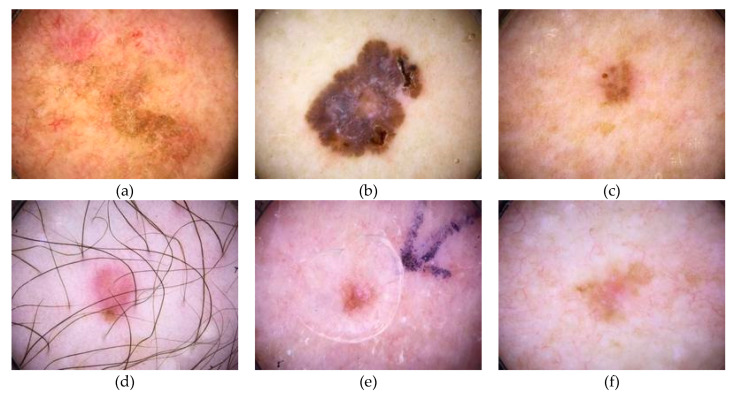
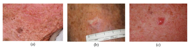
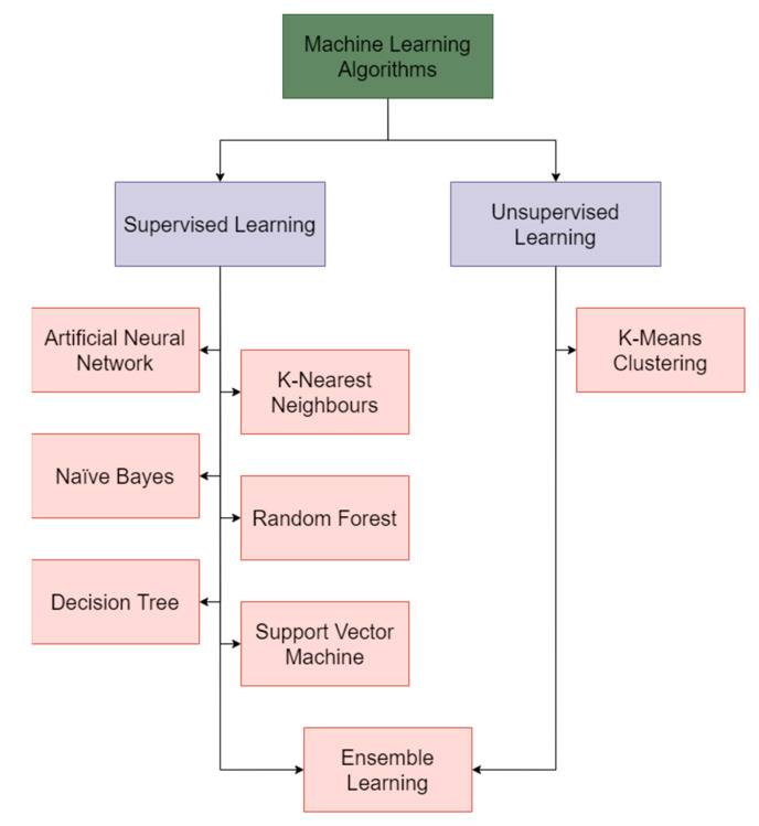
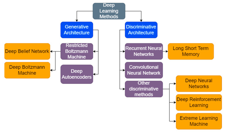
Verification
Claims the paper makes
The claims and statistics of this paper haven’t been checked yet.
Run the check — free, no account needed. It reads the open-access text and checks the sources it cites.
Claims other papers make about this one
Each is checked against this paper’s own text: does it say what the citing paper says it does?
In the citing paper
“Artificial intelligence (AI) for medical diagnosis holds potential, demonstrating high performance in detecting various diseases.57”
From Using artificial intelligence to improve human performance: efficient retinal disease detection training with synthetic images (2024) · cites this paper as [5]
Self-check
Percentages, sums and reported differences re-computed from the paper’s own numbers.
The self-check needs the paper’s full text.
Citations
Cites
2 retracted
RETRACTED ARTICLE: Skin lesion classification using decision trees and random forest algorithms
Dhivyaa, C. R., Sangeetha, K., -, M. B., Amaran, S., Vetriselvi, T., & Johnpaul, P. (2020) · Journal of Ambient Intelligence and Humanized Computing · doi:10.1007/s12652-020-02675-8 · Cited by 90
RetractedRETRACTED: Computer aided intelligent medical system and nursing of breast surgery infection
Sun, J., & Huang, Y. R. (2020) · Microprocessors and Microsystems · doi:10.1016/j.micpro.2020.103769 · Cited by 3
RetractedDermatologist-level classification of skin cancer with deep neural networks
Esteva, A., Kuprel, B., Novoa, R. A., Ko, J., Swetter, S. M., Blau, H. M., & Thrun, S. (2017) · Nature · doi:10.1038/nature21056 · Cited by 14,362
Overview of Next‐Generation Sequencing Technologies
Slatko, B. E., Gardner, A. F., & Ausubel, F. M. (2018) · Current Protocols in Molecular Biology · doi:10.1002/cpmb.59 · Cited by 968
Next-generation sequencing technologies: An overview
Hu, T., Chitnis, N., Monos, D., & Dinh, A. (2021) · Human Immunology · doi:10.1016/j.humimm.2021.02.012 · Cited by 949
Classification of Skin Disease Using Deep Learning Neural Networks with MobileNet V2 and LSTM
Srinivasu, P. N., SivaSai, J. G., Ijaz, M. F., Bhoi, A. K., Kim, W., & Kang, J. J. (2021) · Sensors · doi:10.3390/s21082852 · Cited by 763
Han, S. S., Kim, M. S., Lim, W., Park, G., Park, I., & Chang, S. E. (2018) · Journal of Investigative Dermatology · doi:10.1016/j.jid.2018.01.028 · Cited by 740
Skin Cancer Classification Using Convolutional Neural Networks: Systematic Review
Brinker, T. J., Hekler, A., Utikal, J. S., Grabe, N., Schadendorf, D., Klode, J., Berking, C., Steeb, T., Enk, A. H., & Kalle, C. V. (2018) · Journal of Medical Internet Research · doi:10.2196/11936 · Cited by 528
Goyal, M., Knackstedt, T., Yan, S., & Hassanpour, S. (2020) · Computers in Biology and Medicine · doi:10.1016/j.compbiomed.2020.104065 · Cited by 476
Cancer Diagnosis Using Deep Learning: A Bibliographic Review
Munir, K., Elahi, H., Ayub, A., Frezza, F., & Rizzi, A. (2019) · Cancers · doi:10.3390/cancers11091235 · Cited by 437
Research works printed but not linked: 6
- Nahata, H., and Singh, S.P. (2020). Machine Learning with Health Care Perspective, Springer.
- Alwan, 2022, Skin cancer images classification using naïve bayes, Emergent J. Educ. Discov. Lifelong Learn., 3, 19
- Anas, 2017, Skin cancer classification using K-means clustering, Int. J. Tech. Res. Appl., 5, 62
- Peter Soosai Anandaraj, A., Gomathy, V., Amali Angel Punitha, A., Abitha Kumari, D., Sheeba Rani, S., and Sureshkumar, S. (2021). Cognitive Internet of Medical Things for Smart Healthcare, Springer.
- Gerke, S., Minssen, T., and Cohen, G. (2020). Artificial Intelligence in Healthcare, Academic Press.
- Barata, C., Marques, J.S., and Celebi, M.E. (2014). 2014 IEEE International Conference on Image Processing (ICIP), IEEE.
Cited by
Sorted by citations
Items 101–144 of 144
Dermoskopik Görüntü Sınıflandırmada Temel Veri Artırım Yöntemlerinin Değerlendirilmesi
Onan, A., Bulut, V., & Ezgi, A. (2023) · International Conference on Recent and Innovative Results in Engineering and Technology (ICRIRET) · doi:10.59287/icriret.1376
Pavithra, R., & Samraj, J. (2024) · Lecture notes in electrical engineering · doi:10.1007/978-981-97-2839-8_6
Malaiarasan, S., & Ravi, R. (2024) · International Journal of Imaging Systems and Technology · doi:10.1002/ima.23167
Exploratory Analysis of Skin Cancer Dermatoscopic Image Datasets and Classification Methods
Ghatge, D., Sankpal, P., Gharge, V., Patil, R., & Zanjurne, S. (2024) · International Research Journal on Advanced Engineering Hub (IRJAEH) · doi:10.47392/irjaeh.2024.0385
Artificial Intelligence-Driven Multidirectional Curvelet Analysis for Enganced Skin Cancer DetectionPreprint
RAZAK, M. S. A., Lakshmi, V., V, Theivanathan, G., & Radhakrishnan, K. (2025) · SSRN Electronic Journal · doi:10.2139/ssrn.5127060 · Cited by 5
XAI-Driven Skin Cancer DiagnosisPreprint
Ranjeet, K., Saha, S., & Ratnakumari, M. (2025) · SSRN Electronic Journal · doi:10.2139/ssrn.5237430
Haut-Screening-Apps in der klinischen Praxis
Gerschewski, F. E. (2025) · hautnah · doi:10.1007/s12326-025-00721-w
Hariarasi, A., & Banumathi, J. (2025) · International Conference on Innovative Trends in Information Technology (ICITIIT) · doi:10.1109/icitiit64777.2025.11041049
Petrie, T. C., Samatham, R. V., Webster, D. E., & Leachman, S. A. (2025) · Scientific Data · doi:10.1038/s41597-025-05552-1
Thakur, A., Chowdhury, K., Sharma, V. V., Singh, K., Gupta, J. K., Jain, D., & Sharma, M. C. (2025) · Combinatorial Chemistry & High Throughput Screening · doi:10.2174/0113862073397811250716154921
Comparative Study Of Gradient Optimizers For Enhanced Skin Cancer Image Analysis
Falana, W. O. (2025) · International Symposium on Innovative Approaches in Smart Technologies (ISAS) · doi:10.1109/isas66241.2025.11101868
Enhanced U-Net-Based Deep Learning Model for Accurate Skin Cancer Detection and Prognosis
Kaur, M., Jayan, A., V, Maurya, N., Mahesh, K. S., & Singh, S. (2025) · International Conference on Sensors and Related Networks (SENNET) · doi:10.1109/sennet64220.2025.11136074
Unifying Classifiers: A Stacking-Based Strategy for Skin Disease Prediction
Tamanna, T., & Kumari, R. (2025) · Lecture notes in networks and systems · doi:10.1007/978-981-96-7131-1_34
Roadmap for Representative Artificial Intelligence Models for Thyroid Cancer
Ramchandani, R., Guo, E., Biglou, S. G., Sabbah, S. G., Mostowy, M., Mahiny, D., Hurtubise, C., Anicho‐Okereke, G., Shorr, R., Caulley, L., et al. (2025) · The Laryngoscope · doi:10.1002/lary.70169
Generative AI Framework for Asian Healthcare Organizations
Suchitra, M., Cheriyan, B. V., Shaik, A., & Panguluru, M. (2025) · Green Computational Intelligence · doi:10.1002/9781394383658.ch10
Exploring Skin Cancer Types and Stages: A Comprehensive Review
Kadia, V. M., & Degadwala, S. (2025) · Algorithms for intelligent systems · doi:10.1007/978-981-96-5265-5_3
Utilization of Feature Fusion in Diagnostic Applications
Abir, S. I., Shoha, S., Khan, N. I., & Mousumi, J. (2025) · Springer eBooks · doi:10.1007/978-3-031-94386-7_11
Das, D. P., Das, R., & Sahoo, A. (2025) · Algorithms for intelligent systems · doi:10.1007/978-981-95-0493-0_28
A Web-based Intelligent Framework for Early Facial Melanoma Detection using Deep Learning
Prakash, P. R., Rajasekaran, T., Sobika, M., Sabarish, M., Sivanesh, T., & A, S. S. S. (2025) · International Conference on Intelligent Cyber Physical Systems and Internet of Things (ICoICI) · doi:10.1109/icoici65217.2025.11252532
AI-Driven Assistance for Rapid Dermatology Triage
Sowmya, G. V., Jayasree, N., Chaithanya, V. M., Meena, T., Rani, B. U., & Aishwarya, G. (2025) · International Conference on Intelligent Cyber Physical Systems and Internet of Things (ICoICI) · doi:10.1109/icoici65217.2025.11252685
Skin AI - Virtual Dermatologist
Jyothsna, K., Reddy, V. S. V., Lokesh, D. L., Chaitanya, D. K., Sivakrishna, K., & Priyanka, L. (2025) · International Conference on Intelligent Cyber Physical Systems and Internet of Things (ICoICI) · doi:10.1109/icoici65217.2025.11253360
Deep Learning in Skin Cancer Diagnosis: A Review of Current Techniques and Future Directions
Younus, M. D., Basheer, S. I., Salim, A., & Mashadani, M. (2025) · International Conference on Business Analytics for Technology and Security (ICBATS) · doi:10.1109/icbats66542.2025.11258380
An Effective Model-Based Group Recommender System for Course Registration
Selva Rani, B., Subalakshmi, V., & Prabhakar, V. (2025) · International Conference on Applied Artificial Intelligence and Computing (ICAAIC) · doi:10.1109/icaaic64647.2025.11330973
A Robotic Arm-Based Intelligent Biopsy System
Ullah, Q. Z., Baig, H., Shaukat, M. H. B., Mughal, H., & Hassan, A. (2026) · Infosys science foundation series · doi:10.1007/978-981-95-2212-5_10
Dermafusion-Net: A Hybrid Deep Learning Framework for Automated Skin Cancer Detection
Manikandan, J., Priyanka, M., & Sowmiya, J. (2025) · International Conference on Emerging Research in Electronics, Computer Science and Technology (ICERECT) · doi:10.1109/icerect65215.2025.11377186
Yaswanth, M. S., & Reddy, C. M. K. (2025) · International Conference on Electronics, Communication and Aerospace Technology (ICECA) · doi:10.1109/iceca66444.2025.11382676
AI-Powered Pathology Image Analysis for Cancer Diagnosis
Namdev, M., & Dubey, K. (2025) · International Conference on Artificial Intelligence for Innovations in Healthcare Industries (ICAIIHI) · doi:10.1109/icaiihi67124.2025.11403630
A Comprehensive Review on Skin Lesion Analysis and Prognostic Modeling using Deep Learning
Dandawala, A. F., Jadhav, M. V., Shukla, R., & Kanikar, P. (2025) · International Conference on Artificial Intelligence for Innovations in Healthcare Industries (ICAIIHI) · doi:10.1109/icaiihi67124.2025.11403201
Kong, D., Pan, H., Wu, H. Y., & Chen, J. (2026) · Frontiers in Pediatrics · doi:10.3389/fped.2026.1659176
A brain-inspired computational framework for image-based risk assessment
Zhou, F., Hu, S., Xiaozheng, D. U., Li, N., & Zhou, T. (2026) · Scientific Reports · doi:10.1038/s41598-026-45033-y
Khizar, S. K., Balakrishnan, M., & Reddy, V. L. (2026) · International Conference on Intelligent Data Communication Technologies and Internet of Things (IDCIoT) · doi:10.1109/idciot67589.2026.11455693
Clinical and Regulatory Considerations
Purkayastha, H. D., Pokhrel, M., Bhattacharjee, A., Bhattacharjee, A., Sahu, R. K., & Swain, R. P. (2026) · Springer eBooks · doi:10.1007/978-981-95-7358-5_19
Basics of skin cancer: Types, diagnosis, and current challenges
Khalse, R., Khan, N., Sharma, M., Maru, S., & Patil, P. (2026) · Advances in cancer research · doi:10.1016/bs.acr.2026.03.001
Abofanas, A. L. S. A. K. A. M. M. M., & Abofanas, M. (2026) · IntechOpen eBooks · doi:10.5772/intechopen.1015870
Abedi, A., Wu, Q. M. J., Zhang, N., & Pourpanah, F. (2026) · Artif. Intell. Medicine · doi:10.1016/j.artmed.2026.103465
Nanotechnology-based drug delivery systems for skin cancer treatment
Anand, A., Manjula, S. N., Sharma, H., & Kenganora, M. (2026) · Elsevier eBooks · doi:10.1016/b978-0-443-26564-8.00011-6
Understanding skin cancer: Types, causes, and epidemiology
Mishra, A., Kumar, A., Anand, A., & Kumar, A. (2026) · Elsevier eBooks · doi:10.1016/b978-0-443-26564-8.00010-4
The Promise Vs. Peril of AI in LearningPreprint
Krishan, K. (2026) · SSRN Electronic Journal · doi:10.2139/ssrn.6897021
Al-Younis, I. M., Moosa, S., Bennici, G., Alzahrani, R. A., Alghamdi, W., Amwas, D., Manasra, R., Jaremko, M., & Emwas, A.-H. M. (2026) · IntechOpen eBooks · doi:10.5772/intechopen.1013625
Bhargavi, M., & Balakrishna, S. (2026) · The Review of Socionetwork Strategies · doi:10.1007/s12626-026-00219-8
LesionNet-FCL: Deep Learning Framework for Detecting and Classifying Skin Lesion
Jitya, V., & Bhargavi, M. (2026) · International Conference on Inventive Systems and Control (ICISC) · doi:10.1109/icisc69558.2026.11681652
Kropiwnicka, A., Habirek, K. S., Porażko, M. M., Żurakowska-Zadrożna, A. K., Krupska, A., Czarnecka, J. A., Marcinkowska, Z. M., Gugulska, J. Z., Kieres, M., Wójcik, K. K., et al. (2026) · International Journal of Innovative Technologies in Social Science · doi:10.31435/ijitss.3(51).2026.6064
Skin Lesion Image Classification Using Transformers and Meta Data Fusion Approach
K, R. K. S., Ashok Kumar, T., & Priya, S. (2026) · International Conference on Innovations in Mechanical, Robotics, Computing and Biomedical Engineering (ICIMRBE) · doi:10.1109/icimrbe70745.2026.11709913
Dermoscopy Explainable Intelligence Performance in Evaluating Melanoma and Non-Melanoma Skin Cancer
Holder, K., Maniam, G., Hardway, H., Todd, A., Comfere, N., Damodaran, S. S., Youssef, M., Demer, A., & Link, J. (2026) · Mayo Clinic Proceedings · doi:10.1016/j.mayocp.2026.07.031
Metadata
- Authors
- Melarkode, N., Srinivasan, K., Qaisar, S. M., & Plawiak, P.
- Affiliations
- Vellore Institute of Technology University (IN)
- Effat University (SA)
- Department of Computer Science, Faculty of Computer Science and Telecommunications, Cracow University of Technology, Warszawska 24, 31-155 Krakow, Poland
- Corresponding author
- Srinivasan, Kathiravan
- Venue
- Cancers
- Publisher
- MDPI AG
- Published
- 2023-02-13
- Volume
- 15
- Issue
- 4
- Pages
- 1183
- ISSN
- 2072-6694 (ISSN-L)
- PMC
- PMC9953963
- OpenAlex
- W4320492826
- Cited by
- 144
- References
- 131
- Citations by year
- 2026: 26 · 2025: 62 · 2024: 46 · 2023: 10
- FWCI
- 4.65
- Type
- journal article
- Language
- en
- Open access
- gold
- doi.org
- Licence
- Locations
- Cancers (publishedVersion, cc-by)
- PubMed Central (publishedVersion, cc-by)
- PubMed (publishedVersion)
- HAL (submittedVersion)
- Funders
- VIT University
- Politechnika Krakowska
- Effat University
- Topics
- Health Sciences › Medicine › Oncology › Cutaneous Melanoma Detection and Management
- Physical Sciences › Computer Science › Artificial Intelligence › AI in cancer detection
- Health Sciences › Health Professions › Health Information Management › Artificial Intelligence in Healthcare
- Keywords
- skin cancer, automated diagnosis, early detection, machine learning, deep learning, dermatological image analysis, skin lesion classification, medical image dataset, access to specialist care
About these checks
Registry, retraction, Cites and statistics results are computed, not judged, with one exception: a printed reference that no index lists is read once by a model before it counts as a citation error. Claims are extracted by a model and kept only when their quote appears verbatim in the text; each cited source is then checked, with a model’s help, for whether it says what the claim relies on. Those verdicts can be wrong — read the claim’s quote and the cited source before relying on one.
Statistics and arithmetic are recomputed, not judged.
Confidence bands are shown only for fields calibrated on our annotated test set. None is calibrated yet, so no band is shown.
What counts as a claim ›How we measure confidence ›How we check this ›
Are you an author, or do you see a problem with this page? Request a review
