

The DOI matches its Crossref record; 3 of the 3 references it cites were found in a registry or an index.
ER Stress-Induced Secretion of Proteins and Their Extracellular Functions in the Heart
Abstract
Endoplasmic reticulum (ER) stress is a result of conditions that imbalance protein homeostasis or proteostasis at the ER, for example ischemia, and is a common event in various human pathologies, including the diseased heart. Cardiac integrity and function depend on the active secretion of mature proteins from a variety of cell types in the heart, a process that requires an intact ER environment for efficient protein folding and trafficking to the secretory pathway. As a consequence of ER stress, most protein secretion by the ER secretory pathway is decreased. Strikingly, there is a select group of proteins that are secreted in greater quantities during ER stress. ER stress resulting from the dysregulation of ER Ca2+ levels, for instance, stimulates the secretion of Ca2+-binding ER chaperones, especially GRP78, GRP94, calreticulin, and mesencephalic astrocyte-derived neurotrophic factor (MANF), which play a multitude of roles outside the cell, strongly depending on the cell type and tissue. Here we review current insights in ER stress-induced secretion of proteins, particularly from the heart, and highlight the extracellular functions of these proteins, ranging from the augmentation of cardiac cell viability to the modulation of pro- and anti-apoptotic, oncogenic, and immune-stimulatory cell signaling, cell invasion, extracellular proteostasis, and more. Many of the roles of ER stress-induced protein secretion remain to be explored in the heart. This article is part of a special issue entitled “The Role of Proteostasis Derailment in Cardiac Diseases.”
Overview
The claims and statistics of this paper haven’t been checked yet.
Run the check — free, no account needed. It reads the open-access text and checks the sources it cites.
Integrity signals
Registry
Cites
Statistics
Figures
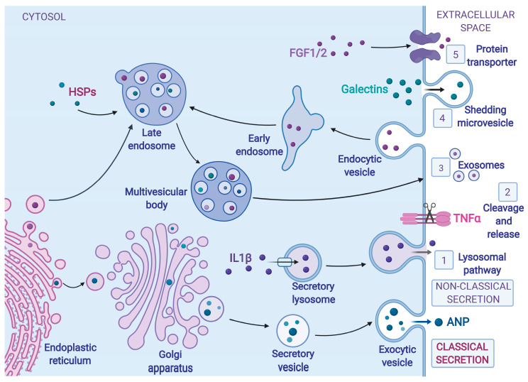
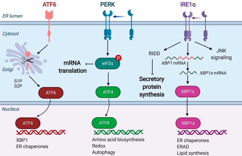
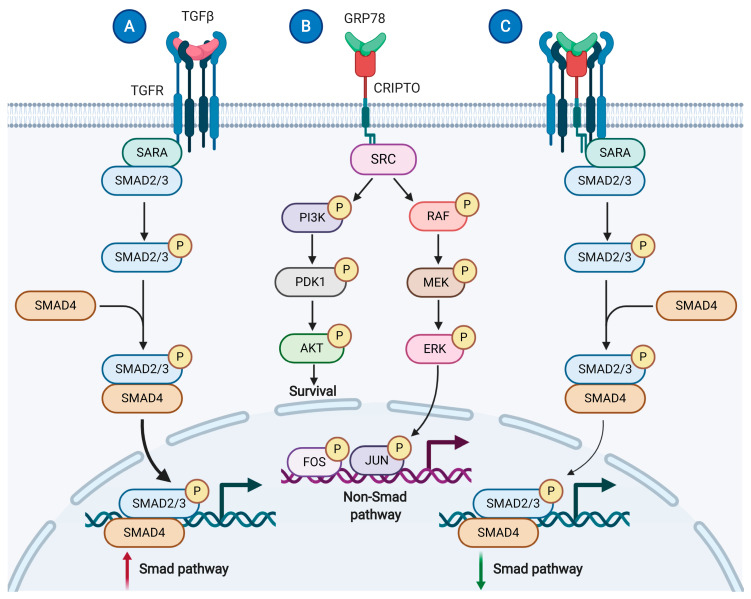
Verification
Claims the paper makes
The claims and statistics of this paper haven’t been checked yet.
Run the check — free, no account needed. It reads the open-access text and checks the sources it cites.
Claims other papers make about this one
Each is checked against this paper’s own text: does it say what the citing paper says it does?
In the citing paper
“others have shown that ER stress can induce the release of calreticulin and other ER-resident chaperones (30, 45).”
From LRPAP1 is released from activated microglia and inhibits microglial phagocytosis and amyloid beta aggregation (2023) · cites this paper as [45]
Self-check
Percentages, sums and reported differences re-computed from the paper’s own numbers.
The self-check needs the paper’s full text.
Citations
Cites
The Biology of Proteostasis in Aging and Disease
Labbadia, J., & Morimoto, R. I. (2015) · Annual Review of Biochemistry · doi:10.1146/annurev-biochem-060614-033955 · Cited by 1,472
Chao, M. P., Jaiswal, S., Weissman-Tsukamoto, R., Alizadeh, A. A., Gentles, A. J., Volkmer, J., Weiskopf, K., Willingham, S. B., Raveh, T., Park, C. Y., et al. (2010) · Science Translational Medicine · doi:10.1126/scitranslmed.3001375 · Cited by 776
Cardiokines: recent progress in elucidating the cardiac secretome
Shimano, M., Ouchi, N., & Walsh, K. (2012) · Circulation · doi:10.1161/circulationaha.112.150656 · Cited by 116
Research works printed but not linked: 134
- Kakkar, 2010, Intramyocardial fibroblast myocyte communication, Circ. Res., 106, 47, 10.1161/CIRCRESAHA.109.207456 · DOI
- Zhou, 2016, Recounting Cardiac Cellular Composition, Circ. Res., 118, 368, 10.1161/CIRCRESAHA.116.308139 · DOI
- Doroudgar, 2011, The cardiokine story unfolds: Ischemic stress-induced protein secretion in the heart, Trends Mol. Med., 17, 207, 10.1016/j.molmed.2010.12.003 · DOI
- Lee, 2004, Bi-Directional Protein Transport Between the Er and Golgi, Annu. Rev. Cell Dev. Biol., 20, 87, 10.1146/annurev.cellbio.20.010403.105307 · DOI · Paper page
- Nickel, 2009, Mechanisms of regulated unconventional protein secretion, Nat. Rev. Mol. Cell Biol., 10, 148, 10.1038/nrm2617 · DOI
- Elvekrog, 2015, Dynamics of co-translational protein targeting, Curr. Opin. Chem. Biol., 29, 79, 10.1016/j.cbpa.2015.09.016 · DOI
- Cabrita, 2010, Protein folding on the ribosome, Curr. Opin. Struct. Biol., 20, 33, 10.1016/j.sbi.2010.01.005 · DOI
- Gerst, 2010, Addressing mRNAs to the ER: Cis sequences act up!, Trends Biochem. Sci., 35, 459, 10.1016/j.tibs.2010.02.006 · DOI
- Weis, 2013, Protein targeting to subcellular organelles via mRNA localization, Biochim. Biophys. Acta Mol. Cell Res., 1833, 260, 10.1016/j.bbamcr.2012.04.004 · DOI
- Cohen-Zontag, O., Baez, C., Lim, L.Q.J., Olender, T., Schirman, D., Dahary, D., Pilpel, Y., and Gerst, J.E. (2019). A secretion-enhancing cis regulatory targeting element (SECReTE) involved in mRNA localization and protein synthesis. PLoS Genet., 15. · DOI
- Palade, 1975, Intracellular Aspects of the Process of Protein Synthesis, Science, 189, 867, 10.1126/science.1096303 · DOI
- Bloch, 1986, Neonatal atria and ventricles secrete atrial natriuretic factor via tissue-specific secretory pathways, Cell, 47, 695, 10.1016/0092-8674(86)90512-X · DOI
- Sei, 1992, The cosecretional maturation of atrial natriuretic factor by primary atrial myocytes, Mol. Endocrinol., 6, 309
- Maack, 1996, Role of atrial natriuretic factor in volume control, Kidney Int., 49, 1732, 10.1038/ki.1996.257 · DOI
- Kim, 2018, Unconventional protein secretion— New insights into the pathogenesis and therapeutic targets of human diseases, J. Cell Sci., 131, jcs213686, 10.1242/jcs.213686 · DOI
- Nickel, 2008, Unconventional Mechanisms of Protein Transport to the Cell Surface of Eukaryotic Cells, Annu. Rev. Cell Dev. Biol., 24, 287, 10.1146/annurev.cellbio.24.110707.175320 · DOI
- Most, 2005, Distinct subcellular location of the Ca2+-binding protein S100A1 differentially modulates Ca2+-cycling in ventricular rat cardiomyocytes, J. Cell Sci., 118, 421, 10.1242/jcs.01614 · DOI
- Balch, 2008, Adapting proteostasis for disease intervention, Science, 319, 916, 10.1126/science.1141448 · DOI · Paper page
- Hipp, 2019, The proteostasis network and its decline in ageing, Nat. Rev. Mol. Cell Biol., 20, 421, 10.1038/s41580-019-0101-y · DOI · Paper page
- Treaster, S.B., Ridgway, I.D., Richardson, C.A., Gaspar, M.B., Chaudhuri, A.R., and Austad, S.N. (2014). Superior proteome stability in the longest lived animal. Age (Omaha). · DOI
- Henning, 2017, Proteostasis in cardiac health and disease, Nat. Rev. Cardiol., 14, 637, 10.1038/nrcardio.2017.89 · DOI · Paper page
- Christis, 2008, Protein folding includes oligomerization—Examples from the endoplasmic reticulum and cytosol, FEBS J., 275, 4700, 10.1111/j.1742-4658.2008.06590.x · DOI
- Michalak, 2009, Calreticulin, a multi-process calcium-buffering chaperone of the endoplasmic reticulum, Biochem. J., 417, 651, 10.1042/BJ20081847 · DOI
- Mohorko, 2011, Oligosaccharyltransferase: The central enzyme of N-linked protein glycosylation, J. Inherit. Metab. Dis., 34, 869, 10.1007/s10545-011-9337-1 · DOI
- Hudson, 2015, Oxidative protein folding: From thiol-disulfide exchange reactions to the redox poise of the endoplasmic reticulum, Free Radic. Biol. Med., 80, 171, 10.1016/j.freeradbiomed.2014.07.037 · DOI
- Gidalevitz, 2013, Orchestration of secretory protein folding by ER chaperones, Biochim. Biophys. Acta, 1833, 2410, 10.1016/j.bbamcr.2013.03.007 · DOI
- Brodsky, 2012, Cleaning Up: ER-associated degradation to the rescue, Cell, 151, 1163, 10.1016/j.cell.2012.11.012 · DOI
- Claessen, 2012, Protein quality control in the ER: Balancing the ubiquitin checkbook, Trends Cell Biol., 22, 22, 10.1016/j.tcb.2011.09.010 · DOI
- Chyn, 1979, In vitro synthesis of the Ca2+ transport ATPase by ribosomes bound to sarcoplasmic reticulum membranes, Proc. Natl. Acad. Sci. USA, 76, 1241, 10.1073/pnas.76.3.1241 · DOI
- Rossi, 2006, Sarcoplasmic reticulum: The dynamic calcium governor of muscle, Muscle Nerve, 33, 715, 10.1002/mus.20512 · DOI
- McLendon, 2015, Proteotoxicity and Cardiac Dysfunction, Circ. Res., 116, 1863, 10.1161/CIRCRESAHA.116.305372 · DOI
- Walter, 2011, The Unfolded Protein Response: From Stress Pathway to Homeostatic Regulation, Science, 334, 1081, 10.1126/science.1209038 · DOI
- Blackwood, 2019, Pharmacologic ATF6 activation confers global protection in widespread disease models by reprograming cellular proteostasis, Nat. Commun., 10, 187, 10.1038/s41467-018-08129-2 · DOI
- Kilberg, 2009, ATF4-dependent transcription mediates signaling of amino acid limitation, Trends Endocrinol. Metab., 20, 436, 10.1016/j.tem.2009.05.008 · DOI
- Plemper, 1999, Endoplasmic reticulum degradation. Reverse protein transport and its end in the proteasome, Mol. Biol. Rep., 26, 125, 10.1023/A:1006913215484 · DOI
- Ron, 2007, Signal integration in the endoplasmic reticulum unfolded protein response, Nat. Rev. Mol. Cell Biol., 8, 519, 10.1038/nrm2199 · DOI
- Harding, 1999, Protein translation and folding are coupled by an endoplasmic- reticulum-resident kinase, Nature, 397, 271, 10.1038/16729 · DOI · Paper page
- Shi, 1998, Identification and Characterization of Pancreatic Eukaryotic Initiation Factor 2 α-Subunit Kinase, PEK, Involved in Translational Control, Mol. Cell. Biol., 18, 7499, 10.1128/MCB.18.12.7499 · DOI
- Cox, 1993, Transcriptional induction of genes encoding endoplasmic reticulum resident proteins requires a transmembrane protein kinase, Cell, 73, 1197, 10.1016/0092-8674(93)90648-A · DOI
- Tirasophon, 1998, A stress response pathway from the endoplasmic reticulum to the nucleus requires a novel bifunctional protein kinase/endoribonuclease (Ire1p) in mammalian cells, Genes Dev., 12, 1812, 10.1101/gad.12.12.1812 · DOI
- Wang, 1998, Cloning of mammalian Ire1 reveals diversity in the ER stress responses, EMBO J., 17, 5708, 10.1093/emboj/17.19.5708 · DOI
- Niwa, 1999, A role for presenilin-1 in nuclear accumulation of Ire1 fragments and induction of the mammalian unfolded protein response, Cell, 99, 691, 10.1016/S0092-8674(00)81667-0 · DOI
- Zhu, 1997, Interaction of ATF6 and serum response factor, Mol. Cell. Biol., 17, 4957, 10.1128/MCB.17.9.4957 · DOI
- Yoshida, 1998, Identification of the cis-acting endoplasmic reticulum stress response element responsible for transcriptional induction of mammalian glucose- regulated proteins: Involvement of basic leucine zipper transcription factors, J. Biol. Chem., 273, 33741, 10.1074/jbc.273.50.33741 · DOI
- Haze, 1999, Mammalian transcription factor ATF6 is synthesized as a transmembrane protein and activated by proteolysis in response to endoplasmic reticulum stress, Mol. Biol. Cell, 10, 3787, 10.1091/mbc.10.11.3787 · DOI
- Wang, 2009, Role of the unfolded protein response regulator GRP78/BiP in development, cancer, and neurological disorders, Antioxidants Redox Signal., 11, 2307, 10.1089/ars.2009.2485 · DOI
- Adams, C.J., Kopp, M.C., Larburu, N., Nowak, P.R., and Ali, M.M.U. (2019). Structure and molecular mechanism of ER stress signaling by the unfolded protein response signal activator IRE1. Front. Mol. Biosci., 6. · DOI
- Lin, 2007, IRE1 signaling affects cell fate during the unfolded protein response, Science, 318, 944, 10.1126/science.1146361 · DOI
- Hetz, 2012, The unfolded protein response: Controlling cell fate decisions under ER stress and beyond, Nat. Rev. Mol. Cell Biol., 13, 89, 10.1038/nrm3270 · DOI
- Xu, 2005, Endoplasmic reticulum stress: Cell life and death decisions, J. Clin. Investig., 115, 2656, 10.1172/JCI26373 · DOI
- Chang, 2018, Coordination between Two Branches of the Unfolded Protein Response Determines Apoptotic Cell Fate, Mol. Cell, 71, 629.e5, 10.1016/j.molcel.2018.06.038 · DOI
- Hofmann, 2019, Protein Misfolding in Cardiac Disease, Circulation, 139, 2085, 10.1161/CIRCULATIONAHA.118.037417 · DOI
- Thuerauf, 2006, Activation of the unfolded protein response in infarcted mouse heart and hypoxic cultured cardiac myocytes, Circ. Res., 99, 275, 10.1161/01.RES.0000233317.70421.03 · DOI
- Doroudgar, 2009, Ischemia activates the ATF6 branch of the endoplasmic reticulum stress response, J. Biol. Chem., 284, 29735, 10.1074/jbc.M109.018036 · DOI
- Groenendyk, 2010, Biology of endoplasmic reticulum stress in the heart, Circ. Res., 107, 1185, 10.1161/CIRCRESAHA.110.227033 · DOI
- Buja, 2005, Myocardial ischemia and reperfusion injury, Cardiovasc. Pathol., 14, 170, 10.1016/j.carpath.2005.03.006 · DOI
- Doroudgar, 2013, New concepts of endoplasmic reticulum function in the heart: Programmed to conserve, J. Mol. Cell. Cardiol., 55, 85, 10.1016/j.yjmcc.2012.10.006 · DOI
- Hill, 2008, Cardiac plasticity, N. Engl. J. Med., 358, 1370, 10.1056/NEJMra072139 · DOI
- Blackwood, 2019, ATF6 Regulates Cardiac Hypertrophy by Transcriptional Induction of the mTORC1 Activator, Rheb, Circ. Res., 124, 79, 10.1161/CIRCRESAHA.118.313854 · DOI
- Blackwood, 2020, Proteomic analysis of the cardiac myocyte secretome reveals extracellular protective functions for the ER stress response, J. Mol. Cell. Cardiol., 143, 132, 10.1016/j.yjmcc.2020.04.012 · DOI
- Maurel, 2014, Getting RIDD of RNA: IRE1 in cell fate regulation, Trends Biochem. Sci., 39, 245, 10.1016/j.tibs.2014.02.008 · DOI
- Reid, 2014, The unfolded protein response triggers selective mRNA release from the endoplasmic reticulum, Cell, 158, 1362, 10.1016/j.cell.2014.08.012 · DOI
- Peters, 2011, Endoplasmic Reticulum Calcium Depletion Impacts Chaperone Secretion, Innate Immunity, and Phagocytic Uptake of Cells, J. Immunol., 187, 919, 10.4049/jimmunol.1100690 · DOI
- Genereux, 2015, Unfolded protein response-induced ER dj3 secretion links ER stress to extracellular proteostasis, EMBO J., 34, 4, 10.15252/embj.201488896 · DOI
- Bers, 2008, Calcium Cycling and Signaling in Cardiac Myocytes, Annu. Rev. Physiol., 70, 23, 10.1146/annurev.physiol.70.113006.100455 · DOI
- Davlouros, 2016, Calcium Homeostasis and Kinetics in Heart Failure, Med. Chem., 12, 151, 10.2174/157340641202160209094548 · DOI
- da Silva, D.C., Valentão, P., Andrade, P.B., and Pereira, D.M. (2020). Endoplasmic reticulum stress signaling in cancer and neurodegenerative disorders: Tools and strategies to understand its complexity. Pharmacol. Res., 155. · DOI
- Wetmore, 1996, Roles of the propeptide and metal ions in the folding and stability of the catalytic domain of stromelysin (matrix metalloproteinase 3), Biochemistry, 35, 6549, 10.1021/bi9530752 · DOI
- Halperin, 2014, The many functions of the endoplasmic reticulum chaperones and folding enzymes, IUBMB Life, 66, 318, 10.1002/iub.1272 · DOI
- Corbett, 1999, Ca2+ Regulation of Interactions between Endoplasmic Reticulum Chaperones, J. Biol. Chem., 274, 6203, 10.1074/jbc.274.10.6203 · DOI
- Ou, 1995, Conformational Changes Induced in the Endoplasmic Reticulum Luminal Domain of Calnexin by Mg-ATP and Ca, J. Biol. Chem., 270, 18051, 10.1074/jbc.270.30.18051 · DOI
- Suzuki, 1991, Regulating the retention of T-cell receptor alpha chain variants within the endoplasmic reticulum: Ca(2+)-dependent association with BiP, J. Cell Biol., 114, 189, 10.1083/jcb.114.2.189 · DOI
- Klappa, 2006, The Endoplasmic Reticulum: Folding, Calcium Homeostasis, Signaling, and Redox Control, Antioxid. Redox Signal., 8, 1391, 10.1089/ars.2006.8.1391 · DOI
- Glembotski, 2012, Mesencephalic astrocyte-derived neurotrophic factor protects the heart from ischemic damage and is selectively secreted upon sarco/endoplasmic reticulum calcium depletion, J. Biol. Chem., 287, 25893, 10.1074/jbc.M112.356345 · DOI
- Ni, 2011, Beyond the endoplasmic reticulum: Atypical GRP78 in cell viability, signalling and therapeutic targeting, Biochem. J., 434, 181, 10.1042/BJ20101569 · DOI
- Gold, 2010, Calreticulin: Non-endoplasmic reticulum functions in physiology and disease, FASEB J., 24, 665, 10.1096/fj.09-145482 · DOI
- Marzec, 2012, GRP94: An HSP90-like protein specialized for protein folding and quality control in the endoplasmic reticulum, Biochim. Biophys. Acta Mol. Cell Res., 1823, 774, 10.1016/j.bbamcr.2011.10.013 · DOI · Paper page
- Yang, 2019, The ER-localized Ca2+-binding protein calreticulin couples ER stress to autophagy by associating with microtubule-associated protein 1A/1B light chain 3, J. Biol. Chem., 294, 772, 10.1074/jbc.RA118.005166 · DOI
- Takemoto, 1992, Heavy chain binding protein (BiP/GRP78) and endoplasmin are exported from the endoplasmic reticulum in rat exocrine pancreatic cells, similar to protein disulfide-isomerase, Arch. Biochem. Biophys., 296, 129, 10.1016/0003-9861(92)90554-A · DOI
- Capitani, 2009, The KDEL receptor: New functions for an old protein, FEBS Lett., 583, 3863, 10.1016/j.febslet.2009.10.053 · DOI
- Pfeffer, 2007, Unsolved Mysteries in Membrane Traffic, Annu. Rev. Biochem., 76, 629, 10.1146/annurev.biochem.76.061705.130002 · DOI
- Zhang, 2010, Cell surface relocalization of the endoplasmic reticulum chaperone and unfolded protein response regulator GRP78/BiP, J. Biol. Chem., 285, 15065, 10.1074/jbc.M109.087445 · DOI
- Glembotski, 2011, Functions for the cardiomyokine, MANF, in cardioprotection, hypertrophy and heart failure, J. Mol. Cell. Cardiol., 51, 512, 10.1016/j.yjmcc.2010.10.008 · DOI
- Rangel, 2010, Cripto-1: An embryonic gene that promotes tumorigenesis, Future Oncol., 6, 1127, 10.2217/fon.10.68 · DOI
- Gray, 2012, Cripto/GRP78 modulation of the TGF-β pathway in development and oncogenesis, FEBS Lett., 586, 1836, 10.1016/j.febslet.2012.01.051 · DOI
- Shani, 2008, GRP78 and Cripto Form a Complex at the Cell Surface and Collaborate To Inhibit Transforming Growth Factor β Signaling and Enhance Cell Growth, Mol. Cell. Biol., 28, 666, 10.1128/MCB.01716-07 · DOI
- Kelber, 2009, Blockade of Cripto binding to cell surface GRP78 inhibits oncogenic Cripto signaling via MAPK/PI3K and Smad2/3 pathways, Oncogene, 28, 2324, 10.1038/onc.2009.97 · DOI
- Tsai, 2015, Characterization and mechanism of stress-induced translocation of 78-kilodalton glucose-regulated protein (GRP78) to the cell surface, J. Biol. Chem., 290, 8049, 10.1074/jbc.M114.618736 · DOI
- Selim, 2009, GRP78: A multifunctional receptor on the cell surface, Antioxidants Redox Signal., 11, 2299, 10.1089/ars.2009.2568 · DOI
- Bi, 2018, Endoplasmic Reticulum Chaperone GRP78 Protects Heart From Ischemia/Reperfusion Injury Through Akt Activation, Circ. Res., 122, 1545, 10.1161/CIRCRESAHA.117.312641 · DOI
- Bhattacharjee, 2005, Regulation of tissue factor-mediated initiation of the coagulatiion cascade by cell surface Grp78, Arterioscler. Thromb. Vasc. Biol., 25, 1737, 10.1161/01.ATV.0000173419.31242.56 · DOI
- Zhao, D., Feng, P., Sun, Y., Qin, Z., Zhang, Z., Tan, Y., Gao, E., Lau, W.B., Ma, X., and Yang, J. (2018). Cardiac-derived CTRP9 protects against myocardial ischemia/reperfusion injury via calreticulin-dependent inhibition of apoptosis article. Cell Death Dis., 9. · DOI
- Petrova, 2003, MANF, J. Mol. Neurosci., 20, 173, 10.1385/JMN:20:2:173 · DOI
- Palgi, 2009, Evidence that DmMANF is an invertebrate neurotrophic factor supporting dopaminergic neurons, Proc. Natl. Acad. Sci. USA, 106, 2429, 10.1073/pnas.0810996106 · DOI
- Neves, J., Zhu, J., Sousa-Victor, P., Konjikusic, M., Riley, R., Chew, S., Qi, Y., Jasper, H., and Lamba, D.A. (2016). Immune modulation by MANF promotes tissue repair and regenerative success in the retina. Science, 353. · DOI
- Lindström, R., Lindholm, P., Kallijärvi, J., Palgi, M., Saarma, M., and Heino, T.I. (2016). Exploring the Conserved Role of MANF in the Unfolded Protein Response in Drosophila melanogaster. PLoS ONE, 11. · DOI
- Tadimalla, 2008, Mesencephalic astrocyte-derived neurotrophic factor is an ischemia-inducible secreted endoplasmic reticulum stress response protein in the heart, Circ. Res., 103, 1249, 10.1161/CIRCRESAHA.108.180679 · DOI
- Bai, M., Vozdek, R., Hnízda, A., Jiang, C., Wang, B., Kuchar, L., Li, T., Zhang, Y., Wood, C., and Feng, L. (2018). Conserved roles of C. elegans and human MANFs in sulfatide binding and cytoprotection. Nat. Commun., 9. · DOI
- Araujo, 2018, GRP78 Is a Targetable Receptor on Cancer and Stromal Cells, EBioMedicine, 33, 2, 10.1016/j.ebiom.2018.06.030 · DOI
- Li, 2012, Glucose regulated protein 78: A critical link between tumor microenvironment and cancer hallmarks, Biochim. Biophys. Acta Rev. Cancer, 1826, 13, 10.1016/j.bbcan.2012.02.001 · DOI
- La, 2019, Tumor-secreted GRP78 facilitates the migration of macrophages into tumors by promoting cytoskeleton remodeling, Cell. Signal., 60, 1, 10.1016/j.cellsig.2019.04.004 · DOI
- Louessard, 2017, Activation of cell surface GRP78 decreases endoplasmic reticulum stress and neuronal death, Cell Death Differ., 24, 1518, 10.1038/cdd.2017.35 · DOI
- Vig, S., Buitinga, M., Rondas, D., Crèvecoeur, I., van Zandvoort, M., Waelkens, E., Eizirik, D.L., Gysemans, C., Baatsen, P., and Mathieu, C. (2019). Cytokine-induced translocation of GRP78 to the plasma membrane triggers a pro-apoptotic feedback loop in pancreatic beta cells. Cell Death Dis., 10. · DOI
- Panayi, 2006, BiP regulates autoimmune inflammation and tissue damage, Autoimmun. Rev., 5, 140, 10.1016/j.autrev.2005.08.006 · DOI
- Qin, K., Ma, S., Li, H., Wu, M., Sun, Y., Fu, M., Guo, Z., Zhu, H., Gong, F., and Lei, P. (2017). GRP78 impairs production of lipopolysaccharide-induced cytokines by interaction with CD14. Front. Immunol., 8. · DOI
- Triantafilou, 2002, GRP78, a Coreceptor for Coxsackievirus A9, Interacts with Major Histocompatibility Complex Class I Molecules Which Mediate Virus Internalization, J. Virol., 76, 633, 10.1128/JVI.76.2.633-643.2002 · DOI
- Jindadamrongwech, 2004, Identification of GRP 78 (BiP) as a liver cell expressed receptor element for dengue virus serotype 2, Arch. Virol., 149, 915, 10.1007/s00705-003-0263-x · DOI
- Honda, 2009, Molecular Chaperone BiP Interacts with Borna Disease Virus Glycoprotein at the Cell Surface, J. Virol., 83, 12622, 10.1128/JVI.01201-09 · DOI
- Nain, M., Mukherjee, S., Karmakar, S.P., Paton, A.W., Paton, J.C., Abdin, M.Z., Basu, A., Kalia, M., and Vrati, S. (2017). GRP78 Is an Important Host Factor for Japanese Encephalitis Virus Entry and Replication in Mammalian Cells. J. Virol., 91. · DOI
- Chu, 2018, Middle East respiratory syndrome coronavirus and bat coronavirus HKU9 both can utilize GRP78 for attachment onto host cells, J. Biol. Chem., 293, 11709, 10.1074/jbc.RA118.001897 · DOI
- Ibrahim, 2020, COVID-19 spike-host cell receptor GRP78 binding site prediction, J. Infect., 80, 554, 10.1016/j.jinf.2020.02.026 · DOI
- Somogyi, 2003, Calreticulin—An endoplasmic reticulum protein with calcium-binding activity is also found in the extracellular matrix, Matrix Biol., 22, 179, 10.1016/S0945-053X(02)00117-8 · DOI
- Tufi, 2008, Reduction of endoplasmic reticulum Ca2+ levels favors plasma membrane surface exposure of calreticulin, Cell Death Differ., 15, 274, 10.1038/sj.cdd.4402275 · DOI
- Greives, 2012, Exogenous calreticulin improves diabetic wound healing, Wound Repair Regen., 20, 715, 10.1111/j.1524-475X.2012.00822.x · DOI
- Obeid, 2007, Calreticulin exposure dictates the immunogenicity of cancer cell death, Nat. Med., 13, 54, 10.1038/nm1523 · DOI · Paper page
- Gardai, 2005, Cell-surface calreticulin initiates clearance of viable or apoptotic cells through trans-activation of LRP on the phagocyte, Cell, 123, 321, 10.1016/j.cell.2005.08.032 · DOI · Paper page
- Owusu, 2018, The role of the endoplasmic reticulum protein calreticulin in mediating TGF-β-stimulated extracellular matrix production in fibrotic disease, J. Cell Commun. Signal., 12, 289, 10.1007/s12079-017-0426-2 · DOI
- Han, A., Li, C., Zahed, T., Wong, M., Smith, I., Hoedel, K., Green, D., and Boiko, A.D. (2019). Calreticulin is a critical cell survival factor in malignant neoplasms. PLoS Biol., 17. · DOI
- Schcolnik-Cabrera, A., Oldak, B., Juárez, M., Cruz-Rivera, M., Flisser, A., and Mendlovic, F. (2019). Calreticulin in phagocytosis and cancer: Opposite roles in immune response outcomes. Apoptosis. · DOI
- Bowers, 2010, Re-Examination of CD91 Function in GRP94 (Glycoprotein 96) Surface Binding, Uptake, and Peptide Cross-Presentation, J. Immunol., 185, 6819, 10.4049/jimmunol.1000448 · DOI
- Bruneau, 1998, Participation of GRP94-related protein in secretion of pancreatic bile salt-dependent lipase and in its internalization by the intestinal epithelium, J. Cell Sci., 111, 2665, 10.1242/jcs.111.17.2665 · DOI
- Liu, 2003, Cell surface expression of an endoplasmic reticulum resident heat shock protein gp96 triggers MyD88-dependent systemic autoimmune diseases, Proc. Natl. Acad. Sci. USA, 100, 15824, 10.1073/pnas.2635458100 · DOI
- Danilova, T., and Lindahl, M. (2018). Emerging roles for mesencephalic astrocyte-derived neurotrophic factor (MANF) in pancreatic beta cells and diabetes. Front. Physiol., 9. · DOI
- 127. Kuhn, T.C., Knobel, J., Burkert-Rettenmaier, S., Li, X., Meyer, I.S., Jungmann, A., Sicklinger, F., Backs, J., Lasitschka, F., and Müller, O.J. (2020). Secretome Analysis of Cardiomyocytes Identifies PCSK6 (Proprotein Convertase Subtilisin/Kexin Type 6) as a Novel Player in Cardiac Remodeling After Myocardial Infarction. Circulation, 141, 1628–1644. · DOI
- Shen, 2005, ERdj3, a stress-inducible endoplasmic reticulum DnaJ homologue, serves as a cofactor for BiP’s interactions with unfolded substrates, Mol. Biol. Cell, 16, 40, 10.1091/mbc.e04-05-0434 · DOI
- Nizard, 2007, Stress-induced retrotranslocation of clusterin/ApoJ into the cytosol, Traffic, 8, 554, 10.1111/j.1600-0854.2007.00549.x · DOI
- Rybak, 1987, Angiogenin abolishes cell-free protein synthesis by specific ribonucleolytic inactivation of ribosomes, Proc. Natl. Acad. Sci. USA, 84, 8330, 10.1073/pnas.84.23.8330 · DOI
- Saxena, 1992, Angiogenin is a cytotoxic, tRNA-specific ribonuclease in the RNase A superfamily, J. Biol. Chem., 267, 21982, 10.1016/S0021-9258(19)36710-9 · DOI
- Greenway, 2006, ANG mutations segregate with familial and “sporadic” amyotrophic lateral sclerosis, Nat. Genet., 38, 411, 10.1038/ng1742 · DOI
- Kieran, 2008, Control of motoneuron survival by angiogenin, J. Neurosci., 28, 14056, 10.1523/JNEUROSCI.3399-08.2008 · DOI
- Kieran, 2009, Angiogenin protects motoneurons against hypoxic injury, Cell Death Differ., 16, 1238, 10.1038/cdd.2009.52 · DOI
- Skorupa, 2012, Motoneurons secrete angiogenin to induce RNA cleavage in astroglia, J. Neurosci., 32, 5024, 10.1523/JNEUROSCI.6366-11.2012 · DOI
- Yamasaki, 2009, Angiogenin cleaves tRNA and promotes stress-induced translational repression, J. Cell Biol., 185, 35, 10.1083/jcb.200811106 · DOI
- Mami, 2016, A novel extrinsic pathway for the unfolded protein response in the kidney, J. Am. Soc. Nephrol., 27, 2670, 10.1681/ASN.2015060703 · DOI
Cited by
Sorted by citations
Reid, K. M., & Brown, G. C. (2023) · Frontiers in Immunology · doi:10.3389/fimmu.2023.1286474 · Cited by 13
Metadata
- Authors
- Meyer, B. A., & Doroudgar, S.
- Affiliations
- Department of Internal Medicine III (Cardiology, Angiology, and Pneumology), Heidelberg University Hospital, Im Neuenheimer Feld 669, 69120 Heidelberg, Germany
- ORCID
- Doroudgar, Shirin
- Venue
- Cells
- Publisher
- MDPI AG
- Published
- 2020-09-10
- Volume
- 9
- Issue
- 9
- Pages
- 2066
- ISSN
- 2073-4409
- PMC
- PMC7563782
- Cited by
- 51
- References
- 3
- Language
- en
- Funders
- Deutsches Zentrum für Herz-Kreislaufforschung (FKZ 81X3500109)
About these checks
Registry, retraction, Cites and statistics results are computed, not judged, with one exception: a printed reference that no index lists is read once by a model before it counts as a citation error. Claims are extracted by a model and kept only when their quote appears verbatim in the text; each cited source is then checked, with a model’s help, for whether it says what the claim relies on. Those verdicts can be wrong — read the claim’s quote and the cited source before relying on one.
Statistics and arithmetic are recomputed, not judged.
Confidence bands are shown only for fields calibrated on our annotated test set. None is calibrated yet, so no band is shown.
What counts as a claim ›How we measure confidence ›How we check this ›
Are you an author, or do you see a problem with this page? Request a review
