

The DOI matches its Crossref record; 121 of the 122 references it cites were found in a registry or an index, and 1 are not checked yet.
Nanoclay-Composite Hydrogels for Bone Tissue Engineering
Open accessAbstract
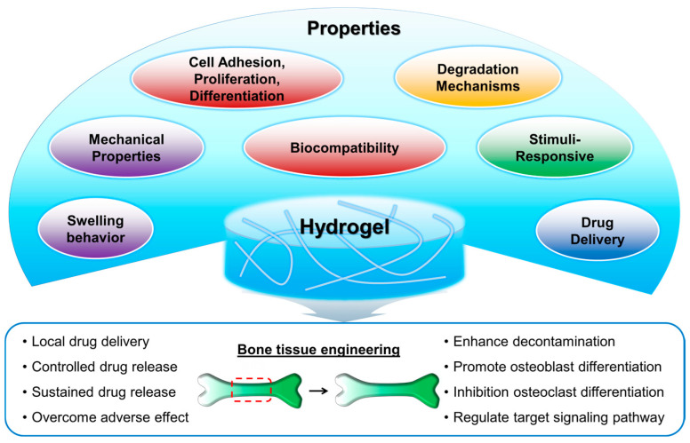
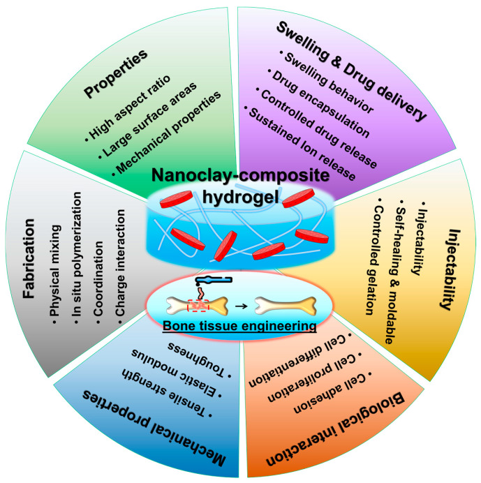
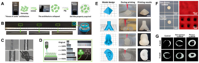
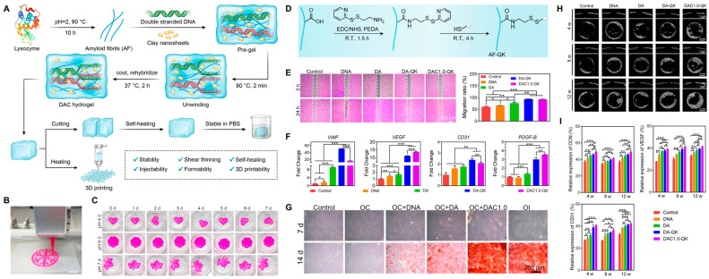
Nanoclay-composite hydrogels represent a promising avenue for advancing bone tissue engineering. Traditional hydrogels face challenges in providing mechanical strength, biocompatibility, and bioactivity necessary for successful bone regeneration. The incorporation of nanoclay into hydrogel matrices offers a potential unique solution to these challenges. This review provides a comprehensive overview of the fabrication, physico-chemical/biological performance, and applications of nanoclay-composite hydrogels in bone tissue engineering. Various fabrication techniques, including in situ polymerization, physical blending, and 3D printing, are discussed. In vitro and in vivo studies evaluating biocompatibility and bioactivity have demonstrated the potential of these hydrogels for promoting cell adhesion, proliferation, and differentiation. Their applications in bone defect repair, osteochondral tissue engineering and drug delivery are also explored. Despite their potential in bone tissue engineering, nanoclay-composite hydrogels face challenges such as optimal dispersion, scalability, biocompatibility, long-term stability, regulatory approval, and integration with emerging technologies to achieve clinical application. Future research directions need to focus on refining fabrication techniques, enhancing understanding of biological interactions, and advancing towards clinical translation and commercialization. Overall, nanoclay-composite hydrogels offer exciting opportunities for improving bone regeneration strategies.
Overview
The claims and statistics of this paper haven’t been checked yet.
Run the check — free, no account needed. It reads the open-access text and checks the sources it cites.
Integrity signals
Registry
Cites
Statistics
Figures
Verification
Claims the paper makes
The claims and statistics of this paper haven’t been checked yet.
Run the check — free, no account needed. It reads the open-access text and checks the sources it cites.
Self-check
Percentages, sums and reported differences re-computed from the paper’s own numbers.
The self-check needs the paper’s full text.
Citations
Cites
1 retracted
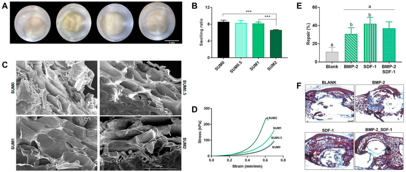
Nano-Silicate-Reinforced and SDF-1α-Loaded Gelatin-Methacryloyl Hydrogel for Bone Tissue Engineering
Shi, Z., Xu, Y., Mulatibieke, R., Zhong, Q., Pan, X., Chen, Y., Lian, Q., Luo, X., Shi, Z., & Zhu, Q. (2020) · International Journal of Nanomedicine · doi:10.2147/ijn.s270681 · Cited by 41
RetractedMechanisms of polymer degradation and erosion
Göpferich, A. (1996) · Biomaterials · doi:10.1016/0142-9612(96)85755-3 · Cited by 1,781
Hydrogels: Properties and Applications in Biomedicine
Ho, T., Chang, C.-C., Chan, H.-P., Chung, T., Shu, C., Chuang, K., Duh, T., Yang, M., & Tyan, Y. (2022) · Molecules · doi:10.3390/molecules27092902 · Cited by 1,239
Stimulus-responsive hydrogels: theory, modern advances, and applications
Koetting, M. C., Peters, J. T., Steichen, S. D., & Peppas, N. A. (2015) · Materials Science and Engineering: R: Reports · doi:10.1016/j.mser.2015.04.001 · Cited by 1,191
Biocompatibility of hydrogel-based scaffolds for tissue engineering applications
Naahidi, S., Jafari, M., Logan, M., Wang, Y., Yuan, Y., Bae, H., Dixon, B., & Chen, P. (2017) · Biotechnology Advances · doi:10.1016/j.biotechadv.2017.05.006 · Cited by 933
Baldwin, P. C., Li, D. J., Auston, D. A., Mir, H., Yoon, R. S., & Koval, K. J. (2019) · Journal of Orthopaedic Trauma · doi:10.1097/bot.0000000000001420 · Cited by 781
Designing degradable hydrogels for orthogonal control of cell microenvironments
Kharkar, P. M., Kiick, K. L., & Kloxin, A. M. (2013) · Chem. Soc. Rev. · doi:10.1039/c3cs60040h · Cited by 717
Hydrogel Bioink Reinforcement for Additive Manufacturing: A Focused Review of Emerging Strategies
Chimene, D., Kaunas, R., & Gaharwar, A. K. (2020) · Advanced Materials · doi:10.1002/adma.201902026 · Cited by 600 · cited 1×
“The mechanical strength of hydrogels is influenced by factors including polymer chain length, cross-linking density, and the presence of reinforcing agents such as nanoparticles and fibers [35–38].”
BackgroundGyles, D. A., Castro, L. D., Júnior, J. O. C. S., & Ribeiro-Costa, R. M. (2017) · European Polymer Journal · doi:10.1016/j.eurpolymj.2017.01.027 · Cited by 557
Stimuli‐Responsive Nanocomposite Hydrogels for Biomedical Applications
Lavrador, P., Esteves, M. R. C., Gaspar, V. M., & Mano, J. F. (2020) · Advanced Functional Materials · doi:10.1002/adfm.202005941 · Cited by 544
Research works printed but not linked: 1
- Wang, A., and Wang, W. (2019). Nanomaterials from Clay Minerals: A New Approach to Green Functional Materials, Elsevier.
Cited by
Sorted by citations
Exosome-Integrated Hydrogels for Bone Tissue Engineering
Hwang, H. S., & Lee, C. (2024) · Gels · doi:10.3390/gels10120762 · Cited by 47
Self-citationHydrogel for bone microenvironment: Strategy and application
Li, G., Zhou, D., Sheng, S., Lin, Q., Jing, Y., Ren, X., & Su, J. (2024) · Chemical Engineering Journal · doi:10.1016/j.cej.2024.156554 · Cited by 30
Mollaheydaralimoazzen, M., Sheikholeslam, M., Poursamar, S. A., Farzan, M., Farzan, M., & Rafienia, M. (2025) · International Journal of Biological Macromolecules · doi:10.1016/j.ijbiomac.2025.144502 · Cited by 23
Bone Tissue Engineering: From Biomaterials to Clinical Trials
Jagadale, S., Damle, M. N., & Joshi, M. G. (2025) · Advances in experimental medicine and biology · doi:10.1007/5584_2024_841 · Cited by 15
Tayeed, M. H., Tehranchi, M., Ehterami, A., Shanei, F., Taleghani, F., Semyari, H., Ganji, S. M., Mehrnia, N., Bozorgzadeh, S., Zamani, S., et al. (2025) · Journal of Biomaterials Applications · doi:10.1177/08853282251329901 · Cited by 14
Jain, S., Dilbaghi, N., Marrazza, G., Hassan, A. A., Kaushik, A. K., Kim, K., & Kumar, S. (2025) · Advances in Colloid and Interface Science · doi:10.1016/j.cis.2025.103529 · Cited by 12
The current status of nano-hydrogel preparations for osteochondral repair: Systematic Review
Amhare, A. F., Qiao, L., Deng, H., Lin, J., Wang, J., Wang, W., & Han, J. (2025) · Frontiers in Bioengineering and Biotechnology · doi:10.3389/fbioe.2025.1611522 · Cited by 12
Song, G. J., Oh, S., Lee, J. H., Lee, M., Hwang, H. S., Koh, J., & Lee, C. (2025) · International Journal of Biological Macromolecules · doi:10.1016/j.ijbiomac.2025.143980 · Cited by 9
Self-citationNature-Based Hydrogels Combined with Nanoparticles for Bone Regeneration
Fernandes, M. M., Vieira, M., Peixoto, D., & Alves, N. M. (2025) · Journal of Functional Biomaterials · doi:10.3390/jfb16090317 · Cited by 8
Valchanov, P., Yaneva, Y., Pavlov, S., Kontny, A., & Dikova, T. D. (2025) · Biomedicines · doi:10.3390/biomedicines13112688 · Cited by 7
Functional Hydrogels in Bone Tissue Engineering: From Material Design to Translational Applications
Petraglia, F. M., Giordano, S., & Santoro, A. (2026) · Biologics · doi:10.3390/biologics6010002 · Cited by 6
Ma, C., Qiu, H., Chen, Y., Mueed, A., & You, L. (2026) · ACS Applied Bio Materials · doi:10.1021/acsabm.5c02248 · Cited by 6
Qu, Q., Zhang, Q., Wang, M., Zhao, S., Li, P., Shang, C., Niu, Y., Zhang, S., & Pan, S. (2025) · Biomedical Physics & Engineering Express · doi:10.1088/2057-1976/adf8ee · Cited by 5
Jin, X., Yue, Y., Hu, H., & Lv, S. (2025) · Pharmaceutics · doi:10.3390/pharmaceutics17040514 · Cited by 4
Ci, H., Jie, J., Zhang, G., Wu, L., Wang, Z., & Sun, J. (2024) · International Journal of Nanomedicine · doi:10.2147/ijn.s498950 · Cited by 4
A Chinese drug-compatibility-based approach to purslane hydrogels for acute eczema therapy
Wang, L., Zhang, Y., Geng, S., Ma, L., Wang, Y., Han, D., Fan, G., Zhang, W., Lv, Y., & Ma, J. (2025) · Frontiers in Pharmacology · doi:10.3389/fphar.2025.1504120 · Cited by 4
Jiang, L., Zhang, Y., Guo, Z., Zhang, Z., & Wang, C. (2025) · ACS Applied Materials & Interfaces · doi:10.1021/acsami.5c02573 · Cited by 4
Saad, I., Fathy, W. A., Amin, R. M., & El-Dek, S. I. (2026) · Beni-Suef University Journal of Basic and Applied Sciences · doi:10.1186/s43088-025-00725-8 · Cited by 3
Gatabi, Z. R., Mirhoseini, M., Abasi, M., Sayyad, M. S., & Ebrahimnejad, P. (2026) · Journal of Drug Delivery Science and Technology · doi:10.1016/j.jddst.2026.108035 · Cited by 3
Zare, S., Eskandani, M., Esfahlan, R. J., & Jaymand, M. (2025) · Scientific Reports · doi:10.1038/s41598-025-30576-3 · Cited by 3
Gum tragacanth-based hemostatic sponge for enhanced hemostasis in dental surgery
Mousavai, M., Khajavi, R., Kalaee, M., & Rahimi, M. K. (2025) · Pharmaceutical Development and Technology · doi:10.1080/10837450.2025.2516238 · Cited by 2
Matthews, C., Tarsitano, E., Odedra, S., Holden, W., Villalan, D. S. T., Kavalakatt, S., Silva, K., Zimmermann, L. A., & Benecke, J. P. V. (2025) · Processes · doi:10.3390/pr13113395 · Cited by 2
Khoshnood, N., Shahrezaee, M., Frampton, J. P., Shahrezaee, M. H., & Zamanian, A. (2025) · Advanced Materials Interfaces · doi:10.1002/admi.202500609 · Cited by 2
Radu, I., Tanasă, E., Dinescu, S., Vlăsceanu, G. M., & Zaharia, C. (2025) · Polymers · doi:10.3390/polym17040536 · Cited by 2
Gao, Y., Dang, X., Zhu, A., & Chen, Z. (2025) · ACS Applied Polymer Materials · doi:10.1021/acsapm.4c04076 · Cited by 2
Elmergawy, F. H., Fathy, I., Amer, M. A., Hamdi, S., & Ibrahim, M. S. (2025) · Journal of Drug Delivery Science and Technology · doi:10.1016/j.jddst.2025.107413 · Cited by 1
Mabrouk, M., Aboelnasr, M. A., Shiomoto, S., Nishimura, S., Beherei, H. H., Alzahrani, E. A., Tanaka, Y., & Tanaka, M. (2025) · Biomaterials Advances · doi:10.1016/j.bioadv.2025.214606 · Cited by 1
Guillén‐Carvajal, K., Valdez, B. S., Beltrán‐Partida, E., Salvador‐Carlos, J., Curiel, M., Castillo-Sáenz, J., González-Mendoza, D., & Cheng, N. (2025) · Gels · doi:10.3390/gels11110910 · Cited by 1
Effect of Sulfated Polysaccharides and Laponite in Composite Porous Scaffolds on Osteogenesis
Karamesouti, A., & Chatzinikolaidou, Μ. (2026) · Biomolecules · doi:10.3390/biom16010080 · Cited by 1
Fooladi, T., Esmaeili, E., & Rad, I. (2026) · Applied Physics A · doi:10.1007/s00339-026-09302-x · Cited by 1
Engineered Nano Polymer-Based Hydrogel
Sharma, A., Dheer, D., & Puri, V. (2026) · Engineering materials · doi:10.1007/978-981-95-3581-1_5 · Cited by 1
Vyas, J., Raytthatha, N., Vyas, P., Shah, N. V., & Modi, D. (2026) · Frontiers in Biomaterials Science · doi:10.3389/fbiom.2026.1861958 · Cited by 1
Nanoclays in tissue engineering
Lokuge, E. L., Appuhamillage, G. A., & Ambagaspitiya, S. S. (2026) · Elsevier eBooks · doi:10.1016/b978-0-443-34290-5.00001-9 · Cited by 1
Anas, M., Younas, M., Isnain, S., Khattak, W. A., Elahi, M., Erkoyuncu, M. T., & Hakki, E. E. (2026) · Journal of soil science and plant nutrition · doi:10.1007/s42729-026-03476-2 · Cited by 1
Hwang, H. S., & Lee, C. (2026) · Biomimetics · doi:10.3390/biomimetics11080593 · Cited by 1
Self-citationShahbazi, P., Abdouss, M., Naeimi, M., Darbemamieh, G., & Jafari‐Nodoushan, M. (2025) · Carbohydrate Polymer Technologies and Applications · doi:10.1016/j.carpta.2025.100771 · Cited by 1
Janmohammadi, M., Bahraminasab, M., & Nourbakhsh, M. S. (2025) · International Journal of Biological Macromolecules · doi:10.1016/j.ijbiomac.2025.148295 · Cited by 1
Antunes, B. S. L., Pedrini, F., Komatsu, D., Asami, J., Dicko, C., & Duek, E. A. R. (2026) · International Journal of Biological Macromolecules · doi:10.1016/j.ijbiomac.2026.154129
Bone Tissue Engineering with Hydrogels Derived from Demineralized Bone Matrix
Ashrafi, F., & Emami, A. (2026) · Regenerative Engineering and Translational Medicine · doi:10.1007/s40883-025-00558-y
Eshkol‐Yogev, I., Kogan, T. H., Levi, I., Salman, M., Gariani, O., & Zilberman, M. (2026) · Journal of Functional Biomaterials · doi:10.3390/jfb17030121
Li, X., Fu, X., Hui, B., Li, X., Su, Z., Zhang, H., & Yu, Z. (2026) · Applied Biochemistry and Biotechnology · doi:10.1007/s12010-026-05715-9
Biopolymer-based hydrogel electrostatic reinforcement for bone regeneration
Khan, A., Walid, B. M., Chowdhury, M. L., Haque, M. A., Chowdhury, M. I. S., & Hoque, M. E. (2026) · Elsevier eBooks · doi:10.1016/b978-0-443-33391-0.00019-4
Solvent-assisted Dual Modification of Chitosan–alginate Scaffolds using Sebacic Acid and Na-MMT
Eghbali, H., Salehi, K., Ranjbar, F. E., & Zarandi, E. R. (2026) · Journal of Polymers and the Environment · doi:10.1007/s10924-026-03854-y
Babu, B. (2026) · Transformative Medical Materials · doi:10.1016/j.trmm.2026.100003
Tantalum‐Based Nanomaterials: From Properties to Biomedical Applications
Jin, Y., Zhang, Y., Yang, C. R., Ren, G., Ding, X., Zhu, S., Gu, Z., & Feng, X. (2026) · Advanced Materials · doi:10.1002/adma.73734
Sharma, P., Saurav, S., Bhanot, R., Sharma, R., Mohan, A., Kumar, A. T. R., Malik, T., & Girdhar, M. (2026) · RSC Advances · doi:10.1039/d6ra00133e
Multifunctional Integrated Hydrogels for Tissue Regeneration and Repair
Liu, Y. (2026) · Tissue Engineering Part B Reviews · doi:10.1177/19373368261469767
Laponite-stabilized pickering polymerization for high-performance latexes and hydrogels
Chen, H., Yao, J., Shen, Z., Kwok, M. W., Ngai, T., & Sun, G. (2026) · Polymer · doi:10.1016/j.polymer.2026.130535
Clay-Based Nanomaterials in Wound Healing: Therapeutic Roles, Mechanisms and Biomedical Applications
Aliyev, A. R., Hasanova, U., Buroni, S., Aliyeva, A., & Israyilova, A. (2026) · Micro · doi:10.3390/micro6030057
Nanoclays in bone remodeling and regeneration
Yogananda, K. A. S. K., Herath, I. S., & Epa, T. W. (2026) · Elsevier eBooks · doi:10.1016/b978-0-443-34290-5.00002-0
Engineering nanoclays to enhance antimicrobial properties
Abubakar, A. A., Sabo, M. J. U., Hashmi, M. Z., & Ali, A. M. (2026) · Elsevier eBooks · doi:10.1016/b978-0-443-34290-5.00019-6
Synthesis, properties, and characterization of nanoclays and their applications in medical therapy
Wijesinghe, J., Madunil, S. L., & Udahapuvida, S. (2026) · Elsevier eBooks · doi:10.1016/b978-0-443-34290-5.00003-2
Nanoclays for targeted drug delivery systems
Jayaweera, Y. U., Khalid, G. A. N., Karunathilake, D. N., Dananjaya, U. M., Attigala, A. D. L., Jayasingha, J. R. P., Gamage, S. P. L., Herath, H. M. L. P., & Manatunga, D. C. (2026) · Elsevier eBooks · doi:10.1016/b978-0-443-34290-5.00017-2
Nanoclay-based nanocomposite surfaces with antimicrobial properties for biomedical applications
Jayakody, J. T. M., & Choi, H. J. (2026) · Elsevier eBooks · doi:10.1016/b978-0-443-34290-5.00014-7
Jain, R., Singhal, N., Vardhan, H., Bist, Y., Chaudhary, P., Sharma, S., Kishore, K., Wagri, N. K., & Gaur, A. (2026) · Frontiers in Sustainable Food Systems · doi:10.3389/fsufs.2026.1941522
Design and Application of Strong and Tough Low-Friction Hydrogels
魏娴, Luo, H., Luo, J., Zhang, D., Huang, B., Liu, Y., Xu, Z., & Kou, R. (2026) · Gels · doi:10.3390/gels12090775
Mugnaini, G., Bonini, M., & Tonelli, M. (2026) · Ceramics International · doi:10.1016/j.ceramint.2026.09.034
Le, T. D. H., Le, T. T., Nguyen, T. –. T., Nguyen, G. T., Vu, B. T., Nguyen, T. H., Tuan, H. N. A., & Le, A. T. (2026) · Journal of Applied Polymer Science · doi:10.1002/app.71454
Kim, D. K., Oh, S., Song, G. J., Lee, J. H., Yoo, A., Lee, J. M., Hwang, H. S., Lee, M., Koh, J., & Lee, C. (2026) · International Journal of Biological Macromolecules · doi:10.1016/j.ijbiomac.2026.154437
Self-citationOrganic–Inorganic Hyaluronic Acid/Sumecton Nanocomposite Hydrogels for Osteogenic Applications
Kim, D. K., Song, G. J., Yoo, A., Hwang, H. S., Lee, M., & Lee, C. (2026) · Gels · doi:10.3390/gels12090851
Self-citationEPS: A potential application in bone tissue engineering for repairing and regeneration
Mandal, S., Jit, T., Sahu, R., Nandi, G., & Palit, P. (2026) · Elsevier eBooks · doi:10.1016/b978-0-443-36619-2.00017-6
Selection of Nanoparticles for Targeted Applications
Mourad, A. I., & Hassan, A. A. (2026) · CRC Press eBooks · doi:10.1201/9781003664390
Injectable hydrogels in bone tissue engineering: from bench to bedside
Sabarees, G., Kanmani, S., Kowsalya, S., Priyadharshini, D., & Solomon, V. R. (2026) · International Journal of Polymeric Materials · doi:10.1080/00914037.2026.2743812
Biomedical and healthcare applications of self-healing polymer nanocomposites
Staszak, K., & Wieszczycka, K. (2026) · Elsevier eBooks · doi:10.1016/b978-0-443-40545-7.00009-6
Metadata
- Authors
- Hwang, H. S., & Lee, C.-S.
- Affiliations
- Dankook University (KR)
- Soonchunhyang University (KR)
- Corresponding author
- Hwang, Hee Sook
- Lee, Chung-Sung
- Venue
- Gels
- Publisher
- MDPI AG
- Published
- 2024-08-03
- Volume
- 10
- Issue
- 8
- Pages
- 513
- ISSN
- 2310-2861 (ISSN-L)
- PMC
- PMC11353637
- OpenAlex
- W4401323500
- Cited by
- 60
- References
- 122
- Citations by year
- 2026: 36 · 2025: 21 · 2024: 3
- FWCI
- 2.15
- Type
- journal article
- Language
- en
- Open access
- gold
- doi.org
- Licence
- Locations
- Gels (publishedVersion, cc-by)
- PubMed Central (publishedVersion, cc-by)
- PubMed (publishedVersion)
- DOAJ (DOAJ: Directory of Open Access Journals) (submittedVersion)
- Funders
- Soonchunhyang University Research Fund
- Topics
- Physical Sciences › Engineering › Biomedical Engineering › Bone Tissue Engineering Materials
- Physical Sciences › Materials Science › Biomaterials › Hydrogels: synthesis, properties, applications
- Physical Sciences › Engineering › Biomedical Engineering › 3D Printing in Biomedical Research
- Keywords
- nanoclay, composite hydrogels, bone tissue engineering, bone regeneration, hydrogel synthesis, in situ polymerization, physical blending, 3D printing, biocompatibility, bioactivity (biomaterials), cell adhesion and differentiation, bone defect repair, osteochondral tissue engineering, drug delivery, nanoclay dispersion
About these checks
Registry, retraction, Cites and statistics results are computed, not judged, with one exception: a printed reference that no index lists is read once by a model before it counts as a citation error. Claims are extracted by a model and kept only when their quote appears verbatim in the text; each cited source is then checked, with a model’s help, for whether it says what the claim relies on. Those verdicts can be wrong — read the claim’s quote and the cited source before relying on one.
Statistics and arithmetic are recomputed, not judged.
Confidence bands are shown only for fields calibrated on our annotated test set. None is calibrated yet, so no band is shown.
What counts as a claim ›How we measure confidence ›How we check this ›
Are you an author, or do you see a problem with this page? Request a review
