

The DOI matches its Crossref record; 121 of the 122 references it cites were found in a registry or an index, and 1 are not checked yet.
Nanoclay-Composite Hydrogels for Bone Tissue Engineering
Open accessAbstract
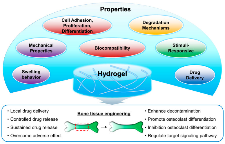
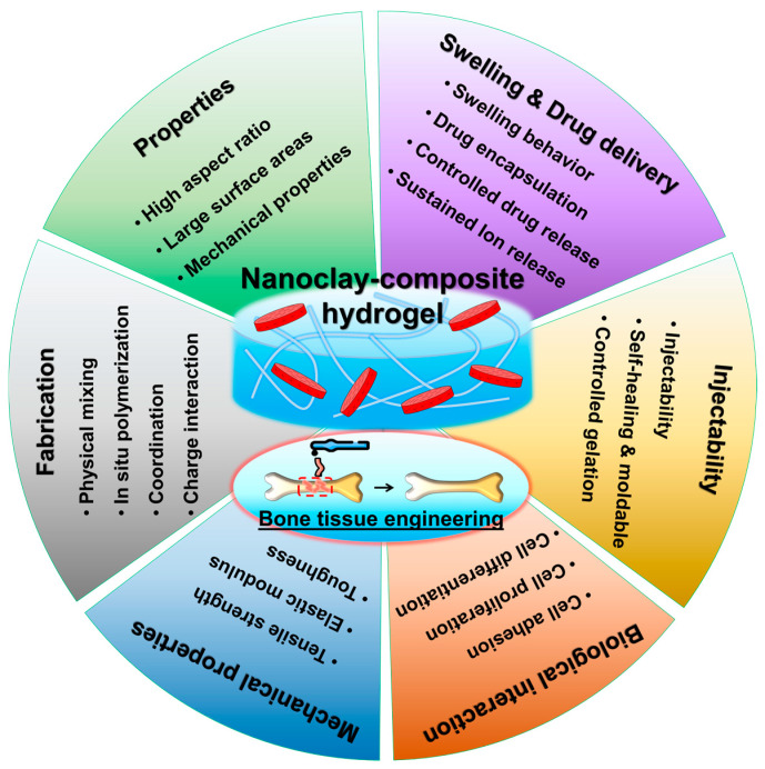
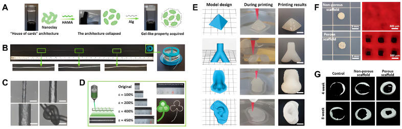
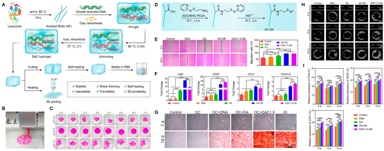
Nanoclay-composite hydrogels represent a promising avenue for advancing bone tissue engineering. Traditional hydrogels face challenges in providing mechanical strength, biocompatibility, and bioactivity necessary for successful bone regeneration. The incorporation of nanoclay into hydrogel matrices offers a potential unique solution to these challenges. This review provides a comprehensive overview of the fabrication, physico-chemical/biological performance, and applications of nanoclay-composite hydrogels in bone tissue engineering. Various fabrication techniques, including in situ polymerization, physical blending, and 3D printing, are discussed. In vitro and in vivo studies evaluating biocompatibility and bioactivity have demonstrated the potential of these hydrogels for promoting cell adhesion, proliferation, and differentiation. Their applications in bone defect repair, osteochondral tissue engineering and drug delivery are also explored. Despite their potential in bone tissue engineering, nanoclay-composite hydrogels face challenges such as optimal dispersion, scalability, biocompatibility, long-term stability, regulatory approval, and integration with emerging technologies to achieve clinical application. Future research directions need to focus on refining fabrication techniques, enhancing understanding of biological interactions, and advancing towards clinical translation and commercialization. Overall, nanoclay-composite hydrogels offer exciting opportunities for improving bone regeneration strategies.
Overview
The claims and statistics of this paper haven’t been checked yet.
Run the check — free, no account needed. It reads the open-access text and checks the sources it cites.
Integrity signals
Registry
Cites
Statistics
Figures
Verification
Claims the paper makes
The claims and statistics of this paper haven’t been checked yet.
Run the check — free, no account needed. It reads the open-access text and checks the sources it cites.
Self-check
Percentages, sums and reported differences re-computed from the paper’s own numbers.
The self-check needs the paper’s full text.
Citations
Cites
1 retracted
Nano-Silicate-Reinforced and SDF-1α-Loaded Gelatin-Methacryloyl Hydrogel for Bone Tissue Engineering
Shi, Z., Xu, Y., Mulatibieke, R., Zhong, Q., Pan, X., Chen, Y., Lian, Q., Luo, X., Shi, Z., & Zhu, Q. (2020) · International Journal of Nanomedicine · doi:10.2147/ijn.s270681 · Cited by 41
RetractedMechanisms of polymer degradation and erosion
Göpferich, A. (1996) · Biomaterials · doi:10.1016/0142-9612(96)85755-3 · Cited by 1,781
Hydrogels: Properties and Applications in Biomedicine
Ho, T., Chang, C.-C., Chan, H.-P., Chung, T., Shu, C., Chuang, K., Duh, T., Yang, M., & Tyan, Y. (2022) · Molecules · doi:10.3390/molecules27092902 · Cited by 1,239
Stimulus-responsive hydrogels: theory, modern advances, and applications
Koetting, M. C., Peters, J. T., Steichen, S. D., & Peppas, N. A. (2015) · Materials Science and Engineering: R: Reports · doi:10.1016/j.mser.2015.04.001 · Cited by 1,191
Biocompatibility of hydrogel-based scaffolds for tissue engineering applications
Naahidi, S., Jafari, M., Logan, M., Wang, Y., Yuan, Y., Bae, H., Dixon, B., & Chen, P. (2017) · Biotechnology Advances · doi:10.1016/j.biotechadv.2017.05.006 · Cited by 933
Baldwin, P. C., Li, D. J., Auston, D. A., Mir, H., Yoon, R. S., & Koval, K. J. (2019) · Journal of Orthopaedic Trauma · doi:10.1097/bot.0000000000001420 · Cited by 781
Designing degradable hydrogels for orthogonal control of cell microenvironments
Kharkar, P. M., Kiick, K. L., & Kloxin, A. M. (2013) · Chem. Soc. Rev. · doi:10.1039/c3cs60040h · Cited by 717
Hydrogel Bioink Reinforcement for Additive Manufacturing: A Focused Review of Emerging Strategies
Chimene, D., Kaunas, R., & Gaharwar, A. K. (2020) · Advanced Materials · doi:10.1002/adma.201902026 · Cited by 600 · cited 1×
“The mechanical strength of hydrogels is influenced by factors including polymer chain length, cross-linking density, and the presence of reinforcing agents such as nanoparticles and fibers [35–38].”
BackgroundGyles, D. A., Castro, L. D., Júnior, J. O. C. S., & Ribeiro-Costa, R. M. (2017) · European Polymer Journal · doi:10.1016/j.eurpolymj.2017.01.027 · Cited by 557
Stimuli‐Responsive Nanocomposite Hydrogels for Biomedical Applications
Lavrador, P., Esteves, M. R. C., Gaspar, V. M., & Mano, J. F. (2020) · Advanced Functional Materials · doi:10.1002/adfm.202005941 · Cited by 544
Recent Advances in Design of Functional Biocompatible Hydrogels for Bone Tissue Engineering
Xu, X. J., Hu, Y., Deng, Y., & Su, J. (2021) · Advanced Functional Materials · doi:10.1002/adfm.202009432 · Cited by 538
Bioactive Silicate Nanoplatelets for Osteogenic Differentiation of Human Mesenchymal Stem Cells
Gaharwar, A. K., Mihaila, S. M., Swami, A., Patel, A. C., Sant, S., Reis, R. L., Marques, A. P., Gomes, M. E., & Khademhosseini, A. (2013) · Advanced Materials · doi:10.1002/adma.201300584 · Cited by 525
A Review of the Synthesis and Applications of Polymer–Nanoclay Composites
Guo, F., Aryana, S. A., Han, Y., & Jiao, Y. (2018) · Applied Sciences · doi:10.3390/app8091696 · Cited by 464
Cui, Z., Kim, S., Baljon, J. J., Wu, B. M., Aghaloo, T. L., & Lee, M. (2019) · Nature Communications · doi:10.1038/s41467-019-11511-3 · Cited by 440
Stimuli-responsive hydrogels in drug delivery and tissue engineering
Sood, N., Bhardwaj, A., Mehta, S., & Mehta, A. (2016) · Drug Delivery · doi:10.3109/10717544.2014.940091 · Cited by 426
Gaharwar, A. K., Cross, L., Peak, C. W., Gold, K. A., Carrow, J. K., Brokesh, A. M., & Singh, K. A. (2019) · Advanced Materials · doi:10.1002/adma.201900332 · Cited by 409
Tissue Engineering Strategies for Bone Regeneration
Mistry, A. S., & Mikos, A. G. (2005) · Advances in biochemical engineering, biotechnology · doi:10.1007/b99997 · Cited by 391
Hydrogel drug delivery system with predictable and tunable drug release and degradation rates
Ashley, G. W., Henise, J., Reid, R. R., & Santi, D. V. (2013) · Proceedings of the National Academy of Sciences · doi:10.1073/pnas.1215498110 · Cited by 386
Bioactuators based on stimulus-responsive hydrogels and their emerging biomedical applications
Shi, Q., Liu, H., Tang, D., Li, Y., Li, X., & Xu, F. (2019) · NPG Asia Materials · doi:10.1038/s41427-019-0165-3 · Cited by 370
Biodegradable Polymers as the Pivotal Player in the Design of Tissue Engineering Scaffolds
Zhang, F., & King, M. W. (2020) · Advanced Healthcare Materials · doi:10.1002/adhm.201901358 · Cited by 320
Montmorillonite nanoclay as a multifaceted drug-delivery carrier: A review
Sarvaiya, J., Shankar, G., Agrawal, Y. K., & Bakre, L. G. (2017) · Journal of Drug Delivery Science and Technology · doi:10.1016/j.jddst.2017.03.023 · Cited by 302
Hydrogel as a Biomaterial for Bone Tissue Engineering: A Review
Yue, S., He, H. J., Li, B., & Hou, T. (2020) · Nanomaterials · doi:10.3390/nano10081511 · Cited by 283
Transparent, elastomeric and tough hydrogels from poly(ethylene glycol) and silicate nanoparticles
Gaharwar, A. K., Rivera, C. P., Wu, C., & Schmidt, G. (2011) · Acta Biomaterialia · doi:10.1016/j.actbio.2011.07.023 · Cited by 252
Zhai, X., Ma, Y., Hou, C., Gao, F., Zhang, Y., Ruan, C., Pan, H., Lu, W. W., & Liu, W. (2017) · ACS Biomaterials Science & Engineering · doi:10.1021/acsbiomaterials.7b00224 · Cited by 247
Gehrke, S. H. (2005) · Springer eBooks · doi:10.1007/bfb0021130 · Cited by 243
Liquid, glass, gel: The phases of colloidal Laponite
Cummins, H. Z. (2007) · Journal of Non-Crystalline Solids · doi:10.1016/j.jnoncrysol.2007.02.066 · Cited by 235
Andrade, F., Roca-Melendres, M. M., Durán‐Lara, E. F., Rafael, D. F. D. S., & Schwartz, S. (2021) · Cancers · doi:10.3390/cancers13051164 · Cited by 231
A review on nanocomposite hydrogels and their biomedical applications
Rafieian, S., Mirzadeh, H., Mahdavi, H., & Masoumi, M. E. (2018) · Science and Engineering of Composite Materials · doi:10.1515/secm-2017-0161 · Cited by 218
Nanoclay-based drug delivery systems and their therapeutic potentials
Khatoon, N., Chu, M. Q., & Zhou, C. H. (2020) · Journal of Materials Chemistry B · doi:10.1039/d0tb01031f · Cited by 209
Nanocomposite Hydrogels and Their Applications in Tissue Engineering
Motealleh, A., & Kehr, N. S. (2016) · Advanced Healthcare Materials · doi:10.1002/adhm.201600938 · Cited by 173
Halloysite nanotubes as a nature’s boon for biomedical applications
Satish, S., Tharmavaram, M., & Rawtani, D. (2019) · Nanobiomedicine · doi:10.1177/1849543519863625 · Cited by 143
Stimuli-Controlled Hydrogels and Their Applications
Willner, I. (2017) · Accounts of Chemical Research · doi:10.1021/acs.accounts.7b00142 · Cited by 140
Controlled release of ofloxacin from chitosan–montmorillonite hydrogel
Hua, S., Yang, H., Wang, W., & Wang, A. (2010) · Applied Clay Science · doi:10.1016/j.clay.2010.07.012 · Cited by 139
Lee, C., Hwang, H. S., Kim, S., Fan, J., Aghaloo, T. L., & Lee, M. (2020) · Advanced Functional Materials · doi:10.1002/adfm.202003717 · Cited by 137
Self-citationEnvironmentally Adaptive Polymer Hydrogels: Maintaining Wet‐Soft Features in Extreme Conditions
Lei, K., Chen, M., Guo, P., Fang, J., Zhang, J., Liu, X., Wang, W., Li, Y., Hu, Z., Ma, Y., et al. (2023) · Advanced Functional Materials · doi:10.1002/adfm.202303511 · Cited by 133
Su, D., Jiang, L., Chen, X., Dong, J., & Shao, Z. (2016) · ACS Applied Materials & Interfaces · doi:10.1021/acsami.6b00891 · Cited by 131
Nanoparticle–Polymer Synergies in Nanocomposite Hydrogels: From Design to Application
Chen, T., Hou, K., Ren, Q., Chen, G., Wei, P., & Zhu, M. (2018) · Macromolecular Rapid Communications · doi:10.1002/marc.201800337 · Cited by 129
Gaharwar, A. K., Kishore, V., Rivera, C. P., Bullock, W. A., Wu, C., Akkuş, O., & Schmidt, G. (2012) · Macromolecular Bioscience · doi:10.1002/mabi.201100508 · Cited by 126
Huang, B., Liu, M., Long, Z., Shen, Y., & Zhou, C. (2016) · Materials Science and Engineering C · doi:10.1016/j.msec.2016.09.001 · Cited by 126
Recent Progress in Hyaluronic-Acid-Based Hydrogels for Bone Tissue Engineering
Hwang, H. S., & Lee, C. (2023) · Gels · doi:10.3390/gels9070588 · Cited by 126
Self-citationMihaila, S. M., Gaharwar, A. K., Reis, R. L., Khademhosseini, A., Marques, A. P., & Gomes, M. E. (2014) · Biomaterials · doi:10.1016/j.biomaterials.2014.07.052 · Cited by 125
Gorgieva, S., & Kokol, V. (2012) · Journal of Biomedical Materials Research Part A · doi:10.1002/jbm.a.34106 · Cited by 125
Ou, Q., Huang, K., Fu, C., Huang, C., Fang, Y., Gu, Z., Wu, J., & Wang, Y. (2019) · Chemical Engineering Journal · doi:10.1016/j.cej.2019.123019 · Cited by 124
Starch-Based Hydrogels as a Drug Delivery System in Biomedical Applications
Lee, C., & Hwang, H. S. (2023) · Gels · doi:10.3390/gels9120951 · Cited by 117
Self-citationHalloysite Nanotube Based Scaffold for Enhanced Bone Regeneration
Huang, K., Ou, Q., Xie, Y., Chen, X., Fang, Y., Huang, C., Wang, Y., Gu, Z., & Wu, J. (2019) · ACS Biomaterials Science & Engineering · doi:10.1021/acsbiomaterials.9b00277 · Cited by 116
Liu, B., Li, J., Lei, X., Cheng, P., Song, Y., Gao, Y., Hu, J., Wang, C., Zhang, S., Li, D., et al. (2020) · Materials Science and Engineering C · doi:10.1016/j.msec.2020.110905 · Cited by 115
Injectable chitosan hydrogel embedding modified halloysite nanotubes for bone tissue engineering
Kazemi-Aghdam, F., Jahed, V., Dehghan-Niri, M., Ganji, F., & Vasheghani‐Farahani, E. (2021) · Carbohydrate Polymers · doi:10.1016/j.carbpol.2021.118311 · Cited by 112
The Application of Hydrogels Based on Natural Polymers for Tissue Engineering
Taghipour, Y. D., Hokmabad, V. R., Bakhshayesh, A. R. D., Asadi, N., Salehi, R., & Nasrabadi, H. T. (2019) · Current Medicinal Chemistry · doi:10.2174/0929867326666190711103956 · Cited by 111
Clay nanocomposites as engineered drug delivery systems
Jafarbeglou, M., Abdouss, M., Shoushtari, A. M., & Jafarbeglou, M. (2016) · RSC Advances · doi:10.1039/c6ra03942a · Cited by 108
The global burden of fracture-related infection: can we do better?
Metsemakers, W., Moriarty, T. F., Morgenstern, M., Marais, L. C., Onsea, J., O’Toole, R. V., Depypere, M., Obremskey, W. T., Verhofstad, M. H. J., McNally, M., et al. (2023) · The Lancet Infectious Diseases · doi:10.1016/s1473-3099(23)00503-0 · Cited by 104
Sohail, K., Khan, I. U., Shahzad, Y., Hussain, T., & Ranjha, N. M. (2014) · Brazilian Journal of Pharmaceutical Sciences · doi:10.1590/s1984-82502011000100018 · Cited by 103
On mechanical properties of nanocomposite hydrogels: Searching for superior properties
Xing, W., & Tang, Y. (2021) · Nano Materials Science · doi:10.1016/j.nanoms.2021.07.004 · Cited by 101
Sustainable and safer nanoclay composites for multifaceted applications
Padil, V. V. T., Kumar, K. P. A., Murugesan, S., Torres–Mendieta, R., Wacławek, S., Cheong, J. Y., Černík, M., & Varma, R. S. (2022) · Green Chemistry · doi:10.1039/d1gc03949k · Cited by 101
Gradient nanocomposite hydrogels for interface tissue engineering
Cross, L., Shah, K. G., Palani, S., Peak, C. W., & Gaharwar, A. K. (2017) · Nanomedicine Nanotechnology Biology and Medicine · doi:10.1016/j.nano.2017.02.022 · Cited by 98
Clay-Based Nanocomposite Hydrogels for Biomedical Applications: A Review
Tipa, C., Cidade, M. T., Borges, J. P., Costa, L. C., Silva, J. A. M. D. C., & Soares, P. I. P. (2022) · Nanomaterials · doi:10.3390/nano12193308 · Cited by 94
Halloysite Clay Nanotubes for Tissue Engineering
Fakhrullin, R. F., & LVOV, Y. M. (2016) · Nanomedicine · doi:10.2217/nnm-2016-0250 · Cited by 94
Hypoxia-mimicking 3D bioglass-nanoclay scaffolds promote endogenous bone regeneration
Zheng, X., Zhang, X., Wang, Y., Liu, Y., Pan, Y., Li, Y., Ji, M. K., Zhao, X., Huang, S., & Yao, Q. (2021) · Bioactive Materials · doi:10.1016/j.bioactmat.2021.03.011 · Cited by 92
Martins, A. M., Alves, C. M., Kasper, F. K., Mikos, A. G., & Reis, R. L. (2009) · Journal of Materials Chemistry · doi:10.1039/b916259n · Cited by 91
Dong, L., Bu, Z., Xiong, Y., Zhang, H., Fang, J., Hu, H., Liu, Z., & Li, X. (2021) · International Journal of Biological Macromolecules · doi:10.1016/j.ijbiomac.2021.07.199 · Cited by 90
Guo, Z., Dong, L., Xia, J., Mi, S., & Sun, W. (2021) · Advanced Healthcare Materials · doi:10.1002/adhm.202100036 · Cited by 89
Man, K., Barroso, I. A., Brunet, M. Y., Peacock, B., Federici, A., Hoey, D. A., & Cox, S. C. (2022) · International Journal of Molecular Sciences · doi:10.3390/ijms23020832 · Cited by 87
Functional Hydrogels and Their Application in Drug Delivery, Biosensors, and Tissue Engineering
Wang, K., Hao, Y., Wang, Y., Chen, J., Mao, L., Deng, Y., Chen, J., Yuan, S., Zhang, T., Ren, J., et al. (2019) · International Journal of Polymer Science · doi:10.1155/2019/3160732 · Cited by 86
Tough Composite Hydrogels with High Loading and Local Release of Biological Drugs
Li, J., Weber, E., Guth, S., Schuleit, M., Kuttler, A., Halleux, C. M., Accart, N., Doelemeyer, A., Basler, A., Tigani, B., et al. (2018) · Advanced Healthcare Materials · doi:10.1002/adhm.201701393 · Cited by 84
Clay nanoparticles as pharmaceutical carriers in drug delivery systems
Dong, J., Cheng, Z., Tan, S., & Zhu, Q. (2020) · Expert Opinion on Drug Delivery · doi:10.1080/17425247.2021.1862792 · Cited by 84
Kim, S., Cui, Z., Fan, J., Fartash, A., Aghaloo, T. L., & Lee, M. (2016) · Journal of Materials Chemistry B · doi:10.1039/c6tb01154c · Cited by 82
Zhang, X., Fan, J., Lee, C., Kim, S., Chen, C., & Lee, M. (2020) · ACS Applied Materials & Interfaces · doi:10.1021/acsami.0c01241 · Cited by 81
Self-citationPalem, R. R., Rao, K. M., Shimoga, G. D., Saratale, R. G., Shinde, S., Ghodake, G. S., & Lee, S. (2021) · International Journal of Biological Macromolecules · doi:10.1016/j.ijbiomac.2021.02.195 · Cited by 78
Nanoparticle-reinforced polyacrylamide hydrogel composites for clinical applications: a review
Awasthi, S., Gaur, J. K., Bobji, M. S., & Srivastava, C. (2022) · Journal of Materials Science · doi:10.1007/s10853-022-07146-3 · Cited by 78
Montmorillonite-based polyacrylamide hydrogel rings for controlled vaginal drug delivery
Sharifzadeh, G., Hezaveh, H., Muhamad, I. I., Hashim, S., & Khairuddin, N. (2019) · Materials Science and Engineering C · doi:10.1016/j.msec.2019.110609 · Cited by 77
Goycoolea, F. M., Heras, Á., Aranaz, I., Galed, G., Fernández-Valle, M. E., & Argüelles-Monal, W. M. (2003) · Macromolecular Bioscience · doi:10.1002/mabi.200300011 · Cited by 73
Li, Y., Xiao, L., Wei, D., Liu, S., Zhang, Z., Lian, R., Wang, L., Chen, Y., Jiang, J., Xiao, Y., et al. (2023) · Advanced Functional Materials · doi:10.1002/adfm.202213047 · Cited by 73
Yang, Q., Miao, Y., Luo, J., Chen, Y., & Wang, Y. (2023) · ACS Nano · doi:10.1021/acsnano.3c04816 · Cited by 72
Chaudhuri, S. D., Mandal, A., Dey, A., & Chakrabarty, D. (2020) · Applied Clay Science · doi:10.1016/j.clay.2019.105405 · Cited by 72
Au, P., Hassan, S., Liu, J., & Leong, Y. (2015) · Process Safety and Environmental Protection · doi:10.1016/j.cherd.2015.07.023 · Cited by 69
Kerativitayanan, P., & Gaharwar, A. K. (2015) · Acta Biomaterialia · doi:10.1016/j.actbio.2015.08.025 · Cited by 67
Zhang, W., Zhang, Y., Zhang, A., Chen, L., Sheng, R., Li, X., Yao, Q., & Chen, J. (2021) · Materials Science and Engineering C · doi:10.1016/j.msec.2021.112215 · Cited by 67
Bioactive Scaffolds Integrated with Liposomal or Extracellular Vesicles for Bone Regeneration
Kang, M., Lee, C., & Lee, M. (2021) · Bioengineering · doi:10.3390/bioengineering8100137 · Cited by 65
Self-citationHalloysite Nanoclay/Biopolymers Composite Materials in Tissue Engineering
Naumenko, E., & Fakhrullin, R. F. (2019) · Biotechnology Journal · doi:10.1002/biot.201900055 · Cited by 63
Wang, H., Memon, H., Hassan, E. A. M., Elagib, T. H. H., Hassan, F. E. A. A., & Yu, M. (2019) · International Journal of Polymer Science · doi:10.1155/2019/8732520 · Cited by 61
Hydrogel: A potential therapeutic material for bone tissue engineering
Zhang, Y., Li, Z., Guan, J., Mao, Y., & Zhou, P. (2021) · AIP Advances · doi:10.1063/5.0035504 · Cited by 60
Nanoclay/Polymer-Based Hydrogels and Enzyme-Loaded Nanostructures for Wound Healing Applications
Villalba‐Rodríguez, A. M., Martínez-González, S., Sosa‐Hernández, J. E., Parra‐Saldívar, R., Bilal, M., & Iqbal, H. M. N. (2021) · Gels · doi:10.3390/gels7020059 · Cited by 57
Pereira, K. A. B., de Aguiar, K. L. N. P., Oliveira, P. F., Vicente, B. M., Pedroni, L. G., & Mansur, C. R. E. (2020) · ACS Omega · doi:10.1021/acsomega.9b02829 · Cited by 57
Rational Design of Hydrogels to Enhance Osteogenic Potential
Kim, S., & Lee, M. (2020) · Chemistry of Materials · doi:10.1021/acs.chemmater.0c03018 · Cited by 55
Halloysite for Controllable Loading and Release
Abdullayev, E., & LVOV, Y. M. (2016) · Developments in clay science · doi:10.1016/b978-0-08-100293-3.00022-4 · Cited by 50
Panahi, Y., Gharekhani, A., Hamishehkar, H., Zakeri‐Milani, P., & Gharekhani, H. (2019) · Advanced Pharmaceutical Bulletin · doi:10.15171/apb.2019.019 · Cited by 45
Fibre-reinforced hydrogels with high optical transparency
Maranchi, J. P., Trexler, M. M., Guo, Q., & Elisseeff, J. H. (2014) · International Materials Reviews · doi:10.1179/1743280414y.0000000032 · Cited by 43
Epidemiology and economic burden of fragility fractures in Austria
Muschitz, C., Hummer, M., Grillari, J., Hlava, A., Birner, A. H., Hemetsberger, M., & Dimai, H. P. (2021) · Osteoporosis International · doi:10.1007/s00198-021-06152-6 · Cited by 43
Materials-based nanotherapeutics for injured and diseased bone
Lee, C., Singh, R. K., Hwang, H. S., Lee, N., Kurian, A. G., Lee, J., Kim, H., Lee, M., & Kim, H. (2023) · Progress in Materials Science · doi:10.1016/j.pmatsci.2023.101087 · Cited by 41
Self-citationZhai, X., Ruan, C., Shen, J., Zheng, C., Zhao, X., Pan, H., & Lu, W. W. (2021) · Journal of Materials Chemistry B · doi:10.1039/d1tb00184a · Cited by 40
Huang, H., Xu, J., Wei, K., Xu, Y. J., Choi, C. K. K., Zhu, M., & Bian, L. (2016) · Macromolecular Bioscience · doi:10.1002/mabi.201600054 · Cited by 37
Kim, S., Fan, J., Lee, C., Chen, C., Bubukina, K., & Lee, M. (2020) · Journal of Biological Engineering · doi:10.1186/s13036-020-0231-y · Cited by 35
Self-citationMauro, N., Chiellini, E., Bartoli, C., Gazzarri, M., Laus, M., Antonioli, D., Griffiths, P. C., Manfredi, A., Ranucci, E., & Ferruti, P. (2016) · Journal of Tissue Engineering and Regenerative Medicine · doi:10.1002/term.2115 · Cited by 35
Zhai, X., Hou, C., Pan, H., Lu, W. W., Liu, W., & Ruan, C. (2018) · Journal of Biomedical Nanotechnology · doi:10.1166/jbn.2018.2559 · Cited by 35
Moayyeri, A., Warden, J., Han, S., Suh, H. S., Pinedo‐Villanueva, R., Harvey, N. C., Curtis, J. R., Silverman, S. L., Multani, J. K., & Yeh, E. J. (2023) · Osteoporosis International · doi:10.1007/s00198-023-06895-4 · Cited by 34
Pietraszek, A., Ledwójcik, G., Lewandowska-Łańcucka, J., Horak, W., Lach, R., Łatkiewicz, A., & Karewicz, A. (2020) · International Journal of Biological Macromolecules · doi:10.1016/j.ijbiomac.2020.07.045 · Cited by 33
The economic impact of infection and/or nonunion on long-bone shaft fractures: a systematic review
Flores, M. J., Brown, K., O’Marr, J., Adejuyigbe, B., Rodarte, P., Gomez-Alvarado, F., Nwachuku, K., Urva, M., & Shearer, D. (2024) · OTA International The Open Access Journal of Orthopaedic Trauma · doi:10.1097/oi9.0000000000000337 · Cited by 32
A New Long-Term Composite Drug Delivery System Based on Thermo-Responsive Hydrogel and Nanoclay
Tipa, C., Cidade, M. T., Vieira, T., Silva, J. A. M. D. C., Soares, P. I. P., & Borges, J. P. (2020) · Nanomaterials · doi:10.3390/nano11010025 · Cited by 32
Chen, C., Li, Z., Xu, C., Kang, M., Lee, C., Aghaloo, T. L., & Lee, M. (2024) · Advanced Healthcare Materials · doi:10.1002/adhm.202303592 · Cited by 32
Self-citationLi, L., Griebel, M. E., Uroz, M., Bubli, S. Y., Gagnon, K. A., Trappmann, B., Baker, B. M., Eyckmans, J., & Chen, C. S. (2024) · Advanced Functional Materials · doi:10.1002/adfm.202309567 · Cited by 31
Three-dimensional printed sodium alginate clay nanotube composite scaffold for bone regeneration
Zhou, Y., Gao, X., Zhao, M., Li, L., & Liu, M. (2024) · Composites Science and Technology · doi:10.1016/j.compscitech.2024.110537 · Cited by 31
Fabrication and Characterization of Porous PEGDA Hydrogels for Articular Cartilage Regeneration
Gonella, S., Domingues, M. F., Miguel, F., Moura, C. S., Rodrigues, C. A. V., Ferreira, F. C., & Silva, J. C. (2024) · Gels · doi:10.3390/gels10070422 · Cited by 31
Effects of Matrix Stiffness on the Differentiation of Multipotent Stem Cells
Zhang, W., Chu, G., Wang, H., Chen, S., Li, B., & Han, F. (2020) · Current Stem Cell Research & Therapy · doi:10.2174/1574888x15666200408114632 · Cited by 29
Hydrocolloids of Egg White and Gelatin as a Platform for Hydrogel-Based Tissue Engineering
Pele, K. G., Amaveda, H., Mora, M., Marcuello, C., Lostao, A., Alamán‐Díez, P., Peréz‐Huertas, S., Pérez, M. Á., García‐Aznar, J. M., & García‐Gareta, E. (2023) · Gels · doi:10.3390/gels9060505 · Cited by 28
Hydrogel-clay Nanocomposites as Carriers for Controlled Release
Ianchiş, R., Ninciuleanu, C. M., Gîfu, I. C., Alexandrescu, E., Nistor, C. L., Nitu, S. G., & Petcu, C. (2018) · Current Medicinal Chemistry · doi:10.2174/0929867325666180831151055 · Cited by 27
Li, Y., Zhao, D., Wang, Z., Meng, Y., Liu, B., Li, L., Liu, R., Dong, S., & Wei, F. (2023) · International Journal of Biological Macromolecules · doi:10.1016/j.ijbiomac.2023.123232 · Cited by 26
Kheirabadi, M., Bagheri, R., & Kabiri, K. (2015) · Polymer Bulletin · doi:10.1007/s00289-015-1362-z · Cited by 26
Nanoclays: Promising Materials for Vaccinology
Govea‐Alonso, D. O., García-Soto, M. J., Betancourt-Mendiola, L., Padilla-Ortega, É., Rosales‐Mendoza, S., & González‐Ortega, O. (2022) · Vaccines · doi:10.3390/vaccines10091549 · Cited by 24
Zhang, X., Fan, J., Chen, C., Aghaloo, T. L., & Lee, M. (2021) · Journal of Materials Chemistry B · doi:10.1039/d1tb01256h · Cited by 23
Laponite nanoclays for the sustained delivery of therapeutic proteins
Gonzalez‐Pujana, A., Igartúa, M., Hernández, R. M., & Santos‐Vizcaíno, E. (2024) · European Journal of Pharmaceutical Sciences · doi:10.1016/j.ejps.2024.106858 · Cited by 21
Ghanaatian, E., & Entezam, M. (2019) · Journal of Applied Polymer Science · doi:10.1002/app.47843 · Cited by 21
Current Hydrogel Solutions for Repairing and Regeneration of Complex Tissues
Wang, Y., Cai, L.-Q., Nugraha, B., Gao, Y., & Leo, H. L. (2014) · Current Medicinal Chemistry · doi:10.2174/0929867321666131212151855 · Cited by 19
Erezuma, I., Lukin, I., Pimenta-Lopes, C., Ventura, F., García‐García, P., Reyes, R., Arnau, M. R., Delgado, A., Taebnia, N., Kadumudi, F. B., et al. (2022) · International Journal of Pharmaceutics · doi:10.1016/j.ijpharm.2022.121895 · Cited by 17
Nazari, S., Naeimi, M., Rafienia, M., & Monajjemi, M. (2023) · Journal of Polymers and the Environment · doi:10.1007/s10924-023-02966-z · Cited by 17
Sumecton reinforced gelatin-based scaffolds for cell-free bone regeneration
Lukin, I., Erezuma, I., García‐García, P., Reyes, R., Évora, C., Kadumudi, F. B., Dolatshahi‐Pirouz, A., & Orive, G. (2023) · International Journal of Biological Macromolecules · doi:10.1016/j.ijbiomac.2023.126023 · Cited by 17
Nanoclays in drug delivery systems
Barbosa, R. D. M., Ferreira, M. A., Meirelles, L. M. A., Zoratto, N., & Raffin, F. N. (2020) · Elsevier eBooks · doi:10.1016/b978-0-12-816783-0.00008-6 · Cited by 15
The fabrication of halloysite nanotube-based multicomponent hydrogel scaffolds for bone healing
Same, S., Kadkhoda, J., Navidi, G., Abedi, F., Aghazadeh, M., Milani, M., Akbarzadeh, A., & Davaran, S. (2022) · Journal of Applied Biomaterials & Functional Materials · doi:10.1177/22808000221111875 · Cited by 14
The burden of long bone fracture and health system response in Malawi: A scoping review
Abbas, S., Chokotho, L. C., Nyamulani, N., & Oliver, V. (2023) · Injury · doi:10.1016/j.injury.2023.111243 · Cited by 13
Thermodynamic study of the adsorption of acridinium derivatives on the clay surface
Yoshida, Y., Shimada, T., Ishida, T., & Takagi, S. (2020) · RSC Advances · doi:10.1039/d0ra03158e · Cited by 12
Vashist, A., Ghosal, A., Vashist, A., Kaushik, A. K., Gupta, Y. K., Nair, M., & Ahmad, S. (2019) · Gels · doi:10.3390/gels5040044 · Cited by 11
Programmable Control in Extracellular Matrix-mimicking Polymer Hydrogels
Hof, K. S., & Bastings, M. M. C. (2017) · CHIMIA · doi:10.2533/chimia.2017.342 · Cited by 11
Laponite® nanoclay gel based microenvironment for plant probiotic rhizobacterial delivery
Snigdha, S., Jishma, P., Kalarikkal, N., Sylas, V. P., Thomas, S., & Radhakrishnan, E. K. (2021) · Rhizosphere · doi:10.1016/j.rhisph.2021.100346 · Cited by 5
Gated Organonanoclays for Large Biomolecules: Controlled Release Triggered by Surfactant Stimulus
Poyatos‐Racionero, E., Pérez‐Esteve, É., Medaglia, S., Aznar, E., Baviera, J. M. B., Martínez‐Máñez, R., Marcos, M. D., & Bernardos, A. (2022) · Nanomaterials · doi:10.3390/nano12152694 · Cited by 3
Research works printed but not linked: 1
- Wang, A., and Wang, W. (2019). Nanomaterials from Clay Minerals: A New Approach to Green Functional Materials, Elsevier.
Cited by
Sorted by citations
Exosome-Integrated Hydrogels for Bone Tissue Engineering
Hwang, H. S., & Lee, C. (2024) · Gels · doi:10.3390/gels10120762 · Cited by 47
Self-citationHydrogel for bone microenvironment: Strategy and application
Li, G., Zhou, D., Sheng, S., Lin, Q., Jing, Y., Ren, X., & Su, J. (2024) · Chemical Engineering Journal · doi:10.1016/j.cej.2024.156554 · Cited by 30
Mollaheydaralimoazzen, M., Sheikholeslam, M., Poursamar, S. A., Farzan, M., Farzan, M., & Rafienia, M. (2025) · International Journal of Biological Macromolecules · doi:10.1016/j.ijbiomac.2025.144502 · Cited by 23
Bone Tissue Engineering: From Biomaterials to Clinical Trials
Jagadale, S., Damle, M. N., & Joshi, M. G. (2025) · Advances in experimental medicine and biology · doi:10.1007/5584_2024_841 · Cited by 15
Tayeed, M. H., Tehranchi, M., Ehterami, A., Shanei, F., Taleghani, F., Semyari, H., Ganji, S. M., Mehrnia, N., Bozorgzadeh, S., Zamani, S., et al. (2025) · Journal of Biomaterials Applications · doi:10.1177/08853282251329901 · Cited by 14
Metadata
- Authors
- Hwang, H. S., & Lee, C.-S.
- Affiliations
- Dankook University (KR)
- Soonchunhyang University (KR)
- Corresponding author
- Hwang, Hee Sook
- Lee, Chung-Sung
- Venue
- Gels
- Publisher
- MDPI AG
- Published
- 2024-08-03
- Volume
- 10
- Issue
- 8
- Pages
- 513
- ISSN
- 2310-2861 (ISSN-L)
- PMC
- PMC11353637
- OpenAlex
- W4401323500
- Cited by
- 60
- References
- 122
- Citations by year
- 2026: 36 · 2025: 21 · 2024: 3
- FWCI
- 2.15
- Type
- journal article
- Language
- en
- Open access
- gold
- doi.org
- Licence
- Locations
- Gels (publishedVersion, cc-by)
- PubMed Central (publishedVersion, cc-by)
- PubMed (publishedVersion)
- DOAJ (DOAJ: Directory of Open Access Journals) (submittedVersion)
- Funders
- Soonchunhyang University Research Fund
- Topics
- Physical Sciences › Engineering › Biomedical Engineering › Bone Tissue Engineering Materials
- Physical Sciences › Materials Science › Biomaterials › Hydrogels: synthesis, properties, applications
- Physical Sciences › Engineering › Biomedical Engineering › 3D Printing in Biomedical Research
- Keywords
- nanoclay, composite hydrogels, bone tissue engineering, bone regeneration, hydrogel synthesis, in situ polymerization, physical blending, 3D printing, biocompatibility, bioactivity (biomaterials), cell adhesion and differentiation, bone defect repair, osteochondral tissue engineering, drug delivery, nanoclay dispersion
About these checks
Registry, retraction, Cites and statistics results are computed, not judged, with one exception: a printed reference that no index lists is read once by a model before it counts as a citation error. Claims are extracted by a model and kept only when their quote appears verbatim in the text; each cited source is then checked, with a model’s help, for whether it says what the claim relies on. Those verdicts can be wrong — read the claim’s quote and the cited source before relying on one.
Statistics and arithmetic are recomputed, not judged.
Confidence bands are shown only for fields calibrated on our annotated test set. None is calibrated yet, so no band is shown.
What counts as a claim ›How we measure confidence ›How we check this ›
Are you an author, or do you see a problem with this page? Request a review
