

The DOI matches its Crossref record; 147 of the 147 references it cites were found in a registry or an index.
Molecular Mechanisms Underlying β-Adrenergic Receptor-Mediated Cross-Talk between Sympathetic Neurons and Immune Cells
Open accessAbstract
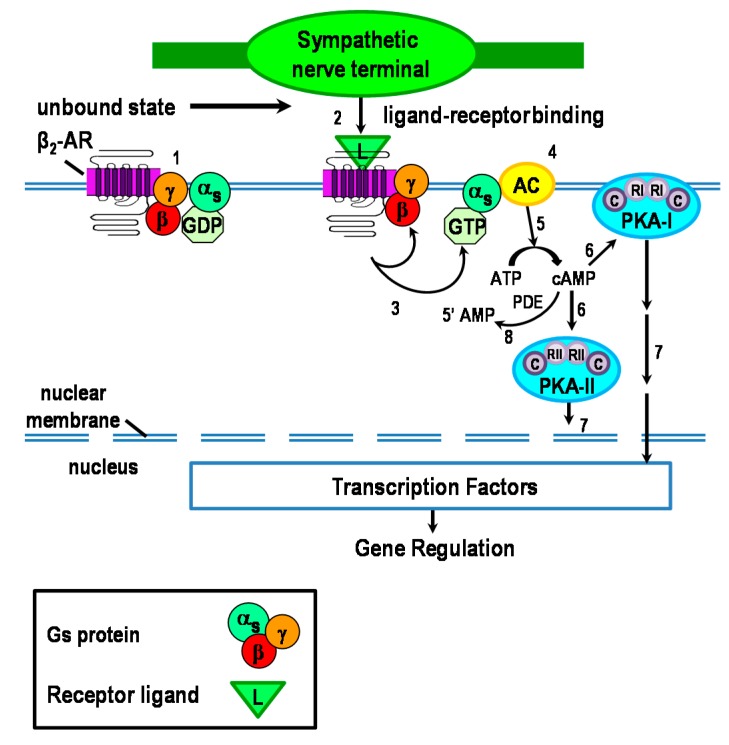
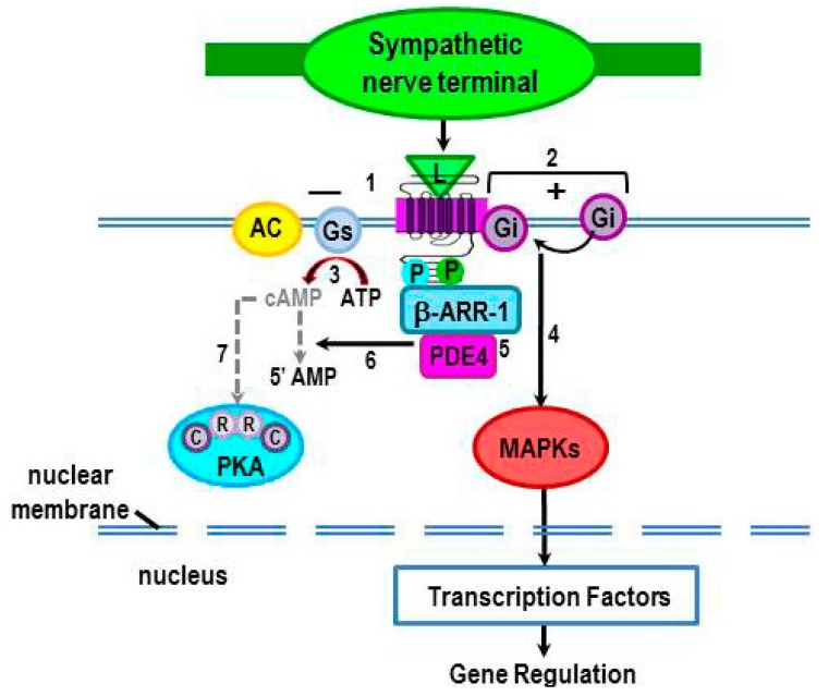
Cross-talk between the sympathetic nervous system (SNS) and immune system is vital for health and well-being. Infection, tissue injury and inflammation raise firing rates of sympathetic nerves, increasing their release of norepinephrine (NE) in lymphoid organs and tissues. NE stimulation of β2-adrenergic receptors (ARs) in immune cells activates the cAMP-protein kinase A (PKA) intracellular signaling pathway, a pathway that interfaces with other signaling pathways that regulate proliferation, differentiation, maturation and effector functions in immune cells. Immune–SNS cross-talk is required to maintain homeostasis under normal conditions, to develop an immune response of appropriate magnitude after injury or immune challenge, and subsequently restore homeostasis. Typically, β2-AR-induced cAMP is immunosuppressive. However, many studies report actions of β2-AR stimulation in immune cells that are inconsistent with typical cAMP–PKA signal transduction. Research during the last decade in non-immune organs, has unveiled novel alternative signaling mechanisms induced by β2-AR activation, such as a signaling switch from cAMP–PKA to mitogen-activated protein kinase (MAPK) pathways. If alternative signaling occurs in immune cells, it may explain inconsistent findings of sympathetic regulation of immune function. Here, we review β2-AR signaling, assess the available evidence for alternative signaling in immune cells, and provide insight into the circumstances necessary for “signal switching” in immune cells.
Overview
The claims and statistics of this paper haven’t been checked yet.
Run the check — free, no account needed. It reads the open-access text and checks the sources it cites.
Integrity signals
Registry
Cites
Statistics
Figures
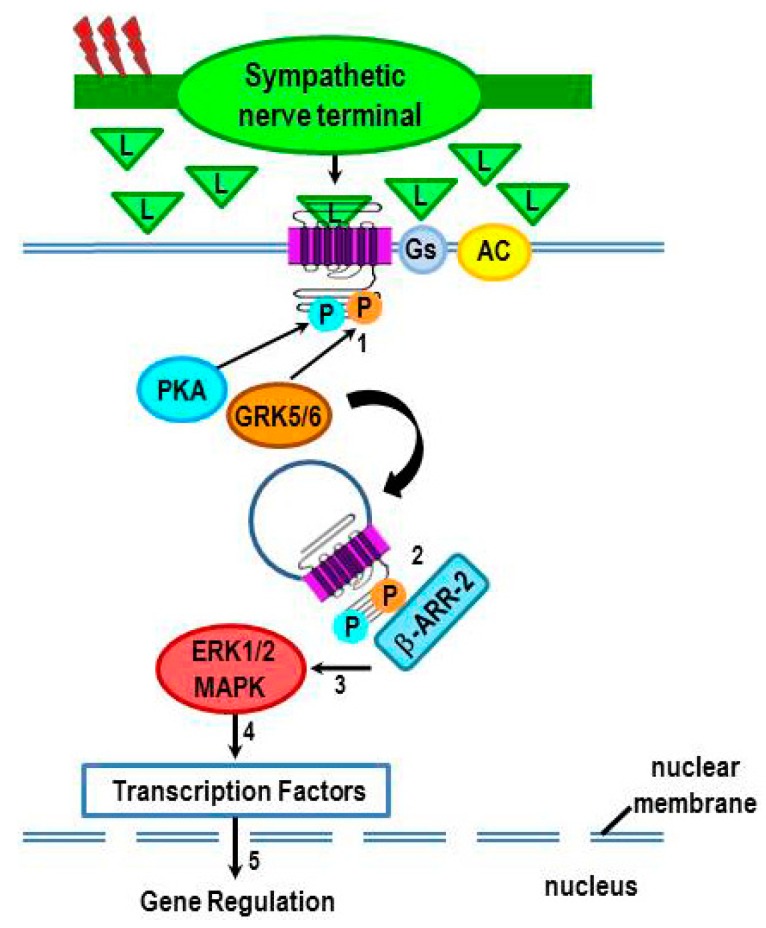
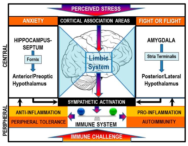
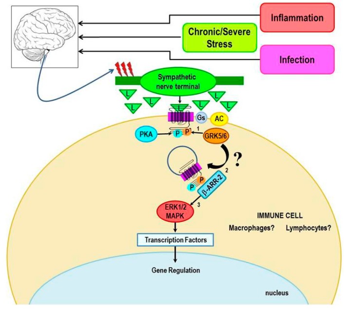
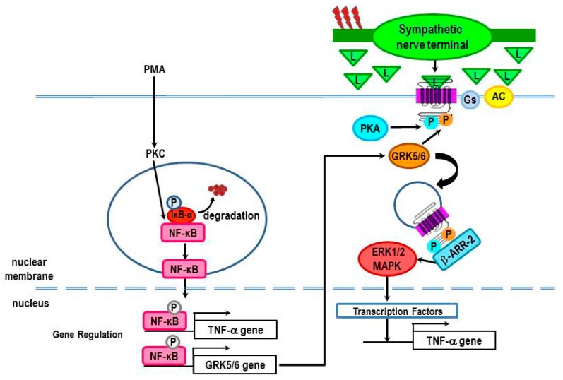
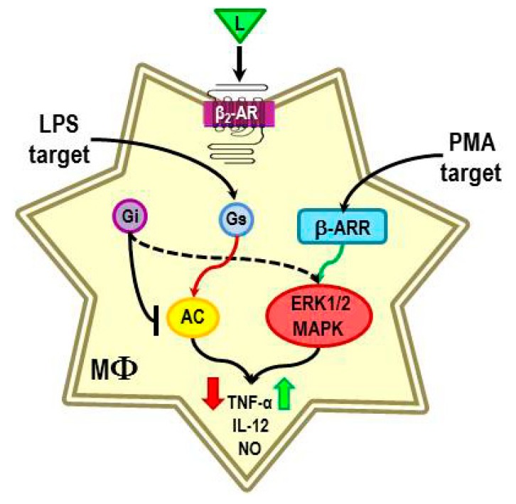
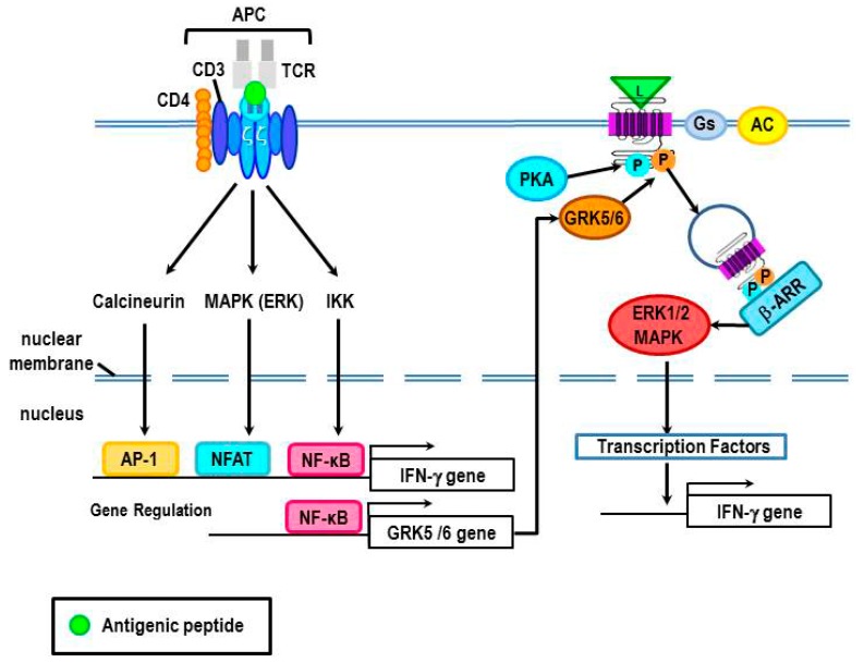
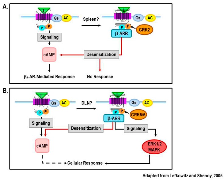
Verification
Claims the paper makes
The claims and statistics of this paper haven’t been checked yet.
Run the check — free, no account needed. It reads the open-access text and checks the sources it cites.
Claims other papers make about this one
Each is checked against this paper’s own text: does it say what the citing paper says it does?
In the citing paper
“Both anti-inflammatory and pro-inflammatory effects of adrenergic signalling have been demonstrated, depending upon the subtype of adrenergic receptors expressed167, the level of activation of specific cell types and the stage of disease progression168.”
From The gut microbiota and the brain–gut–kidney axis in hypertension and chronic kidney disease (2018) · cites this paper as [168]
In the citing paper
“However, persistent activation of the SNS results in changes in signalling within immune organs and cells towards pro-inflammatory pathways168, as observed in hypertension and CKD169,170.”
From The gut microbiota and the brain–gut–kidney axis in hypertension and chronic kidney disease (2018) · cites this paper as [168]
Self-check
Percentages, sums and reported differences re-computed from the paper’s own numbers.
The self-check needs the paper’s full text.
Citations
Cites
1 retracted
Baillie, G. S., Sood, A., McPhee, I., Gall, I., Perry, S. J., Lefkowitz, R. J., & Houslay, M. D. (2003) · Proceedings of the National Academy of Sciences · doi:10.1073/pnas.262787199 · Cited by 358
RetractedFunctional rafts in cell membranes
Simons, K., & Ikonen, E. (1997) · Nature · doi:10.1038/42408 · Cited by 9,757 · cited 2×
“For instance, Ca2+-insensitive AC7 is excluded from lipid rafts [16], but not Ca2+-sensitive AC3 and AC6 [7–9].”
BackgroundMammalian MAP kinase signalling cascades
Chang, L., & Karin, M. (2001) · Nature · doi:10.1038/35065000 · Cited by 5,118
Mitogen-Activated Protein (MAP) Kinase Pathways: Regulation and Physiological Functions*
Pearson, G. W., Robinson, F. L., Gibson, T. B., Xu, B.-E., Karandikar, M. S., Berman, K. S., & Cobb, M. H. (2001) · Endocrine Reviews · doi:10.1210/edrv.22.2.0428 · Cited by 4,111
NF-κB regulation in the immune system
Li, Q., & Verma, I. M. (2002) · Nature Reviews Immunology · doi:10.1038/nri910 · Cited by 4,007
Srimaroeng, C., Cecile, J. P., Walden, R., & Pritchard, J. B. (2013) · Cellular Physiology and Biochemistry · doi:10.1159/000350077 · Cited by 2,989
LPS induction of gene expression in human monocytes
Guha, M., & Mackman, N. (2001) · Cellular Signalling · doi:10.1016/s0898-6568(00)00149-2 · Cited by 2,333
Mitogen-activated protein kinases in innate immunity
Arthur, J. S. C., & Ley, S. C. (2013) · Nature reviews. Immunology · doi:10.1038/nri3495 · Cited by 1,898
Transduction of Receptor Signals by ß-Arrestins
Lefkowitz, R. J., & Shenoy, S. K. (2005) · Science · doi:10.1126/science.1109237 · Cited by 1,729
Signal transduction by the c-Jun N-terminal kinase (JNK) — from inflammation to development
Ip, Y. T., & Davis, R. J. (1998) · Current Opinion in Cell Biology · doi:10.1016/s0955-0674(98)80143-9 · Cited by 1,538
β-Arrestin-Dependent Formation of β 2 Adrenergic Receptor-Src Protein Kinase Complexes
Luttrell, L. M., Ferguson, S. S. G., Daaka, Y., Miller, W. E., Maudsley, S. R., Rocca, G. J. D., Lin, F., Kawakatsu, H., Owada, K. M., Luttrell, D. K., et al. (1999) · Science · doi:10.1126/science.283.5402.655 · Cited by 1,473
β-Arrestins and Cell Signaling
DeWire, S. M., Ahn, S., Lefkowitz, R. J., & Shenoy, S. K. (2007) · Annual Review of Physiology · doi:10.1146/annurev.physiol.69.022405.154749 · Cited by 1,397
β-Arrestin acts as a clathrin adaptor in endocytosis of the β2-adrenergic receptor
Goodman, O. B., Krupnick, J. G., Santini, F., Gurevich, V. V., Penn, R. B., Gagnon, A. W., Keen, J. H., & Benovic, J. (1996) · Nature · doi:10.1038/383447a0 · Cited by 1,383
G PROTEIN–COUPLED RECEPTOR KINASES
Pitcher, J. A., Freedman, N. J., & Lefkowitz, R. J. (1998) · Annual Review of Biochemistry · doi:10.1146/annurev.biochem.67.1.653 · Cited by 1,370
Ji, T. H., Grossmann, M., & Ji, I. (1998) · Journal of Biological Chemistry · doi:10.1074/jbc.273.28.17299 · Cited by 1,281
β-Arrestin: a Protein that Regulates β-adrenergic Receptor Function
Lohse, M. J., Benovic, J., Codina, J., Caron, M. G., & Lefkowitz, R. J. (1990) · Science · doi:10.1126/science.2163110 · Cited by 1,222
THE ROLE OF RECEPTOR KINASES AND ARRESTINS IN G PROTEIN–COUPLED RECEPTOR REGULATION
Krupnick, J. G., & Benovic, J. (1998) · The Annual Review of Pharmacology and Toxicology · doi:10.1146/annurev.pharmtox.38.1.289 · Cited by 1,121
Lefkowitz, R. J. (1998) · Journal of Biological Chemistry · doi:10.1074/jbc.273.30.18677 · Cited by 1,067
Seven-transmembrane-spanning receptors and heart function
Rockman, H. A., Koch, W. J., & Lefkowitz, R. J. (2002) · Nature · doi:10.1038/415206a · Cited by 937
Autonomic innervation and regulation of the immune system (1987–2007)
Nance, D. M., & Sanders, V. M. (2007) · Brain, Behavior, and Immunity · doi:10.1016/j.bbi.2007.03.008 · Cited by 855
Activation and targeting of extracellular signal-regulated kinases by β-arrestin scaffolds
Luttrell, L. M., Roudabush, F. L., Choy, E. W., Miller, W. E., Field, M. E., Pierce, K. L., & Lefkowitz, R. J. (2001) · Proceedings of the National Academy of Sciences · doi:10.1073/pnas.041604898 · Cited by 832
β-Arrestin 2: A Receptor-Regulated MAPK Scaffold for the Activation of JNK3
McDonald, P. H., Chow, C.-W., Miller, W. E., Laporte, S. A., Field, M. E., Lin, F., Davis, R. J., & Lefkowitz, R. J. (2000) · Science · doi:10.1126/science.290.5496.1574 · Cited by 818
β-Arrestin-dependent, G Protein-independent ERK1/2 Activation by the β2 Adrenergic Receptor
Shenoy, S. K., Drake, M. T., Nelson, C. D., Houtz, D. A., Xiao, K., Madabushi, S., Reiter, É., Premont, R. T., Lichtarge, O., & Lefkowitz, R. J. (2005) · Journal of Biological Chemistry · doi:10.1074/jbc.m506576200 · Cited by 758
GRKs and β-arrestins: roles in receptor silencing, trafficking and signaling
Reiter, É., & Lefkowitz, R. J. (2006) · Trends in Endocrinology and Metabolism · doi:10.1016/j.tem.2006.03.008 · Cited by 680
Mammalian Cyclic Nucleotide Phosphodiesterases: Molecular Mechanisms and Physiological Functions
Francis, S. H., Blount, M. A., & Corbin, J. D. (2011) · Physiological Reviews · doi:10.1152/physrev.00030.2010 · Cited by 644
Nobles, K. N., Xiao, K., Ahn, S., Shukla, A. K., Lam, C. M., Rajagopal, S., Strachan, R. T., Huang, T.-Y., Bressler, E. A., Hara, M. R., et al. (2011) · Science Signaling · doi:10.1126/scisignal.2001707 · Cited by 623
Desensitization of G-protein-coupled Receptors
Xiao, Z., Yao, Y., Yu, L., & Devreotes, P. N. (1999) · Journal of Biological Chemistry · doi:10.1074/jbc.274.3.1440 · Cited by 521
Claing, A. (2002) · Progress in Neurobiology · doi:10.1016/s0301-0082(01)00023-5 · Cited by 519
Cyclic nucleotide phosphodiesterases
Essayan, D. M. (2001) · Journal of Allergy and Clinical Immunology · doi:10.1067/mai.2001.119555 · Cited by 503
Bjorn, S. S. (2000) · Frontiers in bioscience · doi:10.2741/skalhegg · Cited by 495
Rockman, H. A., Chien, K. R., Choi, D.-J., Iaccarino, G., Hunter, J. J., Ross, J., Lefkowitz, R. J., & Koch, W. J. (1998) · Proceedings of the National Academy of Sciences · doi:10.1073/pnas.95.12.7000 · Cited by 488
Benovic, J., Kühn, H., Weyand, I., Codina, J., Caron, M. G., & Lefkowitz, R. J. (1987) · Proceedings of the National Academy of Sciences · doi:10.1073/pnas.84.24.8879 · Cited by 482
Structure and mechanism of the G protein-coupled receptor kinases.
Inglese, J., Freedman, N. J., Koch, W. J., & Lefkowitz, R. J. (1993) · Journal of Biological Chemistry · doi:10.1016/s0021-9258(20)80439-6 · Cited by 479
Classical and new roles of β-arrestins in the regulation of G-PROTEIN-COUPLED receptors
Pierce, K. L., & Lefkowitz, R. J. (2001) · Nature reviews. Neuroscience · doi:10.1038/35094577 · Cited by 478
G protein-coupled receptor kinases: More than just kinases and not only for GPCRs
Gurevich, E. V., Tesmer, J. J. G., Mushegian, A. R., & Gurevich, V. V. (2011) · Pharmacology & Therapeutics · doi:10.1016/j.pharmthera.2011.08.001 · Cited by 435
Emerging paradigms of β-arrestin-dependent seven transmembrane receptor signaling
Shukla, A. K., Xiao, K., & Lefkowitz, R. J. (2011) · Trends in Biochemical Sciences · doi:10.1016/j.tibs.2011.06.003 · Cited by 418
Serezani, C. H., Ballinger, M. N., Aronoff, D. M., & Peters-Golden, M. L. (2008) · American Journal of Respiratory Cell and Molecular Biology · doi:10.1165/rcmb.2008-0091tr · Cited by 415
Hausdorff, W. P., Bouvier, M., O’Dowd, B. F., Irons, G. P., Caron, M. G., & Lefkowitz, R. J. (1989) · Journal of Biological Chemistry · doi:10.1016/s0021-9258(18)63907-9 · Cited by 414
Kohm, A. P., & Sanders, V. M. (2001) · Pharmacological Reviews · doi:10.1016/s0031-6997(24)01510-2 · Cited by 399
Quantifying Ligand Bias at Seven-Transmembrane Receptors
Rajagopal, S., Ahn, S., Rominger, D. H., Gowen-MacDonald, W., Lam, C. M., DeWire, S. M., Violin, J. D., & Lefkowitz, R. J. (2011) · Molecular Pharmacology · doi:10.1124/mol.111.072801 · Cited by 391
Beyond Desensitization: Physiological Relevance of Arrestin-Dependent Signaling
Luttrell, L. M., Gesty‐Palmer, D., & Sibley, D. R. (2010) · Pharmacological Reviews · doi:10.1124/pr.109.002436 · Cited by 387
Protein kinase C switches the Raf kinase inhibitor from Raf-1 to GRK-2
Lorenz, K., Lohse, M. J., & Quitterer, U. (2003) · Nature · doi:10.1038/nature02158 · Cited by 384
Sun, Y., Cheng, Z.-J. J., Ma, L., & Pei, G. (2002) · Journal of Biological Chemistry · doi:10.1074/jbc.m207294200 · Cited by 382
The many paths to p38 mitogen-activated protein kinase activation in the immune system
Ashwell, J. D. (2006) · Nature reviews. Immunology · doi:10.1038/nri1865 · Cited by 370
Kohm, A. P., & Sanders, V. M. (2001) · PubMed · Cited by 357
Balabanian, K. (2004) · Blood · doi:10.1182/blood-2004-06-2289 · Cited by 321
Seven transmembrane receptors: something old, something new
Lefkowitz, R. J. (2007) · Acta Physiologica · doi:10.1111/j.1365-201x.2007.01693.x · Cited by 308
Adenylyl Cyclase–A-kinase Anchoring Protein Complexes: The Next Dimension in cAMP Signaling
DESSAUER, C. W. (2009) · Molecular Pharmacology · doi:10.1124/mol.109.059345 · Cited by 263
It takes nerve to tell T and B cells what to do
Kin, N. W., & Sanders, V. M. (2006) · Journal of Leukocyte Biology · doi:10.1189/jlb.1105625 · Cited by 258
β-Arrestin-biased Agonism at the β2-Adrenergic Receptor
Drake, M. T., Violin, J. D., Whalen, E. J., Wisler, J. W., Shenoy, S. K., & Lefkowitz, R. J. (2007) · Journal of Biological Chemistry · doi:10.1074/jbc.m708118200 · Cited by 254
Skålhegg, B. S. (1997) · Frontiers in bioscience · doi:10.2741/a195 · Cited by 246
Lipid rafts: now you see them, now you don't
Shaw, A. S. (2006) · Nature Immunology · doi:10.1038/ni1405 · Cited by 240
Adrenal GRK2 upregulation mediates sympathetic overdrive in heart failure
Lymperopoulos, A., Rengo, G., Funakoshi, H., Eckhart, A. D., & Koch, W. J. (2007) · Nature Medicine · doi:10.1038/nm1553 · Cited by 236
G-protein-coupled receptor kinase activity is increased in hypertension.
Gros, R., Benovic, J., Tan, C. M., & Feldman, R. D. (1997) · Journal of Clinical Investigation · doi:10.1172/jci119381 · Cited by 218
Autonomic regulation of cellular immune function
Bellinger, D. L., & Lorton, D. (2014) · Autonomic Neuroscience · doi:10.1016/j.autneu.2014.01.006 · Cited by 217
Self-citationRole of Phosphorylation in Agonist-promoted β2-Adrenergic Receptor Sequestration
Ferguson, S. S. G., Ménard, L., Barak, L. S., Koch, W. J., Colapietro, A.-M., & Caron, M. G. (1995) · Journal of Biological Chemistry · doi:10.1074/jbc.270.42.24782 · Cited by 216
Cyclic AMP: a selective modulator of NF-κB action
Gerlo, S., Kooijman, R., Beck, I. M. E., Kolmus, K., Spooren, A., & Haegeman, G. (2011) · Cellular and Molecular Life Sciences · doi:10.1007/s00018-011-0757-8 · Cited by 209
Lombardi, M. S., Kavelaars, A., Schedlowski, M., Bijlsma, J. W. J., Okihara, K. L., van de Pol, M., Ochsmann, S., Pawlak, C. R., Schmidt, R. E., & Heijnen, C. J. (1999) · The FASEB Journal · doi:10.1096/fasebj.13.6.715 · Cited by 208
IFN-γ Production by Th1 Cells Generated from Naive CD4+ T Cells Exposed to Norepinephrine
Swanson-Mungerson, M. A., Lee, W. T., & Sanders, V. M. (2001) · The Journal of Immunology · doi:10.4049/jimmunol.166.1.232 · Cited by 207
Tan, K. S., Nackley, A. G., Satterfield, K., Maixner, W., Diatchenko, L., & Flood, P. M. (2007) · Cellular Signalling · doi:10.1016/j.cellsig.2006.06.007 · Cited by 204
Location of cAMP-Dependent Protein Kinase Type I with the TCR-CD3 Complex
Skålhegg, B. S., Taskén, K., Hansson, V., Huitfeldt, H. S., Jahnsen, T., & Lea, T. (1994) · Science · doi:10.1126/science.8272870 · Cited by 196
Pitcher, J. A., Lohse, M. J., Codina, J., Caron, M. G., & Lefkowitz, R. J. (1992) · Biochemistry · doi:10.1021/bi00127a021 · Cited by 194
Desensitization of G protein-coupled receptors.
Freedman, N. J., & Lefkowitz, R. J. (1996) · PubMed · Cited by 190
Oppermann, M., Mack, M., Proudfoot, A. E. I., & Olbrich, H. (1999) · Journal of Biological Chemistry · doi:10.1074/jbc.274.13.8875 · Cited by 185
Elenkov, I. J., Haskó, G., Kovács, K., & Vizi, E. S. (1995) · Journal of Neuroimmunology · doi:10.1016/0165-5728(95)00080-l · Cited by 180
Benczik, M., & Gaffen, S. L. (2004) · Immunological Investigations · doi:10.1081/imm-120030732 · Cited by 165
Ivashkiv, L. B. (2011) · European Journal of Immunology · doi:10.1002/eji.201141783 · Cited by 162
GRKs and arrestins: regulators of migration and inflammation
Vroon, A., Heijnen, C. J., & Kavelaars, A. (2006) · Journal of Leukocyte Biology · doi:10.1189/jlb.0606373 · Cited by 155
PDEs create local domains of cAMP signaling
Mika, D., Leroy, J., Vandecasteele, G., & Fischmeister, R. (2011) · Journal of Molecular and Cellular Cardiology · doi:10.1016/j.yjmcc.2011.08.016 · Cited by 153
Tran, T. M., Friedman, J., Qunaibi, E. A., Baameur, F., Moore, R. H., & Clark, R. B. (2004) · Molecular Pharmacology · doi:10.1124/mol.65.1.196 · Cited by 153
Partial agonists and G protein-coupled receptor desensitization
Clark, R. B., Knoll, B. J., & Barber, R. (1999) · Trends in Pharmacological Sciences · doi:10.1016/s0165-6147(99)01351-6 · Cited by 145
Phosphodiesterase Inhibitors in the Treatment of Inflammatory Diseases
Page, C. P., & Spina, D. (2011) · Handbook of experimental pharmacology · doi:10.1007/978-3-642-17969-3_17 · Cited by 145
TCR- and CD28-Mediated Recruitment of Phosphodiesterase 4 to Lipid Rafts Potentiates TCR Signaling
Abrahamsen, H., Baillie, G. S., Ngai, J., Vang, T., Nika, K., Ruppelt, A., Mustelin, T., Zaccolo, M., Houslay, M. D., & Taskén, K. (2004) · The Journal of Immunology · doi:10.4049/jimmunol.173.8.4847 · Cited by 143
The Ras/MAPK cascade and the control of positive selection
Alberola‐Ila, J., & Hernández‐Hoyos, G. (2003) · Immunological Reviews · doi:10.1034/j.1600-065x.2003.00012.x · Cited by 143
Reinventing the Wheel of Cyclic AMP
Chin, K., YANG, W., Ravatn, R., KITA, T., Reitman, E., Vettori, D., Cvijic, M. E., Shin, M. J., & Iacono, L. (2002) · Annals of the New York Academy of Sciences · doi:10.1111/j.1749-6632.2002.tb04326.x · Cited by 135
Arrestin times for compartmentalised cAMP signalling and phosphodiesterase-4 enzymes
Baillie, G. S., & Houslay, M. D. (2005) · Current Opinion in Cell Biology · doi:10.1016/j.ceb.2005.01.003 · Cited by 134
Regulation of the G Protein-coupled Receptor Kinase GRK5 by Protein Kinase C
Pronin, A. N., & Benovic, J. (1997) · Journal of Biological Chemistry · doi:10.1074/jbc.272.6.3806 · Cited by 132
Pontier, S. M., Percherancier, Y., Galandrin, S., Breit, A., Galès, C., & Bouvier, M. (2008) · Journal of Biological Chemistry · doi:10.1074/jbc.m800778200 · Cited by 128
The p38 Mitogen-Activated Protein Kinase Is Required for IL-12-Induced IFN-γ Expression
Zhang, S., & Kaplan, M. H. (2000) · The Journal of Immunology · doi:10.4049/jimmunol.165.3.1374 · Cited by 128
Chuang, T. T., Sallese, M., Ambrosini, G., Parruti, G., & Blasi, A. D. (1992) · Journal of Biological Chemistry · doi:10.1016/s0021-9258(19)50511-7 · Cited by 121
Erdoğan, S., & Houslay, M. D. (1997) · Biochemical Journal · doi:10.1042/bj3210165 · Cited by 120
Lombardi, M. S., Kavelaars, A., Cobelens, P. M., Schmidt, R. E., Schedlowski, M., & Heijnen, C. J. (2001) · The Journal of Immunology · doi:10.4049/jimmunol.166.3.1635 · Cited by 117
Bernardin, G., Strosberg, A. D., Bernard, A., Mattei, M., & Marullo, S. (1998) · Intensive Care Medicine · doi:10.1007/s001340050768 · Cited by 111
Skålhegg, B. S. (2000) · Frontiers in bioscience · doi:10.2741/a543 · Cited by 111
Vroon, A., Kavelaars, A., Limmroth, V., Lombardi, M. S., Goebel, M. U., van Dam, A., Caron, M. G., Schedlowski, M., & Heijnen, C. J. (2005) · The Journal of Immunology · doi:10.4049/jimmunol.174.7.4400 · Cited by 108
Blasi, A. D., Parruti, G., & Sallese, M. (1995) · Journal of Clinical Investigation · doi:10.1172/jci117641 · Cited by 108
A dual role for the cAMP-dependent protein kinase in tyrosine hydroxylase gene expression.
Kim, K. S., Park, D. H., Wessel, T. C., Song, B., Wagner, J. A., & Joh, T. H. (1993) · Proceedings of the National Academy of Sciences · doi:10.1073/pnas.90.8.3471 · Cited by 100
Multiple functions of G protein-coupled receptor kinases
Watari, K., Nakaya, M., & Kurose, H. (2014) · Journal of Molecular Signaling · doi:10.1186/1750-2187-9-1 · Cited by 98
Trester-Zedlitz, M., Burlingame, A. L., Kobilka, B. K., & von Zastrow, M. (2005) · Biochemistry · doi:10.1021/bi0475469 · Cited by 95
Smith, K. E., Gu, C., Fagan, K. A., Hu, B., & Cooper, D. M. F. (2002) · Journal of Biological Chemistry · doi:10.1074/jbc.m109615200 · Cited by 93
Yuan, N., Friedman, J. E., Whaley, B. S., & Clark, R. B. (1994) · Journal of Biological Chemistry · doi:10.1016/s0021-9258(17)31615-0 · Cited by 88
Distinct Roles of Adenylyl Cyclase VII in Regulating the Immune Responses in Mice
Duan, B., Davis, R., Sadat, E. L., Collins, J. N. R., Sternweis, P. C., Yuan, D., & Jiang, L. I. (2010) · The Journal of Immunology · doi:10.4049/jimmunol.0903474 · Cited by 88
Cytokine Production by Naive and Primary Effector CD4+ T Cells Exposed to Norepinephrine
Ramer-Quinn, D. S., Swanson-Mungerson, M. A., Lee, W. T., & Sanders, V. M. (2000) · Brain Behavior and Immunity · doi:10.1006/brbi.2000.0603 · Cited by 81
Zeiser, R., & Negrin, R. S. (2008) · Cell Cycle · doi:10.4161/cc.7.4.5454 · Cited by 79
Phosphodiesterase 4 and tolerance to β2-adrenoceptor agonists in asthma
Giembycz, M. A. (1996) · Trends in Pharmacological Sciences · doi:10.1016/0165-6147(96)10039-0 · Cited by 76
O’Brien, R. L. F., & Uíttenbogaart, C. H. (2010) · Immunologic Research · doi:10.1007/s12026-010-8170-1 · Cited by 75
Phosphodiesterase 4 and tolerance to beta 2-adrenoceptor agonists in asthma.
Giembycz, M. A. (1996) · PubMed · Cited by 73
Deiss, K., Kisker, C., Lohse, M. J., & Lorenz, K. (2012) · Journal of Biological Chemistry · doi:10.1074/jbc.m112.363812 · Cited by 69
Bjørgo, E., Solheim, S., Abrahamsen, H., Baillie, G. S., Brown, K. M., Berge, T., Okkenhaug, K., Houslay, M. D., & Taskén, K. (2010) · Molecular and Cellular Biology · doi:10.1128/mcb.00696-09 · Cited by 69
Sivertsen, B., Holliday, N. D., Madsen, A. N., & Holst, B. (2013) · British Journal of Pharmacology · doi:10.1111/bph.12361 · Cited by 68
Tachibana, H., Prasad, S. V. N., Lefkowitz, R. J., Koch, W. J., & Rockman, H. A. (2005) · Circulation · doi:10.1161/01.cir.0000142291.70954.df · Cited by 68
Loudon, R., PERUSSIA, B., & Benovic, J. (1996) · Blood · doi:10.1182/blood.v88.12.4547.bloodjournal88124547 · Cited by 68
Molecular mechanisms of feedback inhibition of protein kinase A on intracellular cAMP accumulation
Vandamme, J., Castermans, D., & Thevelein, J. M. (2012) · Cellular Signalling · doi:10.1016/j.cellsig.2012.04.001 · Cited by 64
Baerwald, C., Graefe, C., von Wichert, P., & Krause, A. (1992) · PubMed · Cited by 63
Zoukos, Y., Kidd, D. P., Woodroofe, M. N., Kendall, B. E., Thompson, A. J., & Cuzner, M. L. (1994) · Brain · doi:10.1093/brain/117.2.307 · Cited by 62
Toll-like receptors differentially regulate GPCR kinases and arrestins in primary macrophages
Loniewski, K. J., Shi, Y., Pestka, J. J., & Parameswaran, N. (2008) · Molecular Immunology · doi:10.1016/j.molimm.2007.11.012 · Cited by 60
Giorelli, M., Livrea, P., & Trojano, M. (2004) · Journal of Neuroimmunology · doi:10.1016/j.jneuroim.2004.05.013 · Cited by 60
Chaudhary, L. R., & Avioli, L. V. (2000) · Journal of Cellular Biochemistry · doi:10.1002/(sici)1097-4644(20000301)76:3 · Cited by 58
Macneil, B. J., Jansen, A., Greenberg, A. H., & Nance, D. M. (1996) · American Journal of Physiology-Regulatory, Integrative and Comparative Physiology · doi:10.1152/ajpregu.1996.270.1.r264 · Cited by 58
Liu, Z., Jiang, Y., Li, Y., Wang, J., Fan, L., Scott, M. J., Xiao, G., Li, S., Billiar, T. R., Wilson, M. A., et al. (2013) · The Journal of Immunology · doi:10.4049/jimmunol.1300790 · Cited by 57
GRK6 deficiency in mice causes autoimmune disease due to impaired apoptotic cell clearance
Nakaya, M., Tajima, M., Kosako, H., Nakaya, T., Hashimoto, A. S., Watari, K., Nishihara, H., Ohba, M., Komiya, S., Tani, N., et al. (2013) · Nature Communications · doi:10.1038/ncomms2540 · Cited by 54
Hausdorff, W. P., Lohse, M. J., Bouvier, M., Liggett, S. B., Caron, M. G., & Lefkowitz, R. J. (1990) · PubMed · Cited by 50
Lubahn, C. L., Lorton, D., Schaller, J. A., Sweeney, S. J., & Bellinger, D. L. (2014) · Frontiers in Immunology · doi:10.3389/fimmu.2014.00346 · Cited by 48
Self-citationGriffiths, G. W., Hollinshead, R., Hemmings, B. A., & Nigg, E. A. (1990) · Journal of Cell Science · doi:10.1242/jcs.96.4.691 · Cited by 45
Johnson, J. A., Clark, R. B., Friedman, J. E., Dixon, R. A. F., & Strader, C. D. (1990) · Molecular Pharmacology · doi:10.1016/s0026-895x(25)09464-7 · Cited by 44
Szelényi, J., Selmeczy, Z., Brózik, A., Medgyesi, D., & Magócsi, M. (2006) · Neurochemistry International · doi:10.1016/j.neuint.2006.01.009 · Cited by 43
Wahle, M., Hanefeld, G., Brunn, S., Straub, R. H., Wagner, U. G., Krause, A., Häntzschel, H., & Baerwald, C. (2006) · Arthritis Research & Therapy · doi:10.1186/ar2028 · Cited by 42
Raghuwanshi, S. K., Smith, N., Rivers, E. J., Thomas, A. J., Sutton, N., Hu, Y., Mukhopadhyay, S., Chen, X. L., Leung, T., & Richardson, R. M. (2013) · The Journal of Immunology · doi:10.4049/jimmunol.1202058 · Cited by 41
Wang, Y., Ding, M., Chaudhari, S. K., Ding, Y., Yuan, J. P., Stankowska, D. L., He, S., Krishnamoorthy, R. R., Cunningham, J. T., & Ma, R. (2013) · Journal of Biological Chemistry · doi:10.1074/jbc.m112.410357 · Cited by 40
Wang, W. C. H., Mihlbachler, K. A., Brunnett, A. C., & Liggett, S. B. (2009) · Proceedings of the National Academy of Sciences · doi:10.1073/pnas.0906034106 · Cited by 35
Islam, K. N., & Koch, W. J. (2012) · Journal of Biological Chemistry · doi:10.1074/jbc.m111.324566 · Cited by 35
Reduced catecholarnine response of lymphocytes from patients with rheumatoid arthritis
Baerwald, C., Wahle, M., Ulrichs, T., Jonas, D., von Bierbrauer, A., von Wichert, P., Burmester, G. R., & Krause, A. (1999) · Immunobiology · doi:10.1016/s0171-2985(99)80034-5 · Cited by 32
Yang, J., New, L., Jiang, Y., Han, J., & Su, B. (1998) · Gene · doi:10.1016/s0378-1119(98)00158-9 · Cited by 32
Meltzer, J. C., Macneil, B. J., Sanders, V., Pylypas, S. P., Jansen, A. H., Greenberg, A. H., & Nance, D. M. (2003) · Brain Behavior and Immunity · doi:10.1016/s0889-1591(03)00084-9 · Cited by 32
Lipopolysaccharide-induced sensitization of adenylyl cyclase activity in murine macrophages
Osawa, Y., Lee, H. T., Hirshman, C. A., Xu, D., & Emala, C. W. (2005) · American Journal of Physiology-Cell Physiology · doi:10.1152/ajpcell.00171.2005 · Cited by 28
Novel aspects of G‐protein‐coupled receptor signalling – different ways to achieve specificity
Schulte, G., & Levy, F. O. (2007) · Acta Physiologica · doi:10.1111/j.1365-201x.2007.01696.x · Cited by 28
Altered Sympathetic-to-Immune Cell Signaling viaβ2-Adrenergic Receptors in Adjuvant Arthritis
Lorton, D., Bellinger, D. L., Schaller, J. A., Shewmaker, E., Osredkar, T., & Lubahn, C. L. (2013) · Clinical and Developmental Immunology · doi:10.1155/2013/764395 · Cited by 28
Self-citationEijkelkamp, N., Heijnen, C. J., Carbajal, A. G., Willemen, H. L. D. M., Wang, H., Minett, M. S., Wood, J. N., Schedlowski, M., Dantzer, R., Kelley, K. W., et al. (2012) · Molecular Medicine · doi:10.2119/molmed.2011.00398 · Cited by 27
Mohede, I. C. M., van Ark, I., Brons, F. M., van Oosterhout, A. J. M., & Nijkamp, F. P. (1996) · International Journal of Immunopharmacology · doi:10.1016/0192-0561(96)00008-2 · Cited by 25
The role of GRK6 in animal models of Parkinson's Disease and L-DOPA treatment
Managò, F., Espinoza, S., Salahpour, A., Sotnikova, T. D., Caron, M. G., Premont, R. T., & Gainetdinov, R. R. (2012) · Scientific Reports · doi:10.1038/srep00301 · Cited by 24
Oyama, N., Urasawa, K., Kaneta, S., Sakai, H., Saito, T., Takagi, C., Yoshida, I., Kitabatake, A., & Tsutsui, H. (2005) · Circulation Journal · doi:10.1253/circj.69.987 · Cited by 23
Zoukos, Y., Thomaides, T., Kidd, D. P., Cuzner, M. L., & Thompson, A. J. (2003) · Journal of Neurology Neurosurgery & Psychiatry · doi:10.1136/jnnp.74.2.197 · Cited by 23
Cyclic Nucleotides and Phosphodiesterases in Monocytic Differentiation
Hertz, A. L., & Beavo, J. A. (2011) · Handbook of experimental pharmacology · doi:10.1007/978-3-642-17969-3_16 · Cited by 20
Role of Epac proteins in mechanisms of cAMP-dependent immunoregulation
Ширшев, С. В. (2011) · Biochemistry (Moscow) · doi:10.1134/s000629791109001x · Cited by 19
van Oosterhout, A. J. M., Stam, W. B., Vanderschueren, R. G. J. R. A., & Nijkamp, F. P. (1992) · Journal of Allergy and Clinical Immunology · doi:10.1016/s0091-6749(05)80013-2 · Cited by 19
Effects of canine endotoxin shock on lymphocytic beta-adrenergic receptors.
SILVERMAN, H. J., Lee, N. H., & El‐Fakahany, E. E. (1990) · PubMed · Cited by 17
Tumor necrosis factor produces homologous desensitization of lymphocyte beta 2-adrenergic responses.
Singh, M., Da, N., & Metakis, L. (1993) · PubMed · Cited by 16
Protein kinase c regulation of the adenylyl cyclase system in rat prostatic epithelium
Carmena, M. J., GARCIA-PARAMIO, P., Solano, R. M., & Prieto, J. C. (1995) · The Prostate · doi:10.1002/pros.2990270405 · Cited by 15
The Role of Protein Kinase A Anchoring via the RIIα Regulatory Subunit in the Murine Immune System
Schillace, R. V., Andrews, S. F., Galligan, S. G., Burton, K. A., Starks, H. J., Bouwer, H. G. A., McKnight, G. S., Davey, M. P., & Carr, D. W. (2005) · The Journal of Immunology · doi:10.4049/jimmunol.174.11.6847 · Cited by 12
Endosome sorting of β2‐adrenoceptors is GRK5 independent
Millman, E. E., Rosenfeld, J. L., Vaughan, D. J., Nguyen, J., Dai, W., Foster, E. A., Clark, R. B., Knoll, B. J., & Moore, R. H. (2004) · British Journal of Pharmacology · doi:10.1038/sj.bjp.0705504 · Cited by 11
Peng, Y., Shan, J., Qi, X., Zhang, S., Ma, S., Wang, N., Li, J., Xue, H., & Wu, M. (2006) · Clinical and Experimental Pharmacology and Physiology · doi:10.1111/j.1440-1681.2006.04350.x · Cited by 11
Decrease in Beta-adrenergic Receptors of Lymphocytes in Spontaneously Occurring Acute Asthma
畑岡, 勲., Okayama, M., Sugi, M., Inoue, H., Takishima, T., & Shirato, K. (1993) · CHEST Journal · doi:10.1378/chest.104.2.508 · Cited by 11
Seyyedi, S., Andalib, A., Rezaei, A., Hosseini, S. M., Mohebbi, S. R., Zali, M. R., Vafai, M., Behboo, R., Tabatabaei, S. A., & Shahabi, S. (2012) · Europe PMC (PubMed Central) · Cited by 9
Iizuka, K., Yoshie, Y., & NAKAZAWA, T. (1991) · PubMed · Cited by 7
GRK6, a gatekeeper of visceral hyperalgesia
Murga, C., & Mayor, F. (2008) · Brain Behavior and Immunity · doi:10.1016/j.bbi.2008.10.002 · Cited by 6
Role of beta-adrenoceptor at different stages of bronchial asthma.
Gao, H., Xue, Q., Lin, Y., Wang, L., Zhu, G., Zhao, Q., & Chen, Y. (2001) · PubMed · Cited by 5
Cited by
Sorted by citations
The gut microbiota and the brain–gut–kidney axis in hypertension and chronic kidney disease
Yang, T., Richards, E. M., Pepine, C. J., & Raizada, M. K. (2018) · Nature Reviews Nephrology · doi:10.1038/s41581-018-0018-2 · Cited by 753
β2 Adrenergic-Neurotrophin Feedforward Loop Promotes Pancreatic Cancer
Renz, B. W., Takahashi, R., Tanaka, T., Macchini, M., Hayakawa, Y., Dantes, Z., Maurer, H. C., Chen, X., Jiang, Z., Westphalen, C. B., et al. (2018) · Cancer Cell · doi:10.1016/j.ccell.2017.11.007 · Cited by 543
Mohammadpour, H., MacDonald, C. R., Qiao, G., Chen, M., Dong, B., Hylander, B. L., McCarthy, P. L., Abrams, S. I., & Repasky, E. A. (2019) · Journal of Clinical Investigation · doi:10.1172/jci129502 · Cited by 328
Cholinergic modulation of the immune system presents new approaches for treating inflammation
Hoover, D. B. (2017) · Pharmacology & Therapeutics · doi:10.1016/j.pharmthera.2017.05.002 · Cited by 306
Autonomic dysreflexia after spinal cord injury: Systemic pathophysiology and methods of management
Eldahan, K. C., & Rabchevsky, A. G. (2017) · Autonomic Neuroscience · doi:10.1016/j.autneu.2017.05.002 · Cited by 286
Metadata
- Authors
- Lorton, D., & Bellinger, D.
- Affiliations
- Kent State University (US)
- Loma Linda University (US)
- Corresponding author
- Lorton, Dianne
- ORCID
- Lorton, Dianne
- Venue
- International Journal of Molecular Sciences
- Publisher
- MDPI AG
- Published
- 2015-03-11
- Volume
- 16
- Issue
- 3
- Pages
- 5635-5665
- ISSN
- 1422-0067 (ISSN-L)
- PMC
- PMC4394497
- OpenAlex
- W1993341098
- Cited by
- 221
- References
- 147
- Citations by year
- 2026: 23 · 2025: 31 · 2024: 26 · 2023: 26 · 2022: 17 · 2021: 17 · 2020: 15 · 2019: 22 · 2018: 14 · 2017: 17 · 2016: 10 · 2015: 3
- FWCI
- 1.42
- Type
- journal article
- Language
- en
- Open access
- gold
- dx.doi.org
- Licence
- Locations
- International Journal of Molecular Sciences (publishedVersion, cc-by)
- Europe PMC (PubMed Central) (publishedVersion, cc-by)
- PubMed Central (publishedVersion, cc-by)
- PubMed (publishedVersion)
- DOAJ (DOAJ: Directory of Open Access Journals) (submittedVersion, cc-by-sa)
- Topics
- Life Sciences › Biochemistry, Genetics and Molecular Biology › Molecular Biology › Receptor Mechanisms and Signaling
- Life Sciences › Neuroscience › Neurology › Vagus Nerve Stimulation Research
- Life Sciences › Biochemistry, Genetics and Molecular Biology › Cancer Research › Cancer, Stress, Anesthesia, and Immune Response
- Keywords
- beta2-adrenergic receptor, sympathetic nervous system, norepinephrine, cAMP/PKA signaling, MAPK signaling, signal switching, sympathetic neurons, immune cells, immunosuppression, lymphoid organs
- MeSH terms
- Cell Communication, Humans, Immunity, Innate, Lymphocytes, Receptors, Adrenergic, beta, Sympathetic Nervous System, Signal Transduction, Cyclic AMP-Dependent Protein Kinases, Mitogen-Activated Protein Kinases, Receptors, G-Protein-Coupled, Adaptive Immunity
About these checks
Registry, retraction, Cites and statistics results are computed, not judged, with one exception: a printed reference that no index lists is read once by a model before it counts as a citation error. Claims are extracted by a model and kept only when their quote appears verbatim in the text; each cited source is then checked, with a model’s help, for whether it says what the claim relies on. Those verdicts can be wrong — read the claim’s quote and the cited source before relying on one.
Statistics and arithmetic are recomputed, not judged.
Confidence bands are shown only for fields calibrated on our annotated test set. None is calibrated yet, so no band is shown.
What counts as a claim ›How we measure confidence ›How we check this ›
Are you an author, or do you see a problem with this page? Request a review
