

The DOI matches its Crossref record; 59 of the 61 references it cites were found in a registry or an index, and 2 are not checked yet.
Biological Mechanisms Underlying the Ultraviolet Radiation-Induced Formation of Skin Wrinkling and Sagging I: Reduced Skin Elasticity, Highly Associated with Enhanced Dermal Elastase Activity, Triggers Wrinkling and Sagging
Open accessAbstract
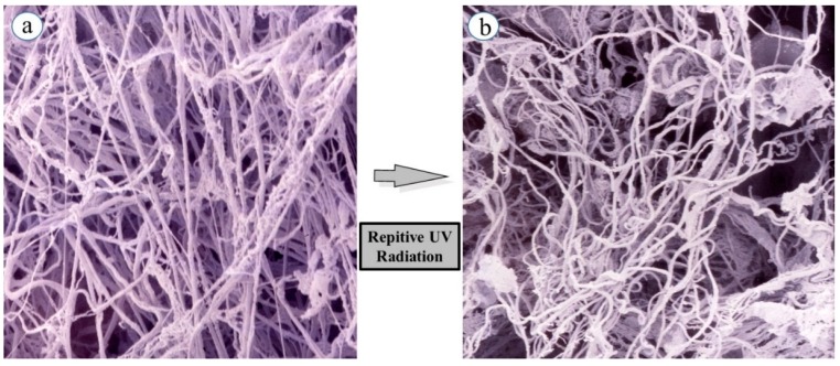
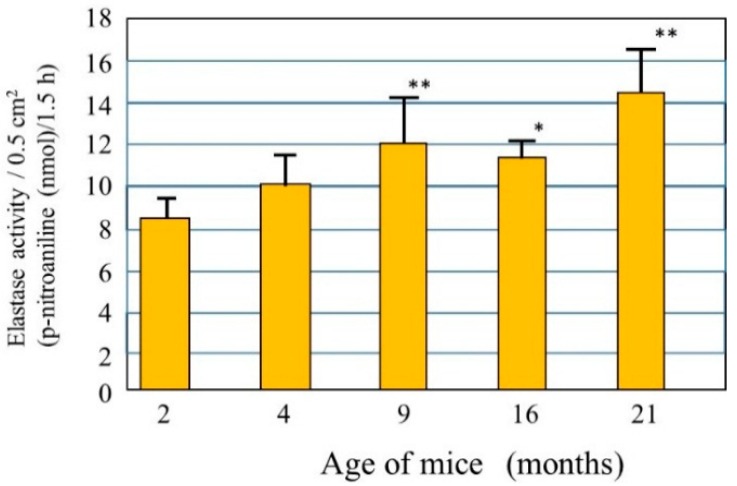
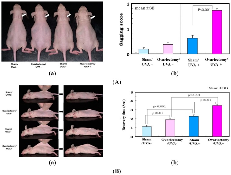
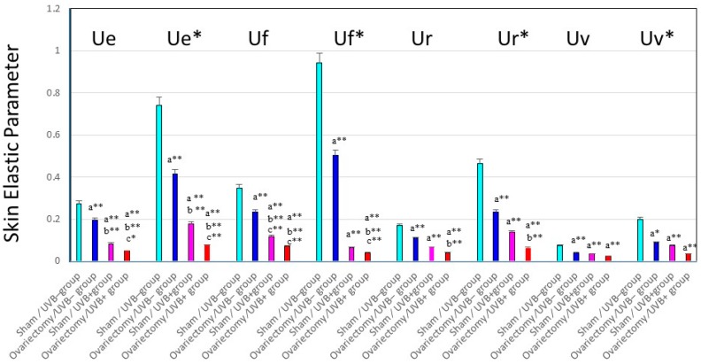
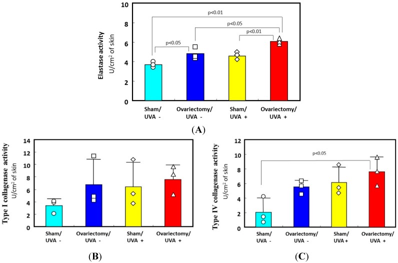
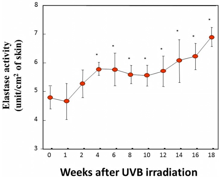
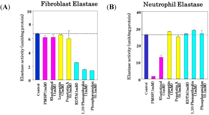
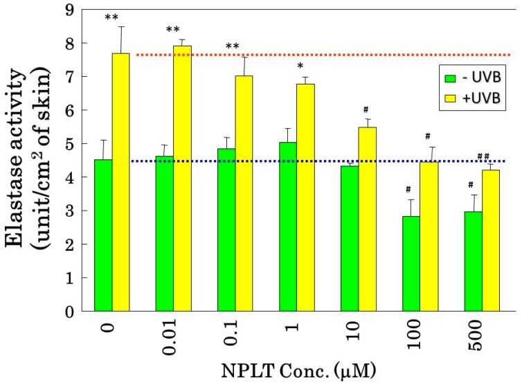
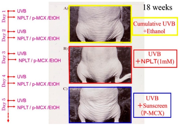
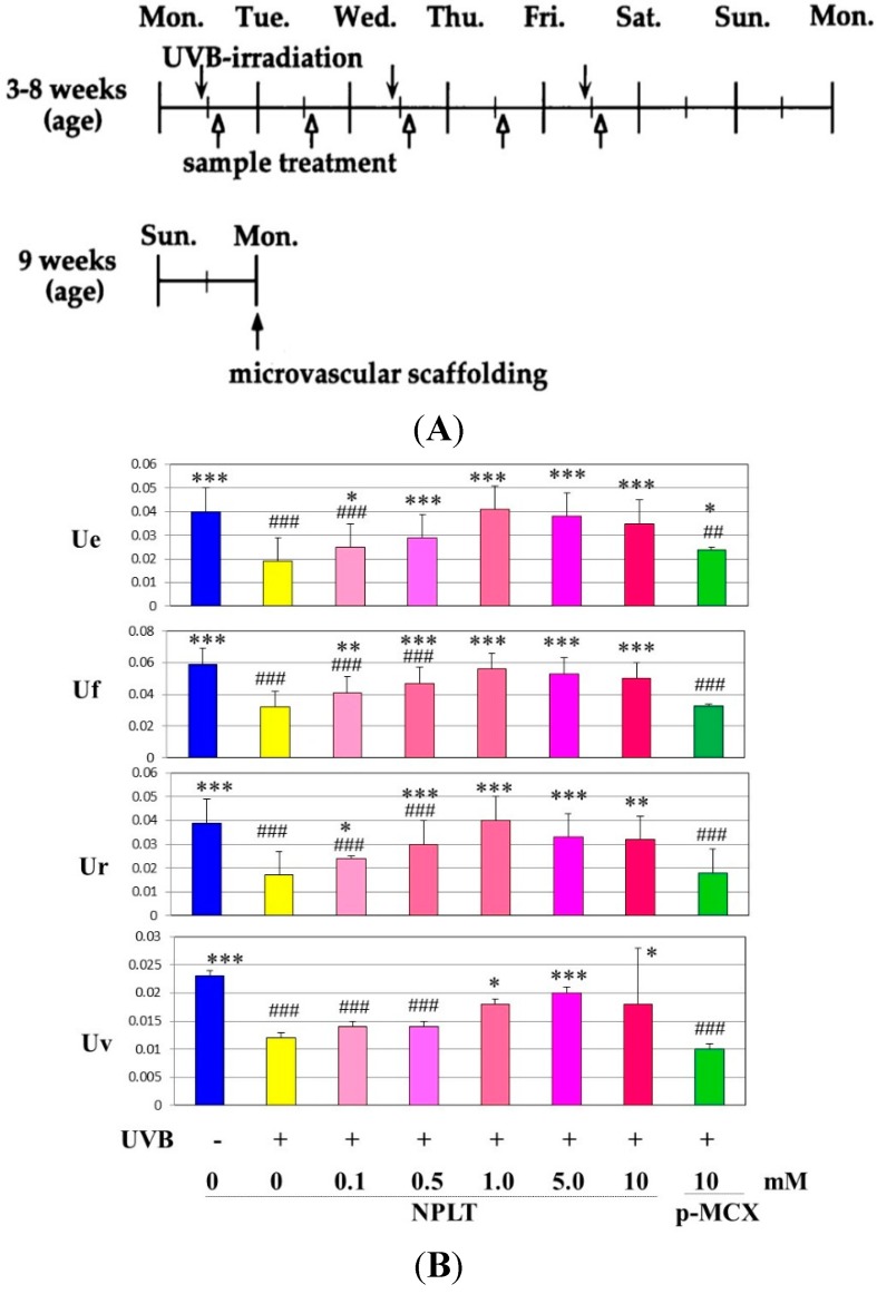
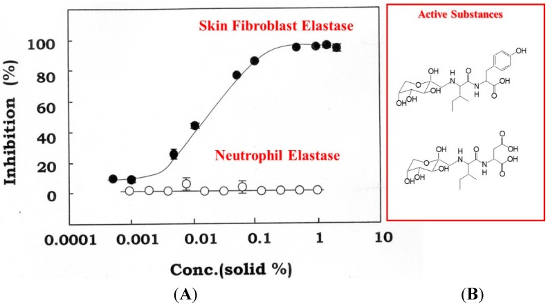
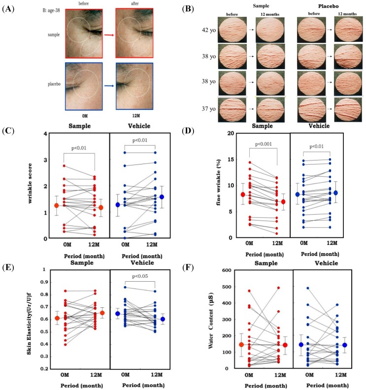
The repetitive exposure of skin to ultraviolet B (UVB) preferentially elicits wrinkling while ultraviolet A (UVA) predominantly elicits sagging. In chronically UVB or UVA-exposed rat skin there is a similar tortuous deformation of elastic fibers together with decreased skin elasticity, whose magnitudes are greater in UVB-exposed skin than in UVA-exposed skin. Comparison of skin elasticity with the activity of matrix metalloproteinases (MMPs) in the dermis of ovariectomized rats after UVB or UVA irradiation demonstrates that skin elasticity is more significantly decreased in ovariectomized rats than in sham-operated rats, which is accompanied by a reciprocal increase in elastase activity but not in the activities of collagenases I or IV. Clinical studies using animal skin and human facial skin demonstrated that topical treatment with a specific inhibitor or an inhibitory extract of skin fibroblast-derived elastase distinctly attenuates UVB and sunlight-induced formation of wrinkling. Our results strongly indicated that the upregulated activity of skin fibroblast-derived elastase plays a pivotal role in wrinkling and/or sagging of the skin via the impairment of elastic fiber configuration and the subsequent loss of skin elasticity.
Overview
The claims and statistics of this paper haven’t been checked yet.
Run the check — free, no account needed. It reads the open-access text and checks the sources it cites.
Integrity signals
Registry
Cites
Statistics
Figures
Verification
Claims the paper makes
The claims and statistics of this paper haven’t been checked yet.
Run the check — free, no account needed. It reads the open-access text and checks the sources it cites.
Claims other papers make about this one
Each is checked against this paper’s own text: does it say what the citing paper says it does?
In the citing paper
“It accelerates the hydrolysis of skin collagen by promoting the production of matrix metalloproteinases (MMPs), leading to tissue destruction and progressive degeneration of dermal extracellular matrix [22,23,24].”
From Diet and Skin Aging—From the Perspective of Food Nutrition (2020) · cites this paper as [22]
Self-check
Percentages, sums and reported differences re-computed from the paper’s own numbers.
The self-check needs the paper’s full text.
Citations
Cites
Molecular Mechanisms of Stress-Responsive Changes in Collagen and Elastin Networks in Skin
Aziz, J., Shezali, H., Radzi, Z., Yahya, N. A., Abu Kassim, N. H., Czernuszka, J., & Rahman, M. T. (2016) · Skin Pharmacology and Physiology · doi:10.1159/000447017 · Cited by 7,323
Tanaka, M., Yamamoto, Y., Misawa, E., Nabeshima, K., Saito, M., Yamauchi, K., Abe, F., & Furukawa, F. (2016) · Skin Pharmacology and Physiology · doi:10.1159/000454718 · Cited by 1,092
Mechanical properties and Young's modulus of human skin in vivo
Agache, P. G., Monneur, C., Lévêque, J., & de Rigal, J. (1980) · Archives of Dermatological Research · doi:10.1007/bf00406415 · Cited by 742
Slominski, A. T., Zmijewski, M. A., Skobowiat, C., Zbytek, B., Slominski, R. M., & Steketee, J. D. (2012) · Advances in anatomy, embryology and cell biology · doi:10.1007/978-3-642-19683-6 · Cited by 505
Slominski (2012) · doi:10.1007/978-3-642-19683-6_1 · Cited by 461
Key role of CRF in the skin stress response system
Slominski, A. T., Zmijewski, M. A., Zbytek, B., Tobin, D. J., Theoharides, T. C., & Rivier, J. (2013) · Endocrine Reviews · doi:10.1210/er.2012-1092 · Cited by 400
Elastic properties of human skin: relation to age, sex, and anatomical region
Cua, A. B., Wilhelm, K., & Maibach, H. I. (1990) · Archives of Dermatological Research · doi:10.1007/bf00375720 · Cited by 383
Age-related changes in the elastic properties and thickness of human facial skin
Takema, Y., Yorimoto, Y., Kawai, M., & Imokawa, G. (1994) · British Journal of Dermatology · doi:10.1111/j.1365-2133.1994.tb04975.x · Cited by 340
Self-citationStructural Changes in Aging Human Skin
Montagna, W., & Carlisle, K. S. (1979) · Journal of Investigative Dermatology · doi:10.1111/1523-1747.ep12532761 · Cited by 323
Alterations in Human Dermal Connective Tissue with Age and Chronic Sun Damage
Smith, J. G., Davidson, E. A., SAMS, W. M., & Clark, R. D. (1962) · Journal of Investigative Dermatology · doi:10.1038/jid.1962.122 · Cited by 293
Research works printed but not linked: 1
- Tsukahara, 2004, A clinical effect of Zingiber officinale (L.) Rose on facial wrinkles and skin elasticity, Jichi Med. School J., 27, 19
Cited by
Sorted by citations
Items 101–187 of 187
Study of skin rigidity variations due to UV radiation using digital holographic interferometry
Acosta, L. S., María del Socorro Hernández, M., Santoyo, F. M., Torre-Ibarra, M. H. D. L., Moreno, J. M. F., Frausto-Rea, G., & Silvino Muñoz, S. (2019) · Optics and Lasers in Engineering · doi:10.1016/j.optlaseng.2019.105909 · Cited by 8
A novel polypeptide inhibitor of MMP-1 attenuates the UVA-mediated skin aging
Tang, P., Li, J., Guo, C., Xia, Z., Zheng, X.-Y., Zhu, S.-Y., Zheng, C., Kang, S.-N., Xu, W., & Li, X.-H. (2025) · Biochemical and Biophysical Research Communications · doi:10.1016/j.bbrc.2025.152681 · Cited by 8
Taibi, M., Elbouzidi, A., Haddou, M., Yahyaoui, M. I., Baraich, A., Allali, A., Khoulati, A., Bellaouchi, R., Asehraou, A., Addi, M., et al. (2024) · Discover Life · doi:10.1007/s11084-024-09666-1 · Cited by 7
Yenny, S. W., Jamsari, J., Hazmi, A. A., Cuandra, K. N., Hanifah, W., Yahono, A. S., Wahyudi, D. P., Buana, G. R., Rahman, A. R., Maharani, A. D., et al. (2024) · Narra J · doi:10.52225/narra.v4i3.1283 · Cited by 7
Liu, Y., Xiong, L. H., Wang, L., Zhou, J., Wang, F., Luo, F., & Shen, X. (2024) · Food & Function · doi:10.1039/d4fo04202f · Cited by 9
Radjah, S. Y., Putri, K. S. S., & Elya, B. (2021) · Pharmacognosy Journal · doi:10.5530/pj.2021.13.97 · Cited by 7
Pasquini, D., Detti, C., Ferrini, F., Brunetti, C., & Gori, A. (2021) · Italus Hortus · doi:10.26353/j.itahort/2021.1.0331 · Cited by 8
Photoprotective activities of Lignosus rhinocerus in UV-irradiated human keratinocytes
Lim, H. S., Simon, S. E., Yow, Y., Saidur, R., & Tan, K. O. (2022) · Journal of Ethnopharmacology · doi:10.1016/j.jep.2022.115621 · Cited by 8
Dora, D. D., Özsoy, U., Yıldırım, Y., Yılmaz, O., Aytaç, S. P., Yılmaz, B., Kurtoğlu, E. D., Akman, A., Tezman, S., Inaloz, H. S., et al. (2024) · Journal of Clinical Medicine · doi:10.3390/jcm13185370 · Cited by 7
Omega-3 fatty acids mitigate skin damage caused by ultraviolet-B radiation
Harauma, A., Enomoto, Y., Endo, S., Hariya, H., & Moriguchi, T. (2024) · Prostaglandins Leukotrienes and Essential Fatty Acids · doi:10.1016/j.plefa.2024.102641 · Cited by 6
Nour, M., Stiger‐Pouvreau, V., Guenneguez, A., Meslet‐Cladière, L., Cérantola, S., Ali, A., Simon, G., Daher, A., & Petek, S. (2024) · Marine Drugs · doi:10.3390/md23010015 · Cited by 6
Melatonin hyalurosomes as a powerful antioxidant for combating skin damage induced by UV radiation
Zewail, M., Abbas, H., Ali, M. E., & Makled, S. (2025) · Journal of Liposome Research · doi:10.1080/08982104.2025.2484732 · Cited by 6
Effect of hyperbaric oxygen treatment on skin elasticity in irradiated patients
Pandey, K., Teguh, D. N., & van Hulst, R. A. (2022) · Diving and Hyperbaric Medicine Journal · doi:10.28920/dhm52.3.208-212 · Cited by 6
Type I Diabetes Mellitus Suppresses Experimental Skin Carcinogenesis
Giakoumaki, M., Lambrou, G. Ι., Vlachodimitropoulos, D. G., Tagka, A., Vitsos, A., Kyriazi, M., Dimakopoulou, A., Anagnostou, V., Karasmani, M., Deli, H., et al. (2024) · Cancers · doi:10.3390/cancers16081507 · Cited by 6
Sinan, K. I., Ferrarese, I., Aktümsek, A., Peron, G., Glamočlija, J., Sokóvić, M., Nenadić, M., Dall’Acqua, S., & Zengin, G. (2021) · Journal of Pharmaceutical and Biomedical Analysis · doi:10.1016/j.jpba.2021.114184 · Cited by 6
Ng, H. Y., Wu, Y. S., Biswas, M., & Sim, M. S. (2025) · Biogerontology · doi:10.1007/s10522-025-10296-x · Cited by 6
REVIEW ARTICLE: POTENTIAL OF NATURAL PRODUCTS IN INHIBITING PREMATURE SKIN AGING
Desmiaty, Y., Faizatun, F., Noviani, Y., Ratih, H., & Ambarwati, N. S. S. (2022) · International Journal of Applied Pharmaceutics · doi:10.22159/ijap.2022.v14s3.05 · Cited by 5
Kısa, D., İmamoğlu, R., Genç, N., Taslımı, P., Kaya, Z., & Taşkın‐Tok, T. (2023) · ChemistrySelect · doi:10.1002/slct.202204196 · Cited by 5
Sun, H., Liu, L., Liu, Y., Wang, S., Hua, Y., Zhao, M., & Su, G. (2024) · eFood · doi:10.1002/efd2.128 · Cited by 5
Tangdilintin, F., Achmad, A. A., Stephanie, S., Sulistiawati, S., Enggi, C. K., Wahyudin, E., Rahman, L., Nainu, F., Manggau, M. A., & Permana, A. D. (2024) · Langmuir · doi:10.1021/acs.langmuir.4c01205 · Cited by 5
Evaluating the need for dermatological care in a postsurgical bariatric sample
Butt, M., Khesroh, E., Simmers, J., Rogers, A. M., Helm, M. F., & Rigby, A. (2021) · Surgery for Obesity and Related Diseases · doi:10.1016/j.soard.2021.03.021 · Cited by 5
Katsuyama, Y., Hiyama, K., Sawamura, A., Kawase, I., Okano, Y., & Masaki, H. (2022) · Biological and Pharmaceutical Bulletin · doi:10.1248/bpb.b22-00379 · Cited by 5
Photodamage and Photoprotection: An In vivo Approach Using Noninvasive Probes
Nawaz, M., Khan, H. M. S., Akhtar, N., Jamshed, T., Qaiser, R., Shoukat, H., & Farooq, M. (2019) · Photochemistry and Photobiology · doi:10.1111/php.13113 · Cited by 5
Ikeda, Y., Nasu, M., Bruxer, J., Díaz‐Puertas, R., Martínez‐Godfrey, J., Bulbiankova, D., Herranz‐López, M., Micol, V., & Álvarez‐Martínez, F. J. (2025) · Food Science & Nutrition · doi:10.1002/fsn3.70631 · Cited by 5
Wang, R., Yang, L., Du, L., Zhao, L., Chen, S., Li, W., Dai, D., Shi, B., & Xie, J. (2025) · Expert Opinion on Drug Discovery · doi:10.1080/17460441.2025.2593382 · Cited by 4
Valentina, I., Achadiyani, A., Adi, S. S., Lesmana, R., & Farenia, R. (2020) · Molecular and Cellular Biomedical Sciences · doi:10.21705/mcbs.v4i3.95 · Cited by 5
Therapeutic potential of elafin in airway inflammatory disease
He, M., Yang, Y., Li, Y., Zhou, X., Xu, L., Zhang, Z., Zhang, H., & Li, Q. (2024) · European Journal of Inflammation · doi:10.1177/1721727x241237437 · Cited by 4
Facial Wrinkling: The Marquee Clinical Sign of Aging Skin
Hillebrand, G. G. (2016) · Springer eBooks · doi:10.1007/978-3-642-27814-3_86-2 · Cited by 4
Green Tea Extract Exerts Anabolic Effects on Extracellular Matrix of the Skin
Türkoğlu, M., Pekmezci, E., & KILIC, S. (2020) · Indian Journal of Pharmaceutical Sciences · doi:10.36468/pharmaceutical-sciences.658 · Cited by 4
Porwal, M., Rastogi, V., Chandra, P., & Shukla, S. (2024) · The Natural Products Journal · doi:10.2174/0122103155273789231122104742 · Cited by 4
Antoniadi, L., Angelis, A., Νίκου, Θ., Michailidis, D., & Skaltsounis, A. (2025) · Molecules · doi:10.3390/molecules30132709 · Cited by 3
Anti-age-потенциал дерматокосметики. Результаты наблюдательной программы
Заславский, Д. В., Викторович, Т. А., & Козлова, Д. В. (2025) · Дерматовенерология Косметология · doi:10.34883/pi.2025.11.3.013 · Cited by 3
Yu, Y., Zhang, Z., Zhang, J., Gao, Y., Li, X., Yang, K., Si, N., Chen, Z., Gu, Z., & Yin, N. (2026) · Microsystems & Nanoengineering · doi:10.1038/s41378-026-01170-y · Cited by 5
The orchestrated network of skin photoaging: From intercellular crosstalk to molecular signaling
Li, B., Ren, C., Zhang, L., & Gu, W. (2026) · BioScience Trends · doi:10.5582/bst.2026.01042 · Cited by 4
Podgórska, A., Kicman, A., Wacewicz-Muczyńska, M., Konończuk, T., & Niczyporuk, M. (2024) · Journal of Clinical Medicine · doi:10.3390/jcm13237439 · Cited by 3
Modern view on pathogenetic mechanisms of age-related skin changes
Потекаев, Н. Н., Borzykh, O. B., Карпова, Е. И., Petrova, M. M., Schneider, N. A., Demina, O. M., & Zatolokina, M. A. (2023) · Medical news of the North Caucasus · doi:10.14300/mnnc.2023.18023 · Cited by 3
Kerna, N. A., Carsrud, N. D. V., Azi, C. I., Jeremiah, S. M., Anderson, J., Ngwu, D. C., Holets, H. M., Flores, J. V., Pruitt, K. D., Alozie, U. G., et al. (2024) · European Journal of Arts Humanities and Social Sciences · doi:10.59324/ejahss.2024.1(3).04 · Cited by 3
Ding, L., Zhang, Q., Wang, C., Yao, C., Shan, F., & Li, Q. (2024) · Microorganisms · doi:10.3390/microorganisms12071472 · Cited by 3
Stamou, P., Mikropoulou, E. V., Chalkiadaki, M., Basdeki, A., Antoniadi, L., Poigny, S., & Halabalaki, M. (2024) · Cosmetics · doi:10.3390/cosmetics11050155 · Cited by 3
Vu, N.-B.-D., Ngo, D., Tran, T.-N.-M., Le, G.-B.-H., Sinh, L. H., & Nguyen, M.-H. (2025) · International Journal of Radiation Biology · doi:10.1080/09553002.2025.2595627 · Cited by 2
Takuathung, M. N., Yaja, K., Aisara, J., Klinjan, P., Anek, P., Inpan, R., Kantasa, T., Chitphan, J., Yeerong, K., Teekachunhatean, S., et al. (2026) · Skin Research and Technology · doi:10.1111/srt.70310 · Cited by 2
Bouffandeau, A., Poisbleau, D., Ouidja, M. O., Karasinski, P., Papy-García, D., Flouzat–Lachaniette, C.-H., Bosc, R., Hersant, B., Meningaud, J., & Haïat, G. (2026) · Journal of the mechanical behavior of biomedical materials/Journal of mechanical behavior of biomedical materials · doi:10.1016/j.jmbbm.2026.107451 · Cited by 2
Whitening, Anti-wrinkle and Skin Protective Effect of Extracts from Different Tea Tree Parts
Im, S. H., & Kim, J. S. (2022) · Korean Journal of Medicinal Crop Science · doi:10.7783/kjmcs.2022.30.5.330 · Cited by 2
Antioxidant and Antiaging Potential of Salak Fruit Extract (Salacca zalacca (Gaert.)Voss)
Widowati, W., Dani, D., Vera, V., & Yuninda, V. A. (2023) · Majalah Obat Tradisional · doi:10.22146/mot.83995 · Cited by 2
Wimmer, B., Schernthaner, J., Edobor, G., Friedrich, A., Poeltner, K., Temaj, G., Wimmer, M., Kronsteiner, E., Pichler, M., Gercke, H., et al. (2024) · International Journal of Molecular Sciences · doi:10.3390/ijms25158430 · Cited by 3
Elhrech, H., Aguerd, O., Omari, N. E., Benali, T., Akhazzane, M., Ullah, R., Alotaibi, A., Chamkhi, I., & Bouyahya, A. (2024) · Chemistry & Biodiversity · doi:10.1002/cbdv.202401654 · Cited by 2
Ahmadi, N. K., & Dinçsoy, A. B. (2025) · Advances in experimental medicine and biology · doi:10.1007/5584_2024_839 · Cited by 5
Skin Elasticity: A Potential Surrogate Marker for Facial Allograft Skin Fibrosis—A Case Series
Stögner, V. A., Boroumand, S., Vafa, A. Z., Huelsboemer, L. F., Kauke‐Navarro, M., Formica, R. N., Leffell, D. J., Haykal, S., & Pomahač, B. (2025) · Clinical Transplantation · doi:10.1111/ctr.70175 · Cited by 2
Facial Wrinkling: The Marquee Clinical Sign of Aging Skin
Hillebrand, G. G. (2016) · Springer eBooks · doi:10.1007/978-3-662-47398-6_86 · Cited by 2
Polyphenol and elastase inhibitors extracted from medicinal plants in Vietnam
Le, X.-T., Pham, M.-U., Huynh-Bao, M.-C., Nguyen-Huynh, H.-T., & Ha, C. A. (2023) · AIP conference proceedings · doi:10.1063/5.0171478 · Cited by 1
Redox state of aging skin: A double-edged sword in wound healing
Chen, Q., Sophabmixay, A., Albertson, T., Ghasb, G., Terzian, A., Yan, L., Young, L. H., & Barsotti, R. (2024) · Advances in biogerontology · doi:10.1016/bs.abger.2024.05.008 · Cited by 1
Takada, M., Pinnawala, U. C., Hirano, S., & Imokawa, G. (2024) · The Journal of Dermatology · doi:10.1111/1346-8138.17520 · Cited by 1
Self-citationNishiyama, Y., Atsumi, S., Tsubouchi, K., & Sezaki, K. (2025) · Proceedings of the ACM on Interactive Mobile Wearable and Ubiquitous Technologies · doi:10.1145/3729463 · Cited by 1
Şener, S. Ö., Shaha, S., Barazandeh, S. S., Şen, Ö., Koçak, E., Subaş, T., Kıraç, Ş. N., & Nemutlu, E. (2025) · Molecules · doi:10.3390/molecules30214191 · Cited by 1
Oral Administration of Edible Snail Extract Powder Prevents UVB ‐Induced Skin Damage
Koo, J., Cho, S., Lim, W., & Lim, T. (2025) · Food Science & Nutrition · doi:10.1002/fsn3.71215 · Cited by 1
González-Rodríguez, Y., Nuez-García, M. A. L., Jiménez, M., Villar‐Suárez, V., & Casado-Santos, A. (2026) · Frontiers in Nutrition · doi:10.3389/fnut.2025.1689701 · Cited by 1
Tammasorn, P., Charoensup, W., Kanjanakawinkul, W., Lin, W.-C., Rades, T., & Chaiyana, W. (2026) · Pharmaceutics · doi:10.3390/pharmaceutics18040443 · Cited by 1
Skin Cancer Prevention and Antiaging: Role of Nicotinamide
Moro, F., Panebianco, A., Bartolocci, V., Capone, A., Guardo, A. D., Hyeraci, M., Gemma, G. P. A., Lella, G. D., Colonna, L., Ricci, F., et al. (2026) · International Journal of Molecular Sciences · doi:10.3390/ijms27114918 · Cited by 1
The effects of Jawoongo soap on skin improvement
Oh, S., Kim, H. M., Kim, M., Jin, X., Zheng, S., & Yi, T. (2024) · Journal of Cosmetic Dermatology · doi:10.1111/jocd.16199 · Cited by 1
Quantifying the Modulation of Elastase Enzyme Activity Through Colorimetric Analysis
Álvarez‐Martínez, F. J., Herranz‐López, M., Barrajón‐Catalán, E., & Micol, V. (2025) · Journal of Visualized Experiments · doi:10.3791/67331 · Cited by 1
Rauap, I. S. B. A., Sugiritama, I. W., Linawati, N. M., & Arijana, I. G. K. N. (2018) · Intisari Sains Medis · doi:10.15562/ism.v9i3.286 · Cited by 1
Nurfitriyawatie, N., Indrayati, A., Rukmana, R. M., & Haryanti, S. (2023) · Journal of Applied Pharmaceutical Science · doi:10.7324/japs.2023.113079 · Cited by 1
Pereira, F. F. (2017) · Digital Library of Theses and Dissertations (Universidade de São Paulo) · doi:10.11606/t.9.2017.tde-26012017-143139
de Moraes, D. C. (2018) · Digital Library of Theses and Dissertations (Universidade de São Paulo) · doi:10.11606/t.60.2018.tde-23052018-095152
Vascular Endothelial Growth Factor as an Immediate-Early Activator of UV-induced Skin InjuryPreprint
Hartono, S. P., Bedell, V. M., Alam, S. K., O’Gorman, M., Serres, M. R., Hall, S. R., Pal, K., Kudgus, R. A., Mukherjee, P., Seelig, D. M., et al. (2020) · bioRxiv (Cold Spring Harbor Laboratory) · doi:10.1101/2020.07.13.198549
Igari, N., Ninomiya, R., & Kawakami, S. (2023) · Cosmetics · doi:10.3390/cosmetics10050135
Gao, D. X., Lim, H. W., & Mohammad, T. F. (2024) · Dermatological Reviews · doi:10.1002/der2.230
Establishing the Inhibition of the Serine Protease Plasmin as a Skin Anti-Aging Pathway
Campiche, R., Imfeld, D., Cuddapah, C., Budel, L., & Gempeler, M. (2024) · Cosmetics · doi:10.3390/cosmetics11030103 · Cited by 1
Lewandowska, J., Zych, M., Szałabska-Rąpała, K., & Kaczmarczyk‐Sedlak, I. (2024) · Annales Academiae Medicae Silesiensis · doi:10.18794/aams/189129
Dwivedi, D., Srivastava, N., Mishra, N., Singh, S. P., & Srivastava, A. (2025) · Journal of Natural Remedies · doi:10.18311/jnr/2025/48209
Liang, Y., Xu, Y., & Kim, J. E. (2026) · Antioxidants · doi:10.3390/antiox15030301 · Cited by 1
Champeau, M. (2020) · theses.fr (ABES) · doi:10.70675/05346a59z3a17z4966z9e2bz2b57a2806336 · Cited by 1
Hwang, S. H., Han, M. H., Hwang, Y. S., Oh, J. H., Lee, J. H., & Jin Woo, K. I. M. (2026) · KSBB Journal · doi:10.7841/ksbbj.2026.41.1.94
Makhlof, M. E. M., El-Sheekh, M. M., Abbas, H., Elsayed, A. M., & Zewail, M. (2026) · Bioorganic Chemistry · doi:10.1016/j.bioorg.2026.109952
Lee, W. I., Kim, T. J., Lee, S., Lee, G., & Park, S. Y. (2026) · Materials Today Bio · doi:10.1016/j.mtbio.2026.103277 · Cited by 1
Zhou, Y., Mu, H., Nie, X., Gao, Y., Wang, H., Fang, L., Luan, T., Ganmanee, M., Qiu, J., Sun, J., et al. (2026) · Advanced Science · doi:10.1002/advs.202522371
Elloumi, W., Maalej, A., Boutefnouchet, S., Karray, F., Mhiri, N., Michel, S., Chamkha, M., & Sayadi, S. (2026) · Journal of Applied Phycology · doi:10.1007/s10811-026-03924-7
Aly, S. H., Kotb, S. S., Refaat, M. M., Nagla, Y. I., Hanna, M. A., Kamel, S. M., Rizk, M. M., Mohamed, A. M., & Gad, H. A. (2026) · New Journal of Chemistry · doi:10.1039/d6nj01514j
Campiche, R., Cherel, M., Francois, G., Peucat, C., Cocogne, F., Camel, E., Fink, B., Shackelford, T. K., Kollias, P., Gempeler, M., et al. (2026) · International Journal of Cosmetic Science · doi:10.1111/ics.70112
Borovik, N. N., & Георгиевна, П. Т. (2026) · Восстановительные биотехнологии, профилактическая, цифровая и предиктивная медицина. · doi:10.17116/rbpdpm202630215
Jeon, S., Lee, J. I., Ss, S., Kim, C. H., Oh, T. J., Gonçalves, M., & Kim, J. W. (2026) · Biomaterials Science · doi:10.1039/d6bm00713a
Addor, F. A. S., Odeimi, J., Rezze, G. G., Fernandes, É., Feiges, M. F., Haykal, D., & Rey, A. B. (2026) · Dermatology and Therapy · doi:10.1007/s13555-026-01862-3
Tauser, R.-G., Ionescu, O.-M., Apotrosoaei, M., Lupaşcu, F., Profire, B.-Ș., Iacob, A.-T., Vasincu, I. M., Lupașcu, D., & Profire, L. (2026) · Cosmetics · doi:10.3390/cosmetics13050241
Metadata
- Authors
- Imokawa, G., & Ishida, K.
- Affiliations
- Chubu University (JP)
- Kao Corporation (Japan) (JP)
- Corresponding author
- Imokawa, Genji
- Venue
- International Journal of Molecular Sciences
- Publisher
- MDPI AG
- Published
- 2015-04-08
- Volume
- 16
- Issue
- 4
- Pages
- 7753-7775
- ISSN
- 1422-0067 (ISSN-L)
- PMC
- PMC4425048
- OpenAlex
- W2089161677
- Cited by
- 206
- References
- 59
- Citations by year
- 2026: 19 · 2025: 19 · 2024: 35 · 2023: 17 · 2022: 40 · 2021: 17 · 2020: 21 · 2019: 14 · 2018: 8 · 2017: 6 · 2016: 9 · 2015: 1
- FWCI
- 1.28
- Type
- journal article
- Language
- en
- Open access
- gold
- dx.doi.org
- Licence
- Locations
- International Journal of Molecular Sciences (publishedVersion, cc-by)
- PubMed Central (publishedVersion, cc-by)
- PubMed (publishedVersion)
- DOAJ (DOAJ: Directory of Open Access Journals) (submittedVersion)
- Topics
- Health Sciences › Medicine › Dermatology › Skin Protection and Aging
- Life Sciences › Biochemistry, Genetics and Molecular Biology › Cancer Research › Protease and Inhibitor Mechanisms
- Health Sciences › Medicine › Dermatology › Dermatologic Treatments and Research
- Keywords
- ultraviolet radiation, skin aging, skin wrinkling, skin sagging, skin elasticity, elastase, matrix metalloproteinases, elastic fibers, UV-B radiation, UVA, ovariectomy, collagenase, dermal fibroblasts, elastase inhibitors, facial skin
- MeSH terms
- Animals, Elasticity, Fibroblasts, Humans, Pancreatic Elastase, Ultraviolet Rays, Skin Aging, Dermis
About these checks
Registry, retraction, Cites and statistics results are computed, not judged, with one exception: a printed reference that no index lists is read once by a model before it counts as a citation error. Claims are extracted by a model and kept only when their quote appears verbatim in the text; each cited source is then checked, with a model’s help, for whether it says what the claim relies on. Those verdicts can be wrong — read the claim’s quote and the cited source before relying on one.
Statistics and arithmetic are recomputed, not judged.
Confidence bands are shown only for fields calibrated on our annotated test set. None is calibrated yet, so no band is shown.
What counts as a claim ›How we measure confidence ›How we check this ›
Are you an author, or do you see a problem with this page? Request a review
