Student or educator? Sign up with your school email and get 6 months of Pro free.
Cited by 27

The DOI matches its Crossref record; 42 of the 42 references it cites were found in a registry or an index.

MicroRNAs Targeting Caspase-3 and -7 in PANC-1 Cells

Park, Jong Kook, Doseff, Andrea I., Schmittgen, Thomas D.·2018·International Journal of Molecular Sciences

Abstract

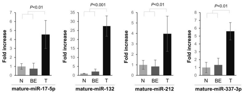
MicroRNAs (miRNAs), a critical part of the RNA silencing machinery, are known to play important regulatory roles in cancer. However, the consequence of miRNA deregulation in cancer is unknown for many miRNAs. Here, we define that miRNAs, miR-17-5p, miR-132-3p/-212-3p, and miR-337-3p are significantly up-regulated in the pancreatic ductal adenocarcinomas (PDAC) compared to the normal and benign tissues. Furthermore, by using PANC-1 cells, we demonstrate that overexpressed miR-337-3p and miR-17-5p/miR-132-3p/-212-3p can regulate executioner caspases-3 and -7, respectively. In addition, over-expression of miRNAs, especially miR-337-3p, attenuates tumor necrosis factor-related apoptosis-inducing ligand (TRAIL) cytotoxicity in PANC-1 cells. Our findings unveil an important biological function for miRNAs up-regulated in PDAC in coordinately regulating caspases, potentially contributing to the malignant progression of PDAC.

Overview

The claims and statistics of this paper haven’t been checked yet.

Run the check — free, no account needed. It reads the open-access text and checks the sources it cites.

A few free checks a day · papers already verified open instantly · results are public

Integrity signals

Registry

Crossref · 10.3390/ijms19041206

Cites

42 of 42 references verified0 citation errors · 0 not checkable

Statistics

Figures

Figure 1 Expression levels of mature miRNAs, miR-17-5p, miR-132-3p, miR-212-3p, and miR-337-3p in normal (N), benign (BE), and malignant (T) human pancreatic tissues. The expression of miRNAs was quantified using the TaqMan MicroRNA Assay. miRNA expression was related to the 18S ribosomal RNA internal control, and data were calculated using the comparative CT method. The relative fold increase of miRNAs with respect to normal tissues are presented.
Figure 1 Expression levels of mature miRNAs, miR-17-5p, miR-132-3p, miR-212-3p, and miR-337-3p in normal (N), benign (BE), and malignant (T) human pancreatic tissues. The expression of miRNAs was quantified using the TaqMan MicroRNA Assay. miRNA expression was related to the 18S ribosomal RNA internal control, and data were calculated using the comparative CT method. The relative fold increase of miRNAs with respect to normal tissues are presented.CC BY · PubMed Central
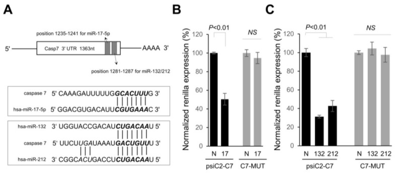
Figure 2 Caspase-7 (casp-7) is directly targeted by miR-17-5p and miR-132-3p/-212-3p in pancreatic tumor cells. (A) Schematic representation showing the location of putative miR-17-5p and -132-3p/-212-3p binding sites harbored in the 3′UTR of casp-7 mRNA. (B) Luciferase reporter plasmids carrying a wild-type (psiC2-C7) or mutant binding sequences in 3′UTR of casp-7 (C7-MUT) were co-transfected in PANC-1 cells with the negative control of pre-miR precursor (N) or miR-17-5p precursor (17) at 50 nM concentration. At 24 h after transfection, luciferase activity was measured using dual-luciferase assays. Normalized Renilla luciferase activity in cells transfected with N was set at 100%. The data are the mean ± s.d. of at least 3 independent experiments. (C) Wild type (psiC2-C7) or mutant plasmids were co-transfected in PANC-1 cells with the negative control of pre-miR precursor (N), miR-132-3p precursor (132) or miR-212-3p precursor (212) at 50 nM concentration. At 24 h after transfection, luciferase activity was measured using dual-luciferase assays. Normalized Renilla luciferase activity in cells transfected with control oligo (N) was set at 100%. The data are the mean ± s.d. of at least 3 independent experiments. NS, not significant.
Figure 2 Caspase-7 (casp-7) is directly targeted by miR-17-5p and miR-132-3p/-212-3p in pancreatic tumor cells. (A) Schematic representation showing the location of putative miR-17-5p and -132-3p/-212-3p binding sites harbored in the 3′UTR of casp-7 mRNA. (B) Luciferase reporter plasmids carrying a wild-type (psiC2-C7) or mutant binding sequences in 3′UTR of casp-7 (C7-MUT) were co-transfected in PANC-1 cells with the negative control of pre-miR precursor (N) or miR-17-5p precursor (17) at 50 nM concentration. At 24 h after transfection, luciferase activity was measured using dual-luciferase assays. Normalized Renilla luciferase activity in cells transfected with N was set at 100%. The data are the mean ± s.d. of at least 3 independent experiments. (C) Wild type (psiC2-C7) or mutant plasmids were co-transfected in PANC-1 cells with the negative control of pre-miR precursor (N), miR-132-3p precursor (132) or miR-212-3p precursor (212) at 50 nM concentration. At 24 h after transfection, luciferase activity was measured using dual-luciferase assays. Normalized Renilla luciferase activity in cells transfected with control oligo (N) was set at 100%. The data are the mean ± s.d. of at least 3 independent experiments. NS, not significant.CC BY · PubMed Central
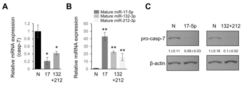
Figure 3 Caspase-7 (casp-7) is downregulated in miR-17-5p and miR-132-3p/-212-3p overexpressing cells. (A) Real time qPCR analyses of casp-7 mRNA levels in PANC-1 cells transfected with pre-miR-negative control (N), pre-miR-17-5p, or a mixture of pre-miR-132-3p and -212-3p (25 nM + 25 nM) at 50 nM for 48 h. The data are the mean ± s.d. for three independent experiments. * p < 0.01. (B) Treatment conditions were the same as in (A) and the expression levels of either miR-17-5p, miR-132-3p, or miR-212-3p in PANC-1 cells were determined by real time qPCR. ** p < 0.001. (C) PANC-1 cells were transfected with pre-miR-negative control (N), pre-miR-17-5p, or a mixture of pre-miR-132-3p and -212-3p (25 nM + 25 nM) at 50 nM for 48 h, and then protein lysates were isolated and subjected to Western blot analysis of pro-casp-7. β-actin served as a loading control and relative expressions are indicated. Western blotting and qPCR were performed on separately treated samples of cells.
Figure 3 Caspase-7 (casp-7) is downregulated in miR-17-5p and miR-132-3p/-212-3p overexpressing cells. (A) Real time qPCR analyses of casp-7 mRNA levels in PANC-1 cells transfected with pre-miR-negative control (N), pre-miR-17-5p, or a mixture of pre-miR-132-3p and -212-3p (25 nM + 25 nM) at 50 nM for 48 h. The data are the mean ± s.d. for three independent experiments. * p < 0.01. (B) Treatment conditions were the same as in (A) and the expression levels of either miR-17-5p, miR-132-3p, or miR-212-3p in PANC-1 cells were determined by real time qPCR. ** p < 0.001. (C) PANC-1 cells were transfected with pre-miR-negative control (N), pre-miR-17-5p, or a mixture of pre-miR-132-3p and -212-3p (25 nM + 25 nM) at 50 nM for 48 h, and then protein lysates were isolated and subjected to Western blot analysis of pro-casp-7. β-actin served as a loading control and relative expressions are indicated. Western blotting and qPCR were performed on separately treated samples of cells.CC BY · PubMed Central
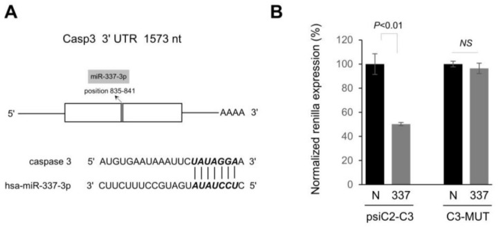
Figure 4 Caspase-3 (casp-3) is directly targeted by miR-337-3p. (A) Schematic diagram of human casp-3 mRNA and the predicted target site of miR-337-3p in the 3′UTR is shown. (B) Luciferase reporter plasmids carrying a wild-type (psiC2-C3) or mutant binding sequences in 3′UTR of casp-3 (C3-MUT) was transiently co-transfected in PANC-1 cells with pre-miR-negative control (N), or miR-337-3p precursor (337) at 50 nM concentration. At 24 h after transfection, luciferase activity was measured using dual-luciferase assays. Normalized Renilla luciferase activity in cells transfected with control oligo (N) was set at 100%. The data are the mean ± s.d. of at least 3 independent experiments. NS, Not significant.
Figure 4 Caspase-3 (casp-3) is directly targeted by miR-337-3p. (A) Schematic diagram of human casp-3 mRNA and the predicted target site of miR-337-3p in the 3′UTR is shown. (B) Luciferase reporter plasmids carrying a wild-type (psiC2-C3) or mutant binding sequences in 3′UTR of casp-3 (C3-MUT) was transiently co-transfected in PANC-1 cells with pre-miR-negative control (N), or miR-337-3p precursor (337) at 50 nM concentration. At 24 h after transfection, luciferase activity was measured using dual-luciferase assays. Normalized Renilla luciferase activity in cells transfected with control oligo (N) was set at 100%. The data are the mean ± s.d. of at least 3 independent experiments. NS, Not significant.CC BY · PubMed Central
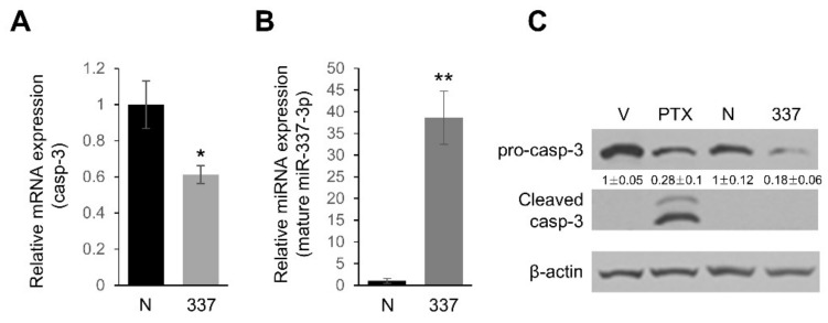
Figure 5 Caspase-3 (casp-3) is downregulated in miR-337-3p overexpressing cells. (A) The level of casp-3 mRNA was determined by real time qPCR and normalized with the 18S RNA level in PANC-1 cells after transfection of pre-miR-negative control (N) or pre-miR-337-3p (337). * p < 0.01. (B) Treatment conditions were the same as in (A) and the expression level of mature miR-337-3p in PANC-1 cells was determined by real time qPCR. ** p < 0.001. (C) Either pre-miR-nega control (N) or pre-miR-337-3p (337) was transfected into PANC-1 cells at 50 nM for 48 h, and then protein lysates were subjected to Western blot analysis of pro-casp-3 and cleaved casp-3. Paclitaxel (PTX) treated (1000 nM for 48 h) PANC-1 cells were used as a positive control of cleaved casp-3 expression. β-actin served as a loading control and relative expression are indicated. V indicates vehicle (DMSO) treated cells. Western blotting and qPCR were performed on separately treated samples of cells.
Figure 5 Caspase-3 (casp-3) is downregulated in miR-337-3p overexpressing cells. (A) The level of casp-3 mRNA was determined by real time qPCR and normalized with the 18S RNA level in PANC-1 cells after transfection of pre-miR-negative control (N) or pre-miR-337-3p (337). * p < 0.01. (B) Treatment conditions were the same as in (A) and the expression level of mature miR-337-3p in PANC-1 cells was determined by real time qPCR. ** p < 0.001. (C) Either pre-miR-nega control (N) or pre-miR-337-3p (337) was transfected into PANC-1 cells at 50 nM for 48 h, and then protein lysates were subjected to Western blot analysis of pro-casp-3 and cleaved casp-3. Paclitaxel (PTX) treated (1000 nM for 48 h) PANC-1 cells were used as a positive control of cleaved casp-3 expression. β-actin served as a loading control and relative expression are indicated. V indicates vehicle (DMSO) treated cells. Western blotting and qPCR were performed on separately treated samples of cells.CC BY · PubMed Central
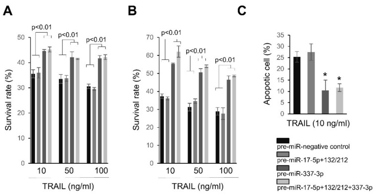
Figure 6 Ectopic expression of miRNAs targeting caspases-3 and caspase-7 renders PANC-1 cells resistant to TRAIL. (A) PANC-1 cells were transfected either pre-miR-nega (100 nM), pre-miR-17-5p (50 nM) + a mixture of pre-miR-132-3p and -212-3p (50 nM), pre-miR-337-3p (100 nM), or pre-miR-17-5p (25 nM) + a mixture of pre-miR-132-3p and -212-3p (25 nM) + pre-miR-337-3p (50 nM) in 96 well plates. At 72 h after transfection, TRAIL was treated for 24 h followed and survival rates were measured using WST1 assay. (B) Treatment conditions were the same as in (A) except that final concentrations of oligos were 150 nM (pre-miR-negative control (150 nM), pre-miR-17-5p (75 nM) + a mixture of pre-miR-132-3p and -212-3p (75 nM), pre-miR-337-3p (150 nM), or pre-miR-17-5p + a mixture of pre-miR-132-3p and -212-3p (75 nM) + pre-miR-337-3p (75 nM)) in 96 well plates. (C) Under same transfection conditions in (B), apoptotic cells were evaluated by Annexin-FITC assay 12 h after treatment 10 ng/mL TRAIL in PANC-1 cells. * p < 0.001.
Figure 6 Ectopic expression of miRNAs targeting caspases-3 and caspase-7 renders PANC-1 cells resistant to TRAIL. (A) PANC-1 cells were transfected either pre-miR-nega (100 nM), pre-miR-17-5p (50 nM) + a mixture of pre-miR-132-3p and -212-3p (50 nM), pre-miR-337-3p (100 nM), or pre-miR-17-5p (25 nM) + a mixture of pre-miR-132-3p and -212-3p (25 nM) + pre-miR-337-3p (50 nM) in 96 well plates. At 72 h after transfection, TRAIL was treated for 24 h followed and survival rates were measured using WST1 assay. (B) Treatment conditions were the same as in (A) except that final concentrations of oligos were 150 nM (pre-miR-negative control (150 nM), pre-miR-17-5p (75 nM) + a mixture of pre-miR-132-3p and -212-3p (75 nM), pre-miR-337-3p (150 nM), or pre-miR-17-5p + a mixture of pre-miR-132-3p and -212-3p (75 nM) + pre-miR-337-3p (75 nM)) in 96 well plates. (C) Under same transfection conditions in (B), apoptotic cells were evaluated by Annexin-FITC assay 12 h after treatment 10 ng/mL TRAIL in PANC-1 cells. * p < 0.001.CC BY · PubMed Central

Verification

Claims the paper makes

The claims and statistics of this paper haven’t been checked yet.

Run the check — free, no account needed. It reads the open-access text and checks the sources it cites.

A few free checks a day · papers already verified open instantly · results are public

Claims other papers make about this one

Each is checked against this paper’s own text: does it say what the citing paper says it does?

Self-check

Percentages, sums and reported differences re-computed from the paper’s own numbers.

The self-check needs the paper’s full text.

Citations

Cites

1 retracted

Show all 42 references

Cited by

See all 27

Metadata

Authors
Park, J. K., Doseff, A. I., & Schmittgen, T. D.
Affiliations
  • Department of Biomedical Science and Research Institute for Bioscience &amp; Biotechnology, Hallym University, Chuncheon 24252, Korea
  • Department of Physiology and Department of Pharmacology and Toxicology, Michigan State University, East Lansing, MI 48824, USA
  • Department of Pharmaceutics, College of Pharmacy, University of Florida, Gainesville, FL 32610, USA
Venue
International Journal of Molecular Sciences
Publisher
MDPI AG
Published
2018-04-16
Volume
19
Issue
4
Pages
1206
ISSN
1422-0067
Cited by
27
References
42
Language
en
Funders
  • National Institutes of Health (CA114304)
  • Basic Science Research Program through the National Research Foundation of Korea (NRF) funded by the Ministry of Education (2017R1D1A3B03035662)
  • Hallym University Research Fund (HRF-201703-003)

About these checks

Registry, retraction, Cites and statistics results are computed, not judged, with one exception: a printed reference that no index lists is read once by a model before it counts as a citation error. Claims are extracted by a model and kept only when their quote appears verbatim in the text; each cited source is then checked, with a model’s help, for whether it says what the claim relies on. Those verdicts can be wrong — read the claim’s quote and the cited source before relying on one.

Statistics and arithmetic are recomputed, not judged.

Confidence bands are shown only for fields calibrated on our annotated test set. None is calibrated yet, so no band is shown.

What counts as a claim ›How we measure confidence ›How we check this ›

Are you an author, or do you see a problem with this page? Request a review

How to cite

Cite CoChat for the checks on this page, and the paper for its own findings.

This page
CoChat. (2026). Citation check: MicroRNAs Targeting Caspase-3 and -7 in PANC-1 Cells. https://app.cochat.ai/papers/10.3390/ijms19041206
The paper
Park, J. K., Doseff, A. I., & Schmittgen, T. D. (2018). MicroRNAs Targeting Caspase-3 and -7 in PANC-1 Cells. International Journal of Molecular Sciences. https://doi.org/10.3390/ijms19041206

Data sources

Bibliographic data: Semantic Scholar, OpenAlex, Crossref.

Contains information from the Semantic Scholar Academic Graph Datasets, made available under the ODC Attribution License (ODC-BY 1.0).

Semantic Scholar’s data is described in: Kinney, R., et al. (2023). The Semantic Scholar Open Data Platform. arXiv:2301.10140.