

The DOI matches its Crossref record; 219 of the 220 references it cites were found in a registry or an index.
Nanoparticle-Mediated Combination Therapy: Two-in-One Approach for Cancer
Open accessAbstract
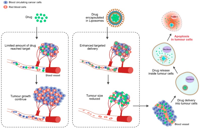
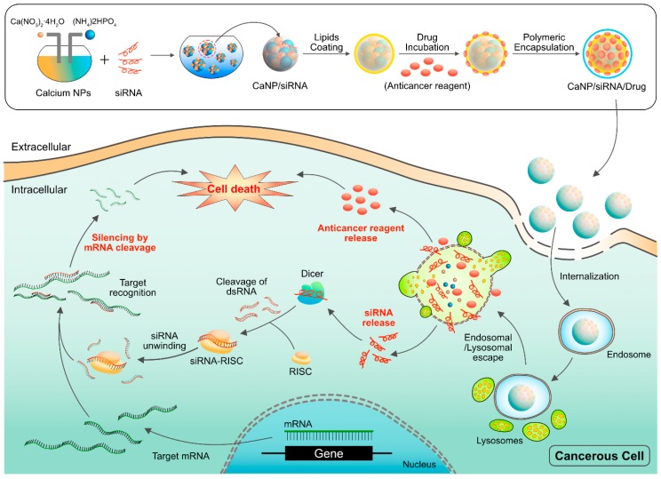
Cancer represents a group of heterogeneous diseases characterized by uncontrolled growth and spread of abnormal cells, ultimately leading to death. Nanomedicine plays a significant role in the development of nanodrugs, nanodevices, drug delivery systems and nanocarriers. Some of the major issues in the treatment of cancer are multidrug resistance (MDR), narrow therapeutic window and undesired side effects of available anticancer drugs and the limitations of anticancer drugs. Several nanosystems being utilized for detection, diagnosis and treatment such as theranostic carriers, liposomes, carbon nanotubes, quantum dots, polymeric micelles, dendrimers and metallic nanoparticles. However, nonbiodegradable nanoparticles causes high tissue accumulation and leads to toxicity. MDR is considered a major impediment to cancer treatment due to metastatic tumors that develop resistance to chemotherapy. MDR contributes to the failure of chemotherapies in various cancers, including breast, ovarian, lung, gastrointestinal and hematological malignancies. Moreover, the therapeutic efficiency of anticancer drugs or nanoparticles (NPs) used alone is less than that of the combination of NPs and anticancer drugs. Combination therapy has long been adopted as the standard first-line treatment of several malignancies to improve the clinical outcome. Combination therapy with anticancer drugs has been shown to generally induce synergistic drug actions and deter the onset of drug resistance. Therefore, this review is designed to report and analyze the recent progress made to address combination therapy using NPs and anticancer drugs. We first provide a comprehensive overview of the angiogenesis and of the different types of NPs currently used in treatments of cancer; those emphasized in this review are liposomes, polymeric NPs, polymeric micelles (PMs), dendrimers, carbon NPs, nanodiamond (ND), fullerenes, carbon nanotubes (CNTs), graphene oxide (GO), GO nanocomposites and metallic NPs used for combination therapy with various anticancer agents. Nanotechnology has provided the convenient tools for combination therapy. However, for clinical translation, we need continued improvements in the field of nanotechnology.
Overview
The claims and statistics of this paper haven’t been checked yet.
Run the check — free, no account needed. It reads the open-access text and checks the sources it cites.
Integrity signals
Registry
Cites
Statistics
Figures
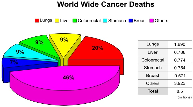
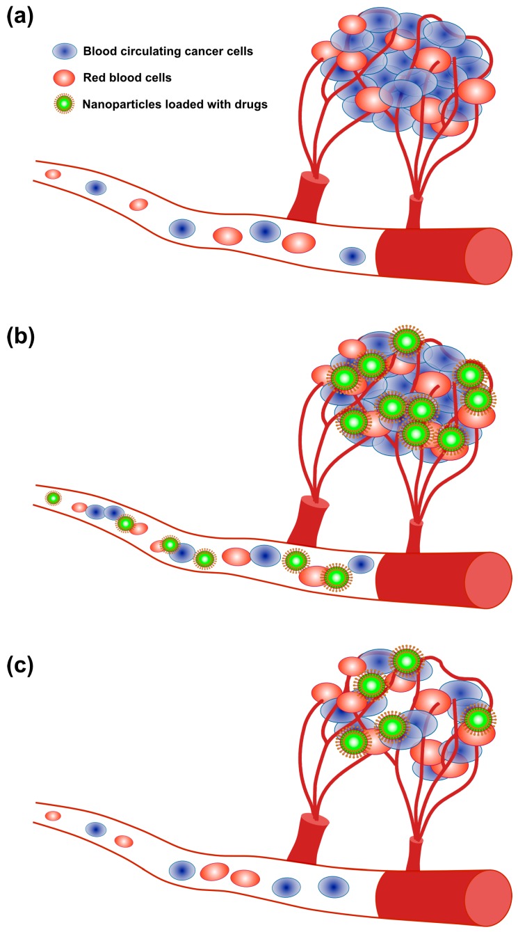
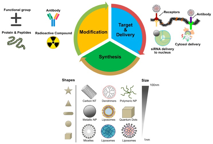
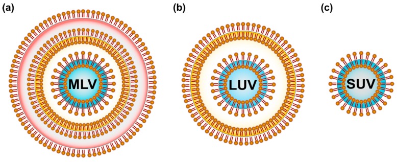
Verification
Claims the paper makes
The claims and statistics of this paper haven’t been checked yet.
Run the check — free, no account needed. It reads the open-access text and checks the sources it cites.
Claims other papers make about this one
Each is checked against this paper’s own text: does it say what the citing paper says it does?
In the citing paper
“Historically, drug combinations have been utilized efficiently to treat various bacterial and viral infections and to fight cancer [18–21].”
From Aprepitant, an antiemetic agent, interferes with metal ion homeostasis of Candida auris and displays potent synergistic interactions with azole drugs (2020) · cites this paper as [21]
Self-check
Percentages, sums and reported differences re-computed from the paper’s own numbers.
The self-check needs the paper’s full text.
Citations
Cites
1 retracted
Yuan, Y., Peng, Q., & Gurunathan, S. (2017) · International Journal of Nanomedicine · doi:10.2147/ijn.s136142 · Cited by 52
Self-citationRetractedJemal, A., Bray, F., Ferlay, J., Ward, E., & Forman, D. (2011) · CA A Cancer Journal for Clinicians · doi:10.3322/caac.20107 · Cited by 55,077
Siegel, R. L., Miller, K. D., & Jemal, A. (2016) · CA: A Cancer Journal for Clinicians · doi:10.3322/caac.21332 · Cited by 16,003
Angiogenesis in cancer and other diseases
Carmeliet, P., & Jain, R. K. (2000) · Nature · doi:10.1038/35025220 · Cited by 9,070
Nanotoxicology: An Emerging Discipline Evolving from Studies of Ultrafine Particles
Oberdörster, G., Oberdörster, E., & Oberdörster, J. (2005) · Environmental Health Perspectives · doi:10.1289/ehp.7339 · Cited by 7,891
Sunitinib versus Interferon Alfa in Metastatic Renal-Cell Carcinoma
Motzer, R. J., Hutson, T. E., Tomczak, P., Michaelson, M. D., Bukowski, R. M., Rixe, O., Oudard, S. M., Négrier, S., Szczylik, C. A., Kim, S. T., et al. (2007) · New England Journal of Medicine · doi:10.1056/nejmoa065044 · Cited by 5,752
Global patterns and trends in colorectal cancer incidence and mortality
Arnold, M., Sierra, M. S., Laversanne, M., Soerjomataram, I., Jemal, A., & Bray, F. (2016) · Gut · doi:10.1136/gutjnl-2015-310912 · Cited by 5,392
Sorafenib in Advanced Clear-Cell Renal-Cell Carcinoma
Escudier, B. J., Eisen, T. G., Stadler, W. M., Szczylik, C. A., Oudard, S. M., Siebels, M., Négrier, S., Chevreau, C., Solska, E., Desai, A. A., et al. (2007) · New England Journal of Medicine · doi:10.1056/nejmoa060655 · Cited by 4,802
Nanoparticle therapeutics: an emerging treatment modality for cancer
Davis, M. E., Chen, Z., & Shin, D. M. (2008) · Nature Reviews Drug Discovery · doi:10.1038/nrd2614 · Cited by 4,233
Drug delivery and nanoparticles: Applications and hazards
de Jong (2008) · International Journal of Nanomedicine · doi:10.2147/ijn.s596 · Cited by 3,854
Research works printed but not linked: 1
- Mewani, 2004, Analysis of gene expression in breast cancer cells treated with antisense raf oligonucleotide using microarray, Proc. Am. Assoc. Cancer Res., 64, 1209
Cited by
Sorted by citations
Magnetic Nanoparticles in Cancer Therapy and Diagnosis
Farzin, A., Etesami, S. A., Quint, J. P., Memić, A., & Tamayol, A. (2020) · Advanced Healthcare Materials · doi:10.1002/adhm.201901058 · Cited by 597
Multifunctional nanoparticle-mediated combining therapy for human diseases
Li, X., Peng, X., Zoulikha, M., Boafo, G. F., Magar, K. T., Ju, Y., & He, W. (2023) · Signal Transduction and Targeted Therapy · doi:10.1038/s41392-023-01668-1 · Cited by 573
Application of Nanomaterials in Biomedical Imaging and Cancer Therapy
Siddique, S., & Chow, J. C. L. (2020) · Nanomaterials · doi:10.3390/nano10091700 · Cited by 411
Gaur, M., Misra, C., Yadav, A. B., Swaroop, S., Maolmhuaidh, F. Ó., Bechelany, M., & Barhoum, A. (2021) · Materials · doi:10.3390/ma14205978 · Cited by 332
Phytosomes as Innovative Delivery Systems for Phytochemicals: A Comprehensive Review of Literature
Barani, M., Sangiovanni, E., Angarano, M., Rajizadeh, M. A., Mehrabani, M., Piazza, S., Gangadharappa, H. V., Pardakhty, A., Mehrbani, M., Dell’Agli, M., et al. (2021) · International Journal of Nanomedicine · doi:10.2147/ijn.s318416 · Cited by 308
A review of nanotechnology-based approaches for breast cancer and triple-negative breast cancer
Jain, V. K., Kumar, H., Anod, H. V., Chand, P., Gupta, N., Dey, S., & Kesharwani, S. S. (2020) · Journal of Controlled Release · doi:10.1016/j.jconrel.2020.07.003 · Cited by 295
Dendrimers: A New Race of Pharmaceutical Nanocarriers
Mittal, P., Saharan, A., Verma, R., Altalbawy, F. M. A., Alfaidi, M., Batiha, G. E., Akter, W., Gautam, R. K., Uddin, M. S., & Rahman, M. S. (2021) · BioMed Research International · doi:10.1155/2021/8844030 · Cited by 269
Antiviral Potential of Nanoparticles—Can Nanoparticles Fight Against Coronaviruses?
Gurunathan, S., Qasim, M., Choi, Y., Tae, J., Park, C., Hong, K., Kim, J., & Song, H. (2020) · Nanomaterials · doi:10.3390/nano10091645 · Cited by 259
Self-citationCobos, M., De-la-Pinta, I., Quindós, G., Fernández, M. J., & Fernández, M. D. (2020) · Nanomaterials · doi:10.3390/nano10020376 · Cited by 218
Aboyewa, J. A., Sibuyi, N. R. S., Meyer, M., & Oguntibeju, O. O. (2021) · Plants · doi:10.3390/plants10091929 · Cited by 200
Miyazawa, T., Itaya, M., Burdeos, G. C., Nakagawa, K., & Miyazawa, T. (2021) · International Journal of Nanomedicine · doi:10.2147/ijn.s298606 · Cited by 198
Bisht, A., Avinash, D., Sahu, K. K., Patel, P., Gupta, G. D., & Kurmi, B. D. (2024) · Drug Delivery and Translational Research · doi:10.1007/s13346-024-01648-0 · Cited by 203
MRI-traceable theranostic nanoparticles for targeted cancer treatment
Anani, T., Rahmati, S., Nayer, S., & David, A. E. (2020) · Theranostics · doi:10.7150/thno.48811 · Cited by 159
Transforming Medicine: Cutting-Edge Applications of Nanoscale Materials in Drug Delivery
Tenchov, R., Hughes, K. J., Ganesan, M., Iyer, K. A., Ralhan, K., Diaz, L. M. L., Bird, R. E., Ivanov, J. M., & Zhou, Q. A. (2025) · ACS Nano · doi:10.1021/acsnano.4c09566 · Cited by 153
Graphene oxide-based hydrogels as a nanocarrier for anticancer drug delivery
Ghawanmeh, A. A., Ali, G. A. M., Algarni, H., Sarkar, S. M., & Chong, K. F. (2019) · Nano Research · doi:10.1007/s12274-019-2300-4 · Cited by 137
Karade, V. C., Sharma, A., Dhavale, R. P., Dhavale, R. P., Shingte, S. R., Patil, P., Kim, J. H., Zahn, D. R. T., Chougale, A. D., Salvan, G., et al. (2021) · Scientific Reports · doi:10.1038/s41598-021-84770-0 · Cited by 126
Self-citationConjugated polymer nano-systems for hyperthermia, imaging and drug delivery
Sarkar, S., & Levi‐Polyachenko, N. H. (2020) · Advanced Drug Delivery Reviews · doi:10.1016/j.addr.2020.01.002 · Cited by 123
Nanomedicine to target multidrug resistant tumors
Lepeltier, É., Rijo, P. M., Rizzolio, F., Popovtzer, R., Petrikaitė, V., Assaraf, Y. G., & Passirani, C. (2020) · Drug Resistance Updates · doi:10.1016/j.drup.2020.100704 · Cited by 116
Combination therapy based on nano codelivery for overcoming cancer drug resistance
Wang, H., & Huang, Y. (2020) · Medicine in Drug Discovery · doi:10.1016/j.medidd.2020.100024 · Cited by 115
Uzma, M., Raghavendra, V. B., Ningaraju, S., Krishna, K. L., Ghosh, A. R., Govindappa, M., & Pugazhendhi, A. (2020) · International Journal of Pharmaceutics · doi:10.1016/j.ijpharm.2020.119729 · Cited by 112
Ghazi, R., Ibrahim, T. K., Nasir, J. A., Gai, S., Ali, G., Boukhris, I., & Rehman, Z. (2025) · RSC Advances · doi:10.1039/d5ra00728c · Cited by 123
Preparation and evaluation of PEG-coated zein nanoparticles for oral drug delivery purposes
Reboredo, C., González-Navarro, C. J., Martínez-Ohárriz, M. C., Martínez‐López, A. L., & Irache, J. M. (2021) · International Journal of Pharmaceutics · doi:10.1016/j.ijpharm.2021.120287 · Cited by 111
Nano-traditional Chinese medicine: a promising strategy and its recent advances
Wei, D., Yang, H. N., Zhang, Y., Zhang, X., Wang, J., Wu, X., & Chang, J. K. (2022) · Journal of Materials Chemistry B · doi:10.1039/d2tb00225f · Cited by 110
Vakilinezhad, M. A., Amini, A., Dara, T., & Alipour, S. (2019) · Colloids and Surfaces B Biointerfaces · doi:10.1016/j.colsurfb.2019.110515 · Cited by 103
Slika, L., & Patra, D. (2019) · Expert Opinion on Drug Delivery · doi:10.1080/17425247.2020.1702644 · Cited by 107
Current Progresses and Challenges of Immunotherapy in Triple-Negative Breast Cancer
Mediratta, K., El‐Sahli, S., D’Costa, V. M., & Wang, L. (2020) · Cancers · doi:10.3390/cancers12123529 · Cited by 102
Metal/metal oxide nanoparticles: A revolution in the biosynthesis and medical applications
de Jesus, R. A., de Assis, G. C., de Oliveira, R. J., Costa, J. A. S., Paranhos, C. M., Iqbal, H. M. N., & Ferreira, L. F. R. (2023) · Nano-Structures & Nano-Objects · doi:10.1016/j.nanoso.2023.101071 · Cited by 99
Truffi, M., Mazzucchelli, S., Bonizzi, A., Sorrentino, L., Allevi, R., Vanna, R., Morasso, C., & Corsi, F. (2019) · International Journal of Molecular Sciences · doi:10.3390/ijms20061263 · Cited by 93
Lim, S. H., Yee, G. T., & Khang, D. (2024) · International Journal of Nanomedicine · doi:10.2147/ijn.s450853 · Cited by 99
Polymeric nanomicelles of curcumin: Potential applications in cancer
Farhoudi, L., Kesharwani, P., Majeed, M. A., Johnston, T. P., & Sahebkar, A. (2022) · International Journal of Pharmaceutics · doi:10.1016/j.ijpharm.2022.121622 · Cited by 91
Nanocarriers Used in Drug Delivery to Enhance Immune System in Cancer Therapy
Lôbo, G. C. N. B., Paiva, K. L. R., Silva, A. L. G., Simões, M. M., Radicchi, M. A., & Báo, S. N. (2021) · Pharmaceutics · doi:10.3390/pharmaceutics13081167 · Cited by 93
Review of Therapeutic Applications of Radiolabeled Functional Nanomaterials
Jeon, J. (2019) · International Journal of Molecular Sciences · doi:10.3390/ijms20092323 · Cited by 87
Tiwari, P., Yadav, K., Shukla, R. P., Gautam, S., Marwaha, D., Sharma, M., & Mishra, P. R. (2023) · Journal of Controlled Release · doi:10.1016/j.jconrel.2023.09.016 · Cited by 88
Breast cancer drug delivery by novel drug-loaded chitosan-coated magnetic nanoparticles
Taherian, A., Esfandiari, N., & Rouhani, S. (2021) · Cancer Nanotechnology · doi:10.1186/s12645-021-00086-8 · Cited by 92
Gurunathan, S., Jeyaraj, M., Kang, M.-H., & Kim, J. (2019) · Nanomaterials · doi:10.3390/nano9081089 · Cited by 80
Self-citationCombining Carbon Nanotubes and Chitosan for the Vectorization of Methotrexate to Lung Cancer Cells
Cirillo, G., Vittorio, O., Kunhardt, D., Valli, E., Voli, F., Farfalla, A., Curcio, M., Spizzirri, U. G., & Hampel, S. (2019) · Materials · doi:10.3390/ma12182889 · Cited by 80
Smart nanocarriers for cancer treatment: Clinical impact and safety
Liao, Z., Wong, S. W., Yeo, H. L., & Zhao, Y. (2020) · NanoImpact · doi:10.1016/j.impact.2020.100253 · Cited by 82
Tuli, H. S., Garg, V. K., Bhushan, S., Uttam, V., Sharma, U., Jain, A., Sak, K., Yadav, V. K., Lorenzo, J. M., Dhama, K., et al. (2022) · Translational Oncology · doi:10.1016/j.tranon.2022.101596 · Cited by 78
Mihailović, V. B., Srećković, N. Z., Nedić, Z., Dimitrijević, S., Matić, M. M., Obradović, A. D., Selaković, D., Rosic, G. L., & Stanković, J. S. K. (2023) · Molecules · doi:10.3390/molecules28020808 · Cited by 74
Innovative nanomaterials for cancer diagnosis, imaging, and therapy: Drug delivery applications
Pourmadadi, M., Eshaghi, M. M., Ostovar, S., Mohammadi, Z. A., Sharma, R. K., Paiva‐Santos, A. C., Rahmani, E., Rahdar, A., & Pandey, S. (2023) · Journal of Drug Delivery Science and Technology · doi:10.1016/j.jddst.2023.104357 · Cited by 72
Epigenetics in Breast Cancer Therapy—New Strategies and Future Nanomedicine Perspectives
Buociková, V., Ríos-Mondragón, I., Pilalis, E. D., Chatziioannou, A. A., Miklikova, S., Mego, M., Pajuste, K., Ruciņš, M., Yamani, N. E., Longhin, E., et al. (2020) · Cancers · doi:10.3390/cancers12123622 · Cited by 69
Ruthenium-based antitumor drugs and delivery systems from monotherapy to combination therapy
Lu, Y., Zhu, D., Le, Q., Wang, Y., & Wang, W. (2022) · Nanoscale · doi:10.1039/d2nr02994d · Cited by 73
Nanotechnology in oncology: advances in biosynthesis, drug delivery, and theranostics
Ammar, M. M., Ali, R., Elaziz, N. A. A., Habib, H., Abbas, F. M., Yassin, M. T., Maniah, K., & Abdelaziz, R. (2025) · Discover Oncology · doi:10.1007/s12672-025-02664-3 · Cited by 64
Eldesouky, H. E., Salama, E. A., Lanman, N. A., Hazbun, T. R., & Seleem, M. N. (2020) · Antimicrobial Agents and Chemotherapy · doi:10.1128/aac.00684-20 · Cited by 56
Polymeric Nanoparticle Delivery of Combination Therapy with Synergistic Effects in Ovarian Cancer
Levit, S. L., & Tang, C. (2021) · Nanomaterials · doi:10.3390/nano11041048 · Cited by 57
Intrinsic and Extrinsic Factors Affecting Microtubule Dynamics in Normal and Cancer Cells
Borys, F., Joachimiak, E., Krawczyk, H., & Fabczak, H. (2020) · Molecules · doi:10.3390/molecules25163705 · Cited by 55
Peripherally acting opioid analgesics and peripherally-induced analgesia
Martı́nez, V., & Abalo, R. (2020) · Behavioural Pharmacology · doi:10.1097/fbp.0000000000000558 · Cited by 55
Insights into Shape-Based Silver Nanoparticles: A Weapon to Cope with Pathogenic Attacks
Karade, V. C., Patil, R. B., Parit, S. B., Kim, J. H., Chougale, A. D., & Dawkar, V. V. (2021) · ACS Sustainable Chemistry & Engineering · doi:10.1021/acssuschemeng.1c03797 · Cited by 53
Self-citationMousavi‐kiasary, S. M. S., Senabreh, A., Zandi, A., Pena, R., Cruz, F., Adibi, A., & Hooshmand, N. (2025) · Pharmaceutics · doi:10.3390/pharmaceutics17060682 · Cited by 56
Nanoformulations of α-Mangostin for Cancer Drug Delivery System
Meylina, L., Muchtaridi, M., Joni, I. M., Mohammed, A. F. A., & Wathoni, N. (2021) · Pharmaceutics · doi:10.3390/pharmaceutics13121993 · Cited by 52
Li, Y., Jiang, Y., Zheng, Z., Du, N., Guan, S., Guo, W., Tang, X., Cui, J., Zhang, L., Liu, K., et al. (2022) · Advanced Materials · doi:10.1002/adma.202110490 · Cited by 49
Micelles as potential drug delivery systems for colorectal cancer treatment
Fatfat, Z., Fatfat, M., & Gali‐Muhtasib, H. U. (2022) · World Journal of Gastroenterology · doi:10.3748/wjg.v28.i25.2867 · Cited by 52
Gurunathan, S., Jeyaraj, M., Kang, M.-H., & Kim, J. (2020) · International Journal of Molecular Sciences · doi:10.3390/ijms21186792 · Cited by 49
Self-citationAnwar, A., Mungroo, M. R., Anwar, A., Sullivan, W. J., Khan, N. A., & Siddiqui, R. (2019) · ACS Infectious Diseases · doi:10.1021/acsinfecdis.9b00263 · Cited by 48
Grześkowiak, B. F., Maziukiewicz, D., Kozłowska, A., Kertmen, A., Coy, E. L., & Mrówczyński, R. (2021) · International Journal of Molecular Sciences · doi:10.3390/ijms22020738 · Cited by 49
Shukla, R. P., Dewangan, J., Urandur, S., Banala, V. T., Diwedi, M., Sharma, S., Agrawal, S., Rath, S. K., Trivedi, R., & Mishra, P. R. (2019) · Biomaterials Science · doi:10.1039/c9bm01246j · Cited by 45
Korupalli, C., Kalluru, P., Nuthalapati, K., Kuthala, N., Thangudu, S., & Vankayala, R. (2020) · Bioengineering · doi:10.3390/bioengineering7030094 · Cited by 45
Buskaran, K., Hussein, M. Z., Moklas, M. A. M., Masarudin, M. J., & Fakurazi, S. (2021) · International Journal of Molecular Sciences · doi:10.3390/ijms22115786 · Cited by 45
Rizki, I. N., Klaypradit, W., & Patmawati, P. (2022) · Sustainable Chemistry and Pharmacy · doi:10.1016/j.scp.2022.100888 · Cited by 43
Nonalcoholic fatty liver disease: Current therapies and future perspectives in drug delivery
Domingues, I., Leclercq, I. A., & Beloqui, A. (2023) · Journal of Controlled Release · doi:10.1016/j.jconrel.2023.09.040 · Cited by 45
Nanomaterials multifunctional behavior for enlightened cancer therapeutics
Raju, G. S. R., Dariya, B., Mungamuri, S. K., Chalikonda, G., Kang, S., Khan, I. N., Sushma, P. S., Nagaraju, G. P., Pavitra, E., & Han, Y. (2019) · Seminars in Cancer Biology · doi:10.1016/j.semcancer.2019.08.013 · Cited by 45
Karuppaiah, A., Siram, K., Selvaraj, D., Ramasamy, M., Babu, D., & Sankar, V. (2019) · Materials Today Communications · doi:10.1016/j.mtcomm.2019.100884 · Cited by 43
Biomarker-guided targeted and immunotherapies in malignant pleural mesothelioma
Yang, H., Xu, D., Schmid, R. A., & Peng, R. (2020) · Therapeutic Advances in Medical Oncology · doi:10.1177/1758835920971421 · Cited by 40
Homing Peptides for Cancer Therapy
Lingasamy, P., & Teesalu, T. (2021) · Advances in experimental medicine and biology · doi:10.1007/978-3-030-58174-9_2 · Cited by 42
Tarannum, M., Hossain, M. A., Holmes, B. M., Yan, S., Mukherjee, P., & Vivero-Escoto, J. L. (2021) · Small · doi:10.1002/smll.202104449 · Cited by 43
Fadeel, D. A. A., Hanafy, M. S., Kelany, N. A., & Elywa, M. (2021) · Heliyon · doi:10.1016/j.heliyon.2021.e07370 · Cited by 39
Elias, L., Taengua, R., Frígols, B., Salesa, B., & Serrano‐Aroca, Á. (2019) · International Journal of Molecular Sciences · doi:10.3390/ijms20143603 · Cited by 38
Impact of Molybdenum Compounds as Anticancer Agents
Odularu, A. T., Ajibade, P. A., & Mbese, J. Z. (2019) · Bioinorganic Chemistry and Applications · doi:10.1155/2019/6416198 · Cited by 38
Purushothaman, B., Lee, J. M., Hong, S., & Song, J. M. (2020) · Journal of Nanobiotechnology · doi:10.1186/s12951-020-00661-y · Cited by 38
Ledezma-Gallegos, F., Jurado, R., Mir, R., Medina, L. A., Mondragon-Fuentes, L., & García‐López, P. (2020) · Pharmaceutics · doi:10.3390/pharmaceutics12090897 · Cited by 38
Eldesouky, H. E., Lanman, N. A., Hazbun, T. R., & Seleem, M. N. (2020) · Virulence · doi:10.1080/21505594.2020.1838741 · Cited by 37
Razavi, Z. S., Razavi, F. S., & Alizadeh, S. S. (2025) · European Journal of Medicinal Chemistry · doi:10.1016/j.ejmech.2025.117357 · Cited by 39
Lopes, J., Rodrigues, C. M. P., Gaspar, M. M., & Reis, C. P. (2022) · Pharmaceutics · doi:10.3390/pharmaceutics14091817 · Cited by 38
Revolutionizing Cancer Treatment: The Promising Horizon of Zein Nanosystems
Preetam, S., Mondal, D. D., Mukerjee, N., Naser, S. S., Tabish, T. A., & Thorat, N. D. (2024) · ACS Biomaterials Science & Engineering · doi:10.1021/acsbiomaterials.3c01540 · Cited by 37
Vitamin K: A novel cancer chemosensitizer
Gul, S., Maqbool, M. F., Maryam, A., Khan, M. N., Shakir, H. A., Irfan, M. I., Ara, C., Li, Y., & Ma, T. (2022) · Biotechnology and Applied Biochemistry · doi:10.1002/bab.2312 · Cited by 36
Tyro3, Axl, Mertk receptor-mediated efferocytosis and immune regulation in the tumor environment
Zhou, L., & Matsushima, G. K. (2021) · International review of cytology · doi:10.1016/bs.ircmb.2021.02.002 · Cited by 35
Heptamethine Cyanine–Based Application for Cancer Theranostics
Zhang, L., Jia, H., Liu, X., Zou, Y., Sun, J., Liu, M., Jia, S., Liu, N., Li, Y., & Wang, Q. (2022) · Frontiers in Pharmacology · doi:10.3389/fphar.2021.764654 · Cited by 35
Voci, S., Gagliardi, A., Molinaro, R., Fresta, M., & Cosco, D. (2021) · Gels · doi:10.3390/gels7020033 · Cited by 32
Wijetunge, S. S., Wen, J., Yeh, C., & Sun, Y. (2019) · Colloids and Surfaces B Biointerfaces · doi:10.1016/j.colsurfb.2019.110572 · Cited by 33
Chen, Q., Sun, S., Lin, H., Li, Z., Wu, A., Liu, X., Wu, F., & Zhang, W. (2021) · ACS Applied Bio Materials · doi:10.1021/acsabm.0c01663 · Cited by 33
Fawzy, M. P., Hassan, H. A. F. M., Sedky, N. K., Nafie, M. S., Youness, R. A., & Fahmy, S. A. (2024) · Nanoscale Advances · doi:10.1039/d4na00488d · Cited by 31
Jhawat, V., Gulia, M., Gupta, S., Maddiboyina, B., & Dutt, R. (2020) · Journal of Controlled Release · doi:10.1016/j.jconrel.2020.08.039 · Cited by 32
Mechanisms of photodynamic therapy
Sharma, D., Singh, S., Kumar, P., Jain, G. K., Aggarwal, G., Almalki, W. H., & Kesharwani, P. (2023) · Elsevier eBooks · doi:10.1016/b978-0-323-85595-2.00017-7 · Cited by 31
Daryanavard, M., Jannesari, Z., Javeri, M., & Abyar, F. (2020) · Spectrochimica Acta Part A Molecular and Biomolecular Spectroscopy · doi:10.1016/j.saa.2020.118175 · Cited by 30
How Far Are We from Prescribing Fasting as Anticancer Medicine?
Deligiorgi, M. V., Liapi, C. A., & Trafalis, D. T. (2020) · International Journal of Molecular Sciences · doi:10.3390/ijms21239175 · Cited by 32
Application of Nanotechnology and Phytochemicals in Anticancer Therapy
Kim, J. H., Dareowolabi, B. O., Thiruvengadam, R., & Moon, E. (2024) · Pharmaceutics · doi:10.3390/pharmaceutics16091169 · Cited by 30
Self-citationChen, Y., Li, N., Xu, B., Wu, M., Yan, X., Zhong, L., Cai, H., Wang, T., Wang, Q., Long, F., et al. (2019) · Biomedicine & Pharmacotherapy · doi:10.1016/j.biopha.2019.109257 · Cited by 31
Attri, A., Thakur, D. S., Kaur, T., Sensale, S., Peng, Z., Kumar, D., & Singh, R. P. (2020) · Molecular Pharmaceutics · doi:10.1021/acs.molpharmaceut.0c00730 · Cited by 29
Emerging concepts in designing next-generation multifunctional nanomedicine for cancer treatment
Chakraborty, K., Tripathi, A., Mishra, S., Mallick, A. M., & Roy, R. S. (2022) · Bioscience Reports · doi:10.1042/bsr20212051 · Cited by 30
Polymer-based nanoparticles for cancer theranostics: advances, challenges, and future perspectives
Shanahan, K., Coen, D., & Nafo, W. (2025) · Exploration of BioMat-X · doi:10.37349/ebmx.2025.101342 · Cited by 29
Gene therapy in wound healing using nanotechnology
Pang, C., Fan, K. S., Wei, L., & Kolar, M. K. (2020) · Wound Repair and Regeneration · doi:10.1111/wrr.12881 · Cited by 29
Current nano-therapeutic approaches ameliorating inflammation in cancer progression
Rehman, M. U., Khan, A., Imtiyaz, Z., Ali, S., Makeen, H. A., Rashid, S., & Arafah, A. M. (2022) · Seminars in Cancer Biology · doi:10.1016/j.semcancer.2022.02.006 · Cited by 29
Targeted nanotherapy for kidney diseases: a comprehensive review
Sabiu, G., Kasinath, V., Jung, S., Li, X., Tsokos, G. C., & Abdi, R. (2022) · Nephrology Dialysis Transplantation · doi:10.1093/ndt/gfac233 · Cited by 28
Nanocarriers as a Tool for the Treatment of Colorectal Cancer
Oliveira, A. L. C. D. S. L., Schomann, T., de Geus‐Oei, L., Kapiteijn, E., Cruz, L. J., & Júnior, R. F. D. A. (2021) · Pharmaceutics · doi:10.3390/pharmaceutics13081321 · Cited by 31
Gagliardi, A., Voci, S., Bonacci, S., Iriti, G., Procopio, A., Fresta, M., & Cosco, D. (2021) · International Journal of Biological Macromolecules · doi:10.1016/j.ijbiomac.2021.10.184 · Cited by 27
Askar, M. A., Thabet, N. M., El-Sayyad, G. S., El-Batal, A. I., Elkodous, M. A., Kawamura, G., Matsuda, A., Elshawi, O. E., Helal, H., & Abdel-Rafei, M. K. (2021) · Cancers · doi:10.3390/cancers13215571 · Cited by 27
Borhaninia, M., Zahiri, M., Abnous, K., Taghdisi, S. M., Ramezani, M., & Alibolandi, M. (2023) · International Journal of Biological Macromolecules · doi:10.1016/j.ijbiomac.2023.125882 · Cited by 27
Antitumor Features of Vegetal Protein-Based Nanotherapeutics
Voci, S., Gagliardi, A., Fresta, M., & Cosco, D. (2020) · Pharmaceutics · doi:10.3390/pharmaceutics12010065 · Cited by 27
Nanoparticle-mediated gene therapy as a novel strategy for the treatment of retinoblastoma
Mandal, M., Banerjee, I., & Mandal, M. (2022) · Colloids and Surfaces B Biointerfaces · doi:10.1016/j.colsurfb.2022.112899 · Cited by 28
Engineered nanoparticles for imaging and targeted drug delivery in hepatocellular carcinoma
Yu, X., Zhang, Q., Wang, L., Zhang, Y., & Zhu, L. (2025) · Experimental Hematology and Oncology · doi:10.1186/s40164-025-00658-z · Cited by 30
Metadata
- Authors
- Gurunathan, S., Kang, M.-H., Qasim, M., & Kim, J.-H.
- Affiliations
- Konkuk University (KR)
- Corresponding author
- Gurunathan, Sangiliyandi
- Kim, Jin-Hoi
- Venue
- International Journal of Molecular Sciences
- Publisher
- MDPI AG
- Published
- 2018-10-20
- Volume
- 19
- Issue
- 10
- Pages
- 3264
- ISSN
- 1422-0067 (ISSN-L)
- PMC
- PMC6214025
- OpenAlex
- W2896053285
- Cited by
- 367
- References
- 220
- Citations by year
- 2026: 26 · 2025: 55 · 2024: 55 · 2023: 51 · 2022: 57 · 2021: 52 · 2020: 47 · 2019: 24
- FWCI
- 2.03
- Type
- journal article
- Language
- en
- Open access
- gold
- dx.doi.org
- Licence
- Locations
- International Journal of Molecular Sciences (publishedVersion, cc-by)
- Europe PMC (PubMed Central) (publishedVersion, other-oa)
- PubMed Central (publishedVersion, cc-by)
- PubMed (publishedVersion)
- Topics
- Physical Sciences › Materials Science › Biomaterials › Nanoparticle-Based Drug Delivery
- Physical Sciences › Engineering › Biomedical Engineering › Nanoplatforms for cancer theranostics
- Health Sciences › Medicine › Oncology › Drug Transport and Resistance Mechanisms
- Keywords
- cancer nanomedicine, multidrug resistance, anticancer drug delivery, liposomes, polymeric micelles, dendrimers, carbon nanotubes, graphene oxide, nanodiamonds, C60, metal nanoparticles, angiogenesis, theranostic nanocarriers, drug synergy
- MeSH terms
- Animals, Antineoplastic Agents, Drug Therapy, Combination, Humans, Nanoparticles
About these checks
Registry, retraction, Cites and statistics results are computed, not judged, with one exception: a printed reference that no index lists is read once by a model before it counts as a citation error. Claims are extracted by a model and kept only when their quote appears verbatim in the text; each cited source is then checked, with a model’s help, for whether it says what the claim relies on. Those verdicts can be wrong — read the claim’s quote and the cited source before relying on one.
Statistics and arithmetic are recomputed, not judged.
Confidence bands are shown only for fields calibrated on our annotated test set. None is calibrated yet, so no band is shown.
What counts as a claim ›How we measure confidence ›How we check this ›
Are you an author, or do you see a problem with this page? Request a review
