

The DOI matches its Crossref record; 211 of the 213 references it cites were found in a registry or an index.
Therapeutic Inhibition of Myc in Cancer. Structural Bases and Computer-Aided Drug Discovery Approaches
Open accessAbstract
Myc (avian myelocytomatosis viral oncogene homolog) represents one of the most sought after drug targets in cancer. Myc transcription factor is an essential regulator of cell growth, but in most cancers it is overexpressed and associated with treatment-resistance and lethal outcomes. Over 40 years of research and drug development efforts did not yield a clinically useful Myc inhibitor. Drugging the “undruggable” is problematic, as Myc inactivation may negatively impact its physiological functions. Moreover, Myc is a disordered protein that lacks effective binding pockets on its surface. It is well established that the Myc function is dependent on dimerization with its obligate partner, Max (Myc associated factor X), which together form a functional DNA-binding domain to activate genomic targets. Herein, we provide an overview of the knowledge accumulated to date on Myc regulation and function, its critical role in cancer, and summarize various strategies that are employed to tackle Myc-driven malignant transformation. We focus on important structure-function relationships of Myc with its interactome, elaborating structural determinants of Myc-Max dimer formation and DNA recognition exploited for therapeutic inhibition. Chronological development of small-molecule Myc-Max prototype inhibitors and corresponding binding sites are comprehensively reviewed and particular emphasis is placed on modern computational drug design methods. On the outlook, technological advancements may soon provide the so long-awaited Myc-Max clinical candidate.
Overview
The claims and statistics of this paper haven’t been checked yet.
Run the check — free, no account needed. It reads the open-access text and checks the sources it cites.
Integrity signals
Registry
Cites
Statistics
Figures
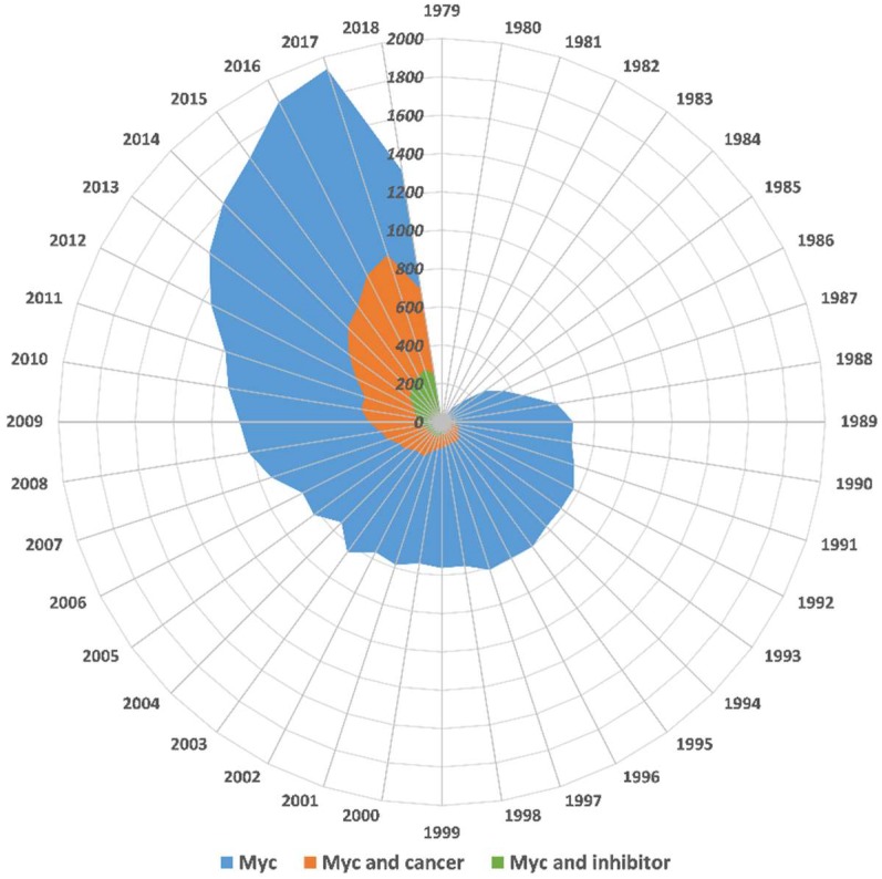
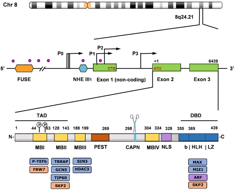
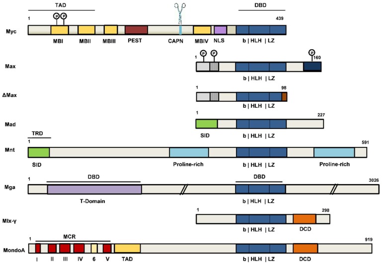
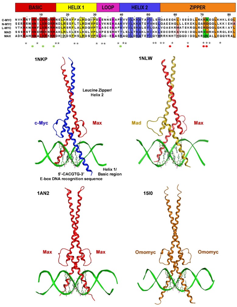
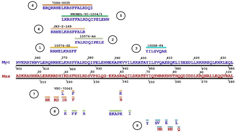
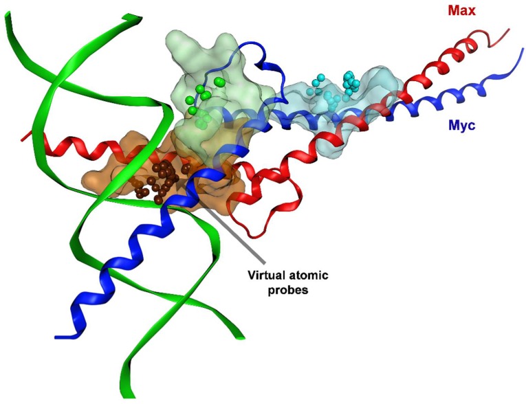
Verification
Claims the paper makes
The claims and statistics of this paper haven’t been checked yet.
Run the check — free, no account needed. It reads the open-access text and checks the sources it cites.
Claims other papers make about this one
Each is checked against this paper’s own text: does it say what the citing paper says it does?
In the citing paper
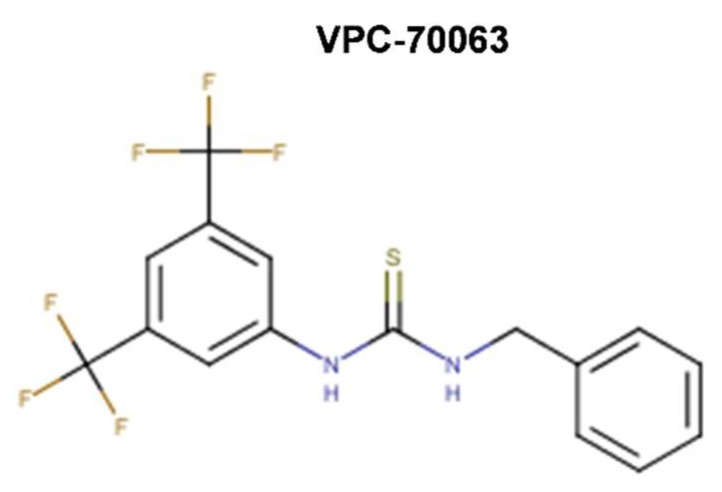
“Two compounds, VPC-70067 (191) and VPC-70063 (192) (Table 3), were ultimately obtained.472”
From Recent advances in targeting the “undruggable” proteins: from drug discovery to clinical trials (2023) · cites this paper as [472]
Self-check
Percentages, sums and reported differences re-computed from the paper’s own numbers.
The self-check needs the paper’s full text.
Citations
Cites
Items 201–211 of 211
Lack of transcriptional repression by max homodimers
Yin, X., Grove, L. E., & Prochownik, E. V. (1998) · Oncogene · doi:10.1038/sj.onc.1201777 · Cited by 19 · cited 1×
“At physiological levels, Max homodimers fail to regulate transcription, but Max overexpression can lead to transcriptional repression [65,66].”
BackgroundWanner, J., Romashko, D., Werner, D. S., May, E. W., Peng, Y., Schulz, R., Foreman, K. W., Russo, S., Arnold, L. D., Pingle, M., et al. (2015) · PLoS ONE · doi:10.1371/journal.pone.0121793 · Cited by 19 · cited 3×
“(2015) [107] generated intracellular self-assembly dimeric inhibitors that showed improved potency and activity in cancer cell lines overexpressing Myc (such as Daudi and Raji Burkitt’s lymphoma human cell lines).”
BackgroundInfluentialCogliati, T., Bk, D., Bar-Ner, M., Cm, C., & Segal, S. (1993) · PubMed · Cited by 18
In silico identification of novel ligands for G-quadruplex in the c-MYC promoter
Kang, H.-J., & Park, H. (2014) · Journal of Computer-Aided Molecular Design · doi:10.1007/s10822-014-9826-z · Cited by 16
Bhat, J., Mondal, S., Sengupta, P., & Chatterjee, S. (2017) · ACS Omega · doi:10.1021/acsomega.6b00531 · Cited by 15
AIMing towards improved antitumor efficacy
Weaver, M. J., Kearns, A. K., Stump, S., Li, C., Gajewski, M. P., Rider, K. C., Backos, D. S., Reigan, P., Beall, H. D., & Natale, N. R. (2015) · Bioorganic & Medicinal Chemistry Letters · doi:10.1016/j.bmcl.2015.02.063 · Cited by 8
Targeting Promoter Quadruplex Nucleic Acids for Cancer Therapy
Paulo, A., Castillo, C. C., & Neidle, S. (2017) · Elsevier eBooks · doi:10.1016/b978-0-12-409547-2.12397-2 · Cited by 7
Crystal structure of the major quadruplex formed in the human c-MYC promoter
Stump, S., Mou, T. C., Sprang, S. R., Natale, N. R., & Beall, H. D. (2017) · Worldwide Protein Data Bank · doi:10.2210/pdb6au4/pdb · Cited by 7
Ma, D., Chan, D. S., Fu, W. C., He, H., Yang, H., Yan, S., & Leung, C. (2012) · PLoS ONE · doi:10.1371/annotation/632f2b99-dd0b-443e-9269-1156d0e7521f · Cited by 6 · cited 1×
“Through virtual screening and NMR spectroscopy, Ma et al. (2012) [198] identified a modular natural compound carbamide 1 (out of five analogues) as a G4 stabilizer that differed from previously described G4 binders, both in terms of chemical structure and binding mode, as it demonstrated G4…”
BackgroundMax protein expression is associated with survival of children with lymphoblastic lymphoma
Yuza, Y., Kawakami, M., TAKAGI, K., Yamazaki, Y., & Urashima, M. (1999) · Pediatrics International · doi:10.1046/j.1442-200x.1999.01140.x · Cited by 6 · cited 1×
“In human cancer, higher Max levels have been associated with better prognosis [69].”
Background
Cited by
Sorted by citations
The MYC oncogene — the grand orchestrator of cancer growth and immune evasion
Dhanasekaran, R., Deutzmann, A., Mahauad‐Fernandez, W. D., Hansen, A. S., Gouw, A. M., & Felsher, D. W. (2021) · Nature Reviews Clinical Oncology · doi:10.1038/s41571-021-00549-2 · Cited by 1,044
Targeting transcription factors in cancer — from undruggable to reality
Bushweller, J. H. (2019) · Nature Reviews Cancer · doi:10.1038/s41568-019-0196-7 · Cited by 919
Therapeutic targeting of “undruggable” MYC
Llombart, V., & Mansour, M. R. (2021) · EBioMedicine · doi:10.1016/j.ebiom.2021.103756 · Cited by 507
Small-Molecule MYC Inhibitors Suppress Tumor Growth and Enhance Immunotherapy
Han, H., Jain, A. D., Truica, M. I., Izquierdo-Ferrer, J., Anker, J. F., Lysy, B., Sagar, V., Luan, Y., Chalmers, Z. R., Unno, K., et al. (2019) · Cancer Cell · doi:10.1016/j.ccell.2019.10.001 · Cited by 479
Recent advances in targeting the “undruggable” proteins: from drug discovery to clinical trials
Xie, X., Yu, T., Li, X., Zhang, N., Foster, L. J., Peng, C., Huang, W., & He, G. (2023) · Signal Transduction and Targeted Therapy · doi:10.1038/s41392-023-01589-z · Cited by 396
Metadata
- Authors
- Carabet, L. A., Rennie, P. S., & Cherkasov, A.
- Affiliations
- University of British Columbia (CA)
- Corresponding author
- Cherkasov, Artem
- ORCID
- Cherkasov, Artem
- Venue
- International Journal of Molecular Sciences
- Publisher
- MDPI AG
- Published
- 2018-12-29
- Volume
- 20
- Issue
- 1
- Pages
- 120
- ISSN
- 1422-0067 (ISSN-L)
- PMC
- PMC6337544
- OpenAlex
- W2907541282
- Cited by
- 172
- References
- 213
- Citations by year
- 2026: 11 · 2025: 23 · 2024: 29 · 2023: 17 · 2022: 23 · 2021: 29 · 2020: 26 · 2019: 14
- FWCI
- 1.81
- Type
- journal article
- Language
- en
- Open access
- gold
- dx.doi.org
- Licence
- Locations
- International Journal of Molecular Sciences (publishedVersion, cc-by)
- Europe PMC (PubMed Central) (publishedVersion, cc-by)
- PubMed Central (publishedVersion, cc-by)
- PubMed (publishedVersion)
- cIRcle (University of British Columbia) (submittedVersion, cc-by)
- DOAJ (DOAJ: Directory of Open Access Journals) (submittedVersion, cc-by-sa)
- Open Collections
- Topics
- Health Sciences › Medicine › Oncology › Cancer-related Molecular Pathways
- Life Sciences › Pharmacology, Toxicology and Pharmaceutics › Drug Discovery › Computational Drug Discovery Methods
- Life Sciences › Biochemistry, Genetics and Molecular Biology › Cancer Research › Cancer Mechanisms and Therapy
- Keywords
- c-Myc, MYC inhibitors, undruggable targets, intrinsically disordered proteins, small molecule inhibitors, structure-function relationship, protein-protein interaction, DNA-binding domain, cancer therapy resistance, computer-aided drug design
- MeSH terms
- Animals, Antineoplastic Agents, Humans, Protein Binding, Proto-Oncogene Proteins c-myc, Quantitative Structure-Activity Relationship, Small Molecule Libraries, Drug Discovery, Molecular Docking Simulation
About these checks
Registry, retraction, Cites and statistics results are computed, not judged, with one exception: a printed reference that no index lists is read once by a model before it counts as a citation error. Claims are extracted by a model and kept only when their quote appears verbatim in the text; each cited source is then checked, with a model’s help, for whether it says what the claim relies on. Those verdicts can be wrong — read the claim’s quote and the cited source before relying on one.
Statistics and arithmetic are recomputed, not judged.
Confidence bands are shown only for fields calibrated on our annotated test set. None is calibrated yet, so no band is shown.
What counts as a claim ›How we measure confidence ›How we check this ›
Are you an author, or do you see a problem with this page? Request a review
