

The DOI matches its Crossref record; 189 of the 191 references it cites were found in a registry or an index.
The Evolving Role of CD8+CD28− Immunosenescent T Cells in Cancer Immunology
Open accessAbstract
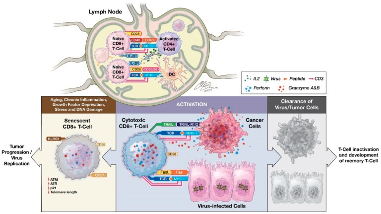
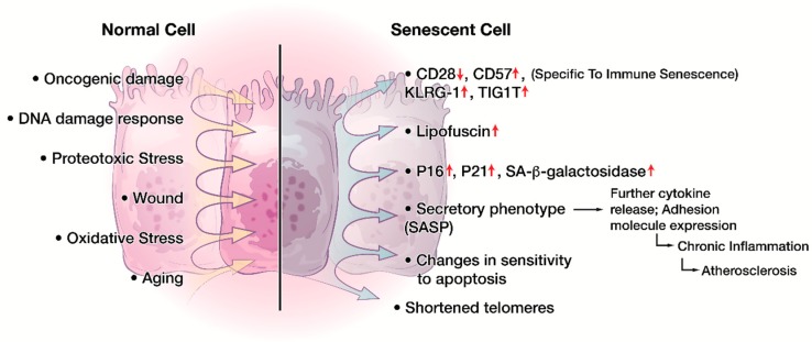
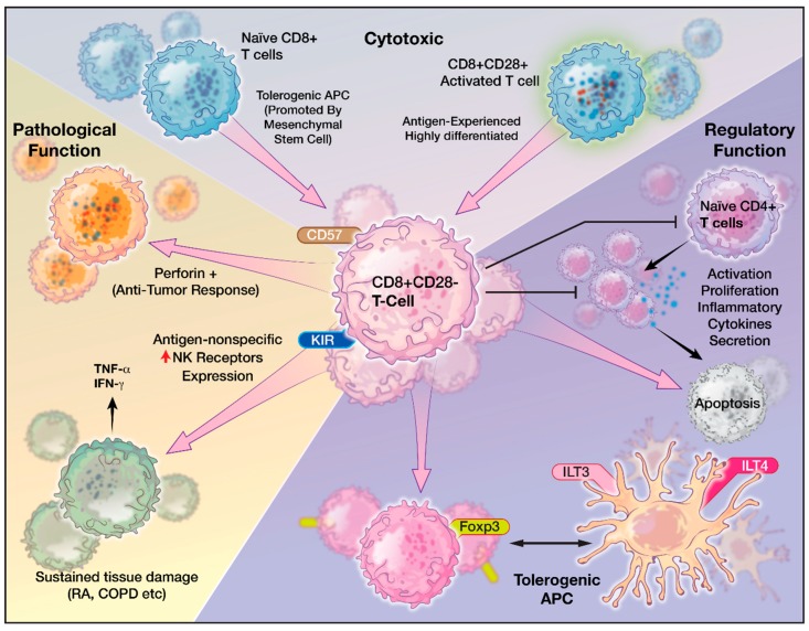
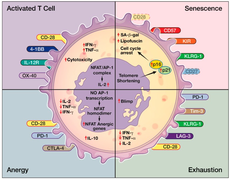
Functional, tumor-specific CD8+ cytotoxic T lymphocytes drive the adaptive immune response to cancer. Thus, induction of their activity is the ultimate aim of all immunotherapies. Success of anti-tumor immunotherapy is precluded by marked immunosuppression in the tumor microenvironment (TME) leading to CD8+ effector T cell dysfunction. Among the many facets of CD8+ T cell dysfunction that have been recognized—tolerance, anergy, exhaustion, and senescence—CD8+ T cell senescence is incompletely understood. Naïve CD8+ T cells require three essential signals for activation, differentiation, and survival through T-cell receptor, costimulatory receptors, and cytokine receptors. Downregulation of costimulatory molecule CD28 is a hallmark of senescent T cells and increased CD8+CD28− senescent populations with heterogeneous roles have been observed in multiple solid and hematogenous tumors. T cell senescence can be induced by several factors including aging, telomere damage, tumor-associated stress, and regulatory T (Treg) cells. Tumor-induced T cell senescence is yet another mechanism that enables tumor cell resistance to immunotherapy. In this paper, we provide a comprehensive overview of CD8+CD28− senescent T cell population, their origin, their function in immunology and pathologic conditions, including TME and their implication for immunotherapy. Further characterization and investigation into this subset of CD8+ T cells could improve the efficacy of future anti-tumor immunotherapy.
Overview
The claims and statistics of this paper haven’t been checked yet.
Run the check — free, no account needed. It reads the open-access text and checks the sources it cites.
Integrity signals
Registry
Cites
Statistics
Figures
Verification
Claims the paper makes
The claims and statistics of this paper haven’t been checked yet.
Run the check — free, no account needed. It reads the open-access text and checks the sources it cites.
Claims other papers make about this one
Each is checked against this paper’s own text: does it say what the citing paper says it does?
In the citing paper
“Therefore, the T cell phenotype of CD27−CD28−CD57+KLRG-1+ is also an indicator of immunosenescence.”
From Immunosenescence: a key player in cancer development (2020) · cites this paper as [41]
Self-check
Percentages, sums and reported differences re-computed from the paper’s own numbers.
The self-check needs the paper’s full text.
Citations
Cites
PD-1 and PD-L1 Immune Checkpoint Blockade to Treat Breast Cancer
Hartkopf, A. D., Taran, F., Wallwiener, M., Walter, C. B., Krämer, B., Grischke, E., & Brucker, S. Y. (2016) · Breast Care · doi:10.1159/000453569 · Cited by 30,989
Improved Survival with Ipilimumab in Patients with Metastatic Melanoma
Hodi, F. S., O'Day, S. J., McDermott, D. F., Weber, R. W., Sosman, J. A., Haanen, J. B., Gonzalez, R., Robert, C., Schadendorf, D., Hassel, J. C., et al. (2010) · New England Journal of Medicine · doi:10.1056/nejmoa1003466 · Cited by 15,155
The blockade of immune checkpoints in cancer immunotherapy
Pardoll, D. M. (2012) · Nature Reviews Cancer · doi:10.1038/nrc3239 · Cited by 13,838
Safety, activity, and immune correlates of anti–PD-1 antibody in cancer
Topalian, S. L., Hodi, F. S., Brahmer, J. R., Gettinger, S. N., Smith, D. C., McDermott, D. F., Powderly, J. D., Carvajal, R. D., Sosman, J. A., Atkins, M. B., et al. (2012) · New England Journal of Medicine · doi:10.1056/nejmoa1200690 · Cited by 12,628
Safety and activity of anti-PD-L1 antibody in patients with advanced cancer
Brahmer, J. R., Tykodi, S. S., Chow, L. Q. M., Hwu, W.-J., Topalian, S. L., Hwu, P., Drake, C. G., Camacho, L. H., Kauh, J., Odunsi, K., et al. (2012) · New England Journal of Medicine · doi:10.1056/nejmoa1200694 · Cited by 7,945 · cited 1×
“For example, CD8+CD28− T ells directly inhibit Ag-presenting function of DCs by inducing inhibitory receptors, such as imm noglobulin like transcript 3 (ILT3) and ILT4, which leads DCs to be immune tolerant than immunogenic [12 ,126].”
BackgroundType, Density, and Location of Immune Cells Within Human Colorectal Tumors Predict Clinical Outcome
Galon, J., Costes, A., Sanchez-Cabo, F., Kirilovsky, A., Mlecnik, B., Lagorce-Pagès, C., Tosolini, M., Camus, M., Berger, A., Wind, P., et al. (2006) · Science · doi:10.1126/science.1129139 · Cited by 6,516
The future of immune checkpoint therapy
Sharma, P., & Allison, J. P. (2015) · Science · doi:10.1126/science.aaa8172 · Cited by 4,878
Neoantigens in cancer immunotherapy
Schumacher, T. N., & Schreiber, R. D. (2015) · Science · doi:10.1126/science.aaa4971 · Cited by 4,783
Molecular and cellular insights into T cell exhaustion
Wherry, E. J., & Kurachi, M. (2015) · Nature Reviews Immunology · doi:10.1038/nri3862 · Cited by 4,652
Wherry, E. J. (2011) · Nature Immunology · doi:10.1038/ni.2035 · Cited by 4,062
Cited by
Sorted by citations
Immunosenescence: molecular mechanisms and diseases
Liu, Z., Liang, Q., Ren, Y., Guo, C., Ge, X., Wang, L., Cheng, Q., Luo, P., Zhang, Y., & Han, X. (2023) · Signal Transduction and Targeted Therapy · doi:10.1038/s41392-023-01451-2 · Cited by 1,082
Immunosenescence: a key player in cancer development
Lian, J., Yue, Y., Yu, W., & Zhang, Y. (2020) · Journal of Hematology & Oncology · doi:10.1186/s13045-020-00986-z · Cited by 510
SIRT1 is downregulated by autophagy in senescence and ageing
Xu, C., Wang, L., Fozouni, P., Evjen, G., Chandra, V., Jiang, J., Lu, C., Nicastri, M. C., Bretz, C. L., Winkler, J. D., et al. (2020) · Nature Cell Biology · doi:10.1038/s41556-020-00579-5 · Cited by 478
Bao, H., Cao, J., Chen, M., Min, C., Chen, W., Chen, X., Chen, Y., Chen, Y., Chen, Y., Chen, Z., et al. (2023) · Science China Life Sciences · doi:10.1007/s11427-023-2305-0 · Cited by 402
Exercise and the immune system: taking steps to improve responses to cancer immunotherapy
Gustafson, M. P., Wheatley, C. M., Rosenthal, A. C., Gastineau, D. A., Katsanis, E., Johnson, B. D., & Simpson, R. J. (2021) · Journal for ImmunoTherapy of Cancer · doi:10.1136/jitc-2020-001872 · Cited by 166
Metadata
- Authors
- Huff, W. X., Kwon, J. H., Henriquez, M., Fetcko, K., & Dey, M.
- Affiliations
- Indiana University School of Medicine (US)
- University of Illinois Chicago (US)
- Corresponding author
- Dey, Mahua
- Venue
- International Journal of Molecular Sciences
- Publisher
- MDPI AG
- Published
- 2019-06-08
- Volume
- 20
- Issue
- 11
- Pages
- 2810
- ISSN
- 1422-0067 (ISSN-L)
- PMC
- PMC6600236
- OpenAlex
- W2948362628
- Cited by
- 217
- References
- 191
- Citations by year
- 2026: 20 · 2025: 35 · 2024: 48 · 2023: 32 · 2022: 33 · 2021: 31 · 2020: 14 · 2019: 2
- FWCI
- 1.73
- Type
- journal article
- Language
- en
- Open access
- gold
- dx.doi.org
- Licence
- Locations
- International Journal of Molecular Sciences (publishedVersion, cc-by)
- PubMed Central (publishedVersion, cc-by)
- PubMed (publishedVersion)
- DOAJ (DOAJ: Directory of Open Access Journals) (submittedVersion, cc-by-sa)
- IUScholarWorks (Indiana University) (submittedVersion, cc-by-nc-nd)
- Purdue University Indianapolis (Indiana University) (submittedVersion, cc-by-nc-nd)
- PMC
- Funders
- National Institutes of Health (K08NS092895)
- Topics
- Life Sciences › Immunology and Microbiology › Immunology › T-cell and B-cell Immunology
- Health Sciences › Medicine › Oncology › Cancer Immunotherapy and Biomarkers
- Life Sciences › Biochemistry, Genetics and Molecular Biology › Cell Biology › Telomeres, Telomerase, and Senescence
- Keywords
- CD8+CD28- T cells, T cell senescence, CD8+ cytotoxic T lymphocytes, tumor microenvironment, immunosuppression, T cell exhaustion, regulatory T cells, costimulatory molecules, telomere damage, anti-tumor immunotherapy, aging
- MeSH terms
- Immunosenescence, Animals, Humans, Immunotherapy, Neoplasms, CD28 Antigens, CD8-Positive T-Lymphocytes
About these checks
Registry, retraction, Cites and statistics results are computed, not judged, with one exception: a printed reference that no index lists is read once by a model before it counts as a citation error. Claims are extracted by a model and kept only when their quote appears verbatim in the text; each cited source is then checked, with a model’s help, for whether it says what the claim relies on. Those verdicts can be wrong — read the claim’s quote and the cited source before relying on one.
Statistics and arithmetic are recomputed, not judged.
Confidence bands are shown only for fields calibrated on our annotated test set. None is calibrated yet, so no band is shown.
What counts as a claim ›How we measure confidence ›How we check this ›
Are you an author, or do you see a problem with this page? Request a review
