

The DOI matches its Crossref record; 41 of the 41 references it cites were found in a registry or an index.
PCSK9 Induces Rat Smooth Muscle Cell Proliferation and Counteracts the Pleiotropic Effects of Simvastatin
Abstract
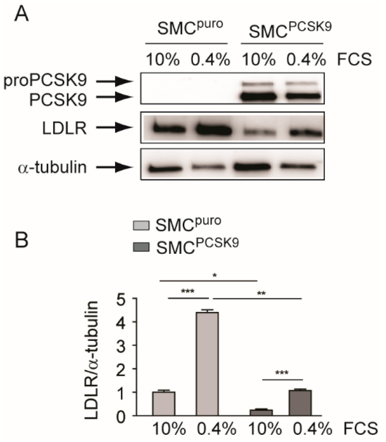
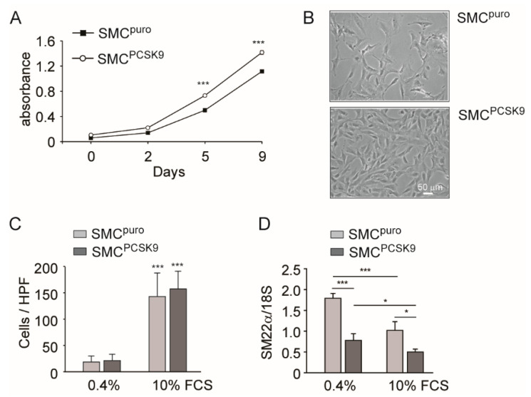
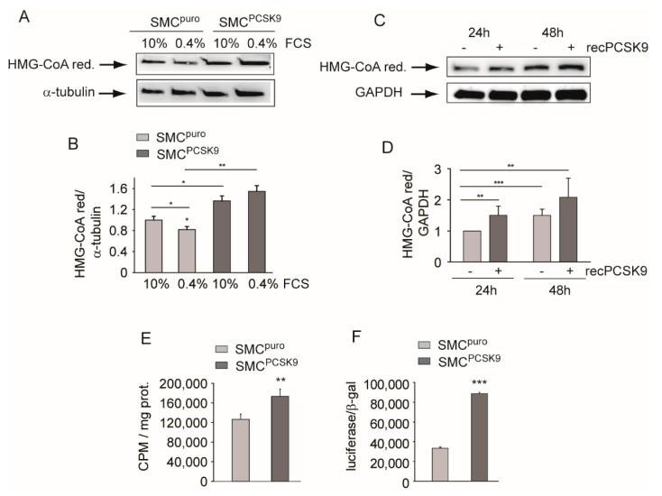
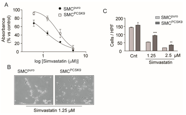
Human atherosclerotic plaque contains smooth muscle cells (SMCs) negative for the contractile phenotype (α-smooth muscle actin) but positive for proprotein convertase subtilisin/kexin type 9 (PCSK9). Thus, we generated rat SMCs which overexpressed human PCSK9 (SMCsPCSK9) with the aim of investigating the role of PCSK9 in the phenotype of SMCs. PCSK9 overexpression in SMCsPCSK9 led to a significant downregulation of the low-density lipoprotein receptor (Ldlr) as well as transgelin (Sm22α), a marker of the contractile phenotype. The cell proliferation rate of SMCsPCSK9 was significantly faster than that of the control SMCs (SMCspuro). Interestingly, overexpression of PCSK9 did not impact the migratory capacity of SMCs in response to 10% FCS, as determined by Boyden’s chamber assay. Expression and activity of 3-hydroxy-3-methylglutaryl-coenzyme A reductase (Hmgcr) was significantly increased in the presence of PCSK9, both in SMCPCSK9 and after treatment with recombinant PCSK9. The transcriptional activity of sterol regulatory element-binding protein (SREBP) was also increased in the presence of PSCK9, suggesting a direct role of PCSK9 in the control of SRE-responsive genes, like HMGCR. We also observed that cholesterol biosynthesis is elevated in SMCPCSK9, potentially explaining the increased proliferation observed in these cells. Finally, concentration-dependent experiments with simvastatin demonstrated that SMCsPCSK9 were partially resistant to the antiproliferative and antimigratory effect of this drug. Taken together, these data further support a direct role of PCSK9 in proliferation, migration, and phenotypic changes in SMCs—pivotal features of atherosclerotic plaque development. We also provide new evidence on the role of PCSK9 in the pharmacological response to statins—gold standard lipid-lowering drugs with pleiotropic action.
Overview
The claims and statistics of this paper haven’t been checked yet.
Run the check — free, no account needed. It reads the open-access text and checks the sources it cites.
Integrity signals
Registry
Cites
Statistics
Figures
Verification
Claims the paper makes
The claims and statistics of this paper haven’t been checked yet.
Run the check — free, no account needed. It reads the open-access text and checks the sources it cites.
Claims other papers make about this one
Each is checked against this paper’s own text: does it say what the citing paper says it does?
The source backs part of the claim, not all of it — or backs it for a different population, condition or comparison.
In the citing paper
“On the other hand, as reported for other cells [44], PCSK9 induced a dose-dependent increase in endogenous cholesterol synthesis, likely as a feedback response to cholesterol reduction mediated by the activation of the transcription factor Sterol Regulatory Element-Binding Protein (SREBP) [44,45].”
From PCSK9 affects astrocyte cholesterol metabolism and reduces neuron cholesterol supplying in vitro: potential implications in Alzheimer’s disease (2022) · cites this paper as [44]
Experimental — accuracy not yet measured
Self-check
Percentages, sums and reported differences re-computed from the paper’s own numbers.
The self-check needs the paper’s full text.
Citations
Cites
Regulating Rho GTPases and their regulators
Hodge, R. G., & Ridley, A. J. (2016) · Nature Reviews Molecular Cell Biology · doi:10.1038/nrm.2016.67 · Cited by 872
LRP: Role in Vascular Wall Integrity and Protection from Atherosclerosis
Boucher, P., Gotthardt, M., Li, W.-P., Anderson, R. G. W., & Herz, J. (2003) · Science · doi:10.1126/science.1082095 · Cited by 581
Dubuc, G., Chamberland, A., Wassef, H., Davignon, J., Seidah, N. G., Bernier, L., & Prat, A. (2004) · Arteriosclerosis, Thrombosis, and Vascular Biology · doi:10.1161/01.atv.0000134621.14315.43 · Cited by 570
Lipid composition of the cancer cell membrane
Szlasa, W., Zendran, I., Zalesińska, A., Tarek, M., & Kulbacka, J. (2020) · Journal of Bioenergetics and Biomembranes · doi:10.1007/s10863-020-09846-4 · Cited by 435
Zaid, A., Roubtsova, A., Essalmani, R., Marcinkiewicz, J., Chamberland, A., Hamelin, J., Tremblay, M., Jacques, H., Jin, W., Davignon, J., et al. (2008) · Hepatology · doi:10.1002/hep.22354 · Cited by 404
Maxwell, K. N., Soccio, R. E., Duncan, E. M., Sehayek, E., & Breslow, J. L. (2003) · Journal of Lipid Research · doi:10.1194/jlr.m300203-jlr200 · Cited by 329
Grünler, J., Ericsson, J., & Dallner, G. (1994) · Biochimica et Biophysica Acta (BBA) - Lipids and Lipid Metabolism · doi:10.1016/0005-2760(94)90200-3 · Cited by 261
Ferri, N., Tibolla, G., Pirillo, A., Cipollone, F., Mezzetti, A., Pacia, S., Corsini, A., & Catapano, A. L. (2012) · Atherosclerosis · doi:10.1016/j.atherosclerosis.2011.11.026 · Cited by 258
Self-citationCross-talk between LOX-1 and PCSK9 in vascular tissues
Ding, Z., Liu, S., Wang, X., Deng, X., Fan, Y., Shahanawaz, J., Reis, R. J. S., Varughese, K. I., Sawamura, T., & Mehta, J. L. (2015) · Cardiovascular Research · doi:10.1093/cvr/cvv178 · Cited by 246
Damask, A., Steg, P. G., Schwartz, G. G., Szarek, M. J., Hagström, E., Badimón, L., Chapman, M. J., Boileau, C., Tsimikas, S., Ginsberg, H. N., et al. (2019) · Circulation · doi:10.1161/circulationaha.119.044434 · Cited by 243
Cited by
Papotti, B., Adorni, M. P., Marchi, C., Zimetti, F., Ronda, N., Panighel, G., Lupo, M. G., Vilella, A., Giuliani, D., Ferri, N., et al. (2022) · International Journal of Molecular Sciences · doi:10.3390/ijms232012192 · Cited by 41
Self-citation
Metadata
- Authors
- Lupo, M. G., Marchianò, S., Adorni, M. P., Zimetti, F., Ruscica, M., Greco, M. F., Corsini, A., & Ferri, N.
- Affiliations
- Dipartimento di Scienze del Farmaco, Università degli Studi di Padova, 35131 Padova, Italy
- Department of Laboratory Medicine and Pathology, University of Washington, Seattle, WA 98195, USA
- Unit of Neurosciences, Department of Medicine and Surgery, Università degli Studi di Parma, 43125 Parma, Italy
- Dipartimento di Scienze degli Alimenti e del Farmaco, Università degli Studi di Parma, 43124 Parma, Italy
- Dipartimento di Scienze Farmacologiche e Biomolecolari, Università degli Studi di Milano, 20133 Milan, Italy
- ORCID
- Venue
- International Journal of Molecular Sciences
- Publisher
- MDPI AG
- Published
- 2021-04-16
- Volume
- 22
- Issue
- 8
- Pages
- 4114
- ISSN
- 1422-0067
- PMC
- PMC8073479
- Cited by
- 15
- References
- 41
- Language
- en
About these checks
Registry, retraction, Cites and statistics results are computed, not judged, with one exception: a printed reference that no index lists is read once by a model before it counts as a citation error. Claims are extracted by a model and kept only when their quote appears verbatim in the text; each cited source is then checked, with a model’s help, for whether it says what the claim relies on. Those verdicts can be wrong — read the claim’s quote and the cited source before relying on one.
Statistics and arithmetic are recomputed, not judged.
Confidence bands are shown only for fields calibrated on our annotated test set. None is calibrated yet, so no band is shown.
What counts as a claim ›How we measure confidence ›How we check this ›
Are you an author, or do you see a problem with this page? Request a review
How to cite
Cite CoChat for the checks on this page, and the paper for its own findings.
- This page
- CoChat. (2026). Citation check: PCSK9 Induces Rat Smooth Muscle Cell Proliferation and Counteracts the Pleiotropic Effects of Simvastatin. https://app.cochat.ai/papers/10.3390/ijms22084114
- The paper
- Lupo, M. G., Marchianò, S., Adorni, M. P., Zimetti, F., Ruscica, M., Greco, M. F., Corsini, A., & Ferri, N. (2021). PCSK9 Induces Rat Smooth Muscle Cell Proliferation and Counteracts the Pleiotropic Effects of Simvastatin. International Journal of Molecular Sciences. https://doi.org/10.3390/ijms22084114
Data sources
Bibliographic data: Semantic Scholar, OpenAlex, Crossref.
Contains information from the Semantic Scholar Academic Graph Datasets, made available under the ODC Attribution License (ODC-BY 1.0).
Semantic Scholar’s data is described in: Kinney, R., et al. (2023). The Semantic Scholar Open Data Platform. arXiv:2301.10140.
