Student or educator? Sign up with your school email and get 6 months of Pro free.
Cited by 11RNA Research and SplicingMitochondrial Function and PathologyPhotosynthetic Processes and Mechanisms

The DOI matches its Crossref record; 107 of the 107 references it cites were found in a registry or an index.

MISF2 Encodes an Essential Mitochondrial Splicing Cofactor Required for nad2 mRNA Processing and Embryo Development in Arabidopsis thaliana

Open access
Nguyen, Tan-Trung, Best, Corinne, Shevtsov, Sofia, Zmudjak, Michal, Quadrado, Martine, Mizrahi, Ron, Zer, Hagit, Mireau, Hakim, Ostersetzer-Biran, Oren·2022·International Journal of Molecular Sciences

Published version of a preprint

Abstract

Mitochondria play key roles in cellular energy metabolism in eukaryotes. Mitochondria of most organisms contain their own genome and specific transcription and translation machineries. The expression of angiosperm mtDNA involves extensive RNA-processing steps, such as RNA trimming, editing, and the splicing of numerous group II-type introns. Pentatricopeptide repeat (PPR) proteins are key players in plant organelle gene expression and RNA metabolism. In the present analysis, we reveal the function of the MITOCHONDRIAL SPLICING FACTOR 2 gene (MISF2, AT3G22670) and show that it encodes a mitochondria-localized PPR protein that is crucial for early embryo development in Arabidopsis. Molecular characterization of embryo-rescued misf2 plantlets indicates that the splicing of nad2 intron 1, and thus respiratory complex I biogenesis, are strongly compromised. Moreover, the molecular function seems conserved between MISF2 protein in Arabidopsis and its orthologous gene (EMP10) in maize, suggesting that the ancestor of MISF2/EMP10 was recruited to function in nad2 processing before the monocot–dicot divergence ~200 million years ago. These data provide new insights into the function of nuclear-encoded factors in mitochondrial gene expression and respiratory chain biogenesis during plant embryo development.

Overview

The claims and statistics of this paper haven’t been checked yet.

Run the check — free, no account needed. It reads the open-access text and checks the sources it cites.

A few free checks a day · papers already verified open instantly · results are public

Integrity signals

Registry

Crossref · 10.3390/ijms23052670

Cites

107 of 107 references verified0 citation errors · 0 not checkable

Statistics

Figures

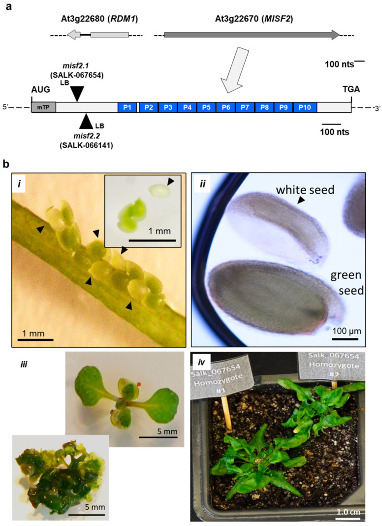
Figure 1 MISF2 (At3g22670) gene topology and misf2 mutant phenotypes. (a) Scheme of the At3g22670 locus and gene structure. The large arrow points toward motif-structure arrangements of the MISF2 coding region. The position of the two T-DNA insertion sites in the coding region of MISF2 (i.e., SALK-line 067654, misf2.1, SALK-line 066141, and misf2.2) are located 324 and 350 nucleotides downstream to the ATG start codon, in a region that corresponds to the N-terminal domain of MISF2, upstream of the PPR motifs. (b) Morphologies of misf2 hetero- and homozygous mutants. Green and white seeds harboring wild-type/heterozygous and homozygous mutant embryos respectively were collected from surface-sterilized immature siliques of heterozygous misf2 plants (i) and sown on MS agar media supplemented with vitamins. Arrows point toward white seeds. Panel B (ii) shows differential interference contrast microscopy images (i.e., Nomarski) of embryos found in green or white seeds. Following germination, a few rescued homozygous misf2 plantlets (iii) were able to survive on soil, although failed to set flowers and viable seeds (iv).
Figure 1 MISF2 (At3g22670) gene topology and misf2 mutant phenotypes. (a) Scheme of the At3g22670 locus and gene structure. The large arrow points toward motif-structure arrangements of the MISF2 coding region. The position of the two T-DNA insertion sites in the coding region of MISF2 (i.e., SALK-line 067654, misf2.1, SALK-line 066141, and misf2.2) are located 324 and 350 nucleotides downstream to the ATG start codon, in a region that corresponds to the N-terminal domain of MISF2, upstream of the PPR motifs. (b) Morphologies of misf2 hetero- and homozygous mutants. Green and white seeds harboring wild-type/heterozygous and homozygous mutant embryos respectively were collected from surface-sterilized immature siliques of heterozygous misf2 plants (i) and sown on MS agar media supplemented with vitamins. Arrows point toward white seeds. Panel B (ii) shows differential interference contrast microscopy images (i.e., Nomarski) of embryos found in green or white seeds. Following germination, a few rescued homozygous misf2 plantlets (iii) were able to survive on soil, although failed to set flowers and viable seeds (iv).CC BY · PubMed Central
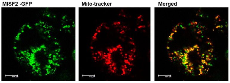
Figure 2 MISF2 is localized to the mitochondria. Arabidopsis plant cells were transformed with a construct expressing the GFP fused in frame to the N-terminal region (i.e., 203 amino acids) of the MISF2 protein. The fluorescence corresponding to the GFP (green, left), the MitoTracker® marker (red, center), and the merged signals (right) are shown. Bars = 10 μm.
Figure 2 MISF2 is localized to the mitochondria. Arabidopsis plant cells were transformed with a construct expressing the GFP fused in frame to the N-terminal region (i.e., 203 amino acids) of the MISF2 protein. The fluorescence corresponding to the GFP (green, left), the MitoTracker® marker (red, center), and the merged signals (right) are shown. Bars = 10 μm.CC BY · PubMed Central
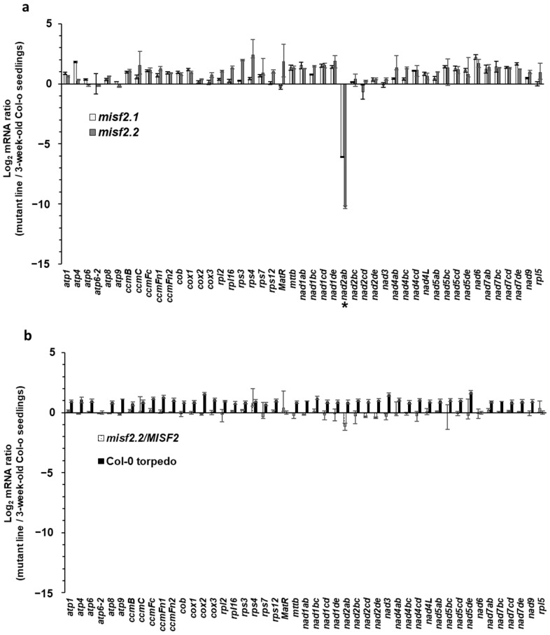
Figure 3 Relative accumulation of mitochondrial mRNAs in misf2 mutants. Analysis of various mt-RNAs levels in Arabidopsis wild-type (Col-0), misf2 mutants, and complemented misf2.2/MISF2 plants by RT-qPCR. RNA extracted from 3-week-old wild-type seedlings (Col-0), 4-months-old rescued misf2 mutants, plantlets derived from immature Col-0 seeds (i.e., at the torpedo stage), and functionally complemented misf2.2/MISF2 mutants were reverse-transcribed and the relative steady-state levels of cDNAs corresponding to mitochondrial mRNAs evaluated by qPCR. Log2 ratios of mt mRNA abundances in misf2.1 and misf2.2 mutant lines (a), plantlets derived from immature Col-0 seeds, and complementation line (b) to those of 3-week-old MS-agar grown wild-type plants are shown. Asterisk indicates to reduced nad2ab transcript. The values are means of three biological replicates (error bars indicate one standard deviation).
Figure 3 Relative accumulation of mitochondrial mRNAs in misf2 mutants. Analysis of various mt-RNAs levels in Arabidopsis wild-type (Col-0), misf2 mutants, and complemented misf2.2/MISF2 plants by RT-qPCR. RNA extracted from 3-week-old wild-type seedlings (Col-0), 4-months-old rescued misf2 mutants, plantlets derived from immature Col-0 seeds (i.e., at the torpedo stage), and functionally complemented misf2.2/MISF2 mutants were reverse-transcribed and the relative steady-state levels of cDNAs corresponding to mitochondrial mRNAs evaluated by qPCR. Log2 ratios of mt mRNA abundances in misf2.1 and misf2.2 mutant lines (a), plantlets derived from immature Col-0 seeds, and complementation line (b) to those of 3-week-old MS-agar grown wild-type plants are shown. Asterisk indicates to reduced nad2ab transcript. The values are means of three biological replicates (error bars indicate one standard deviation).CC BY · PubMed Central
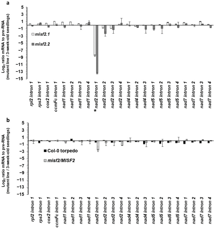
Figure 4 Mitochondrial intron splicing efficiencies in misf2 mutants. The relative accumulation of mRNA and pre-RNA transcripts in wild-type, misf2 mutants, and complemented misf2.2/MISF2 plants, corresponding to the 23 group II intron sequences in Arabidopsis, was evaluated by RT-qPCR. The histogram shows the splicing efficiencies as indicated by the log2 ratios of pre-RNA to mature mRNA transcript abundance in misf2.1 and misf2.2 mutant lines compared with those in wild-type plants (a), as well as germinated wild-type seeds collected at the torpedo stage (Col-0-torpedo), and complemented line compared with those of wild-type plants (b). Asterisk indicates the altered splicing of nad2 intron 1. The values are means of three (misf2.1, misf2.2/MISF2) and five (misf2.2, Col-0) biological replicates (error bars indicate one standard deviation).
Figure 4 Mitochondrial intron splicing efficiencies in misf2 mutants. The relative accumulation of mRNA and pre-RNA transcripts in wild-type, misf2 mutants, and complemented misf2.2/MISF2 plants, corresponding to the 23 group II intron sequences in Arabidopsis, was evaluated by RT-qPCR. The histogram shows the splicing efficiencies as indicated by the log2 ratios of pre-RNA to mature mRNA transcript abundance in misf2.1 and misf2.2 mutant lines compared with those in wild-type plants (a), as well as germinated wild-type seeds collected at the torpedo stage (Col-0-torpedo), and complemented line compared with those of wild-type plants (b). Asterisk indicates the altered splicing of nad2 intron 1. The values are means of three (misf2.1, misf2.2/MISF2) and five (misf2.2, Col-0) biological replicates (error bars indicate one standard deviation).CC BY · PubMed Central
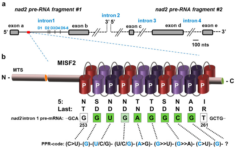
Figure 5 The predicted MISF2 binding site in nad2 intron 1. (a) The expression of nad2 in Arabidopsis mitochondria involves the transcription of two precursor RNA transcripts, which are divided by the second intron. The maturation of nad2 requires the splicing of four introns found in cis (introns 1, 3, and 4) or trans (intron 2) configurations. The first pre-mRNA fragment consists of two exons separated by intron 1, while the second fragment harbors three exons separated by introns 3 and 4. The six typical stem-loop domains (D1–D6) are indicated for nad2 intron 1. (b) MISF2 is a P-type PPR protein, which harbors a mitochondrial targeting sequence (MTS) and 10 PPR motifs. The fifth and the last amino acids of each PPR repeat (Figure S1) are indicated below each PPR repeats. The best corresponding RNA binding site (i.e., 5′-GUGAGGCG-3′) is indicated within the first intron of nad2 pre-RNA fragment #1, with bases marked in green for perfect matches to the proposed binding site, in pale green for partial matches, and white for non-matching or unassigned nucleotides.
Figure 5 The predicted MISF2 binding site in nad2 intron 1. (a) The expression of nad2 in Arabidopsis mitochondria involves the transcription of two precursor RNA transcripts, which are divided by the second intron. The maturation of nad2 requires the splicing of four introns found in cis (introns 1, 3, and 4) or trans (intron 2) configurations. The first pre-mRNA fragment consists of two exons separated by intron 1, while the second fragment harbors three exons separated by introns 3 and 4. The six typical stem-loop domains (D1–D6) are indicated for nad2 intron 1. (b) MISF2 is a P-type PPR protein, which harbors a mitochondrial targeting sequence (MTS) and 10 PPR motifs. The fifth and the last amino acids of each PPR repeat (Figure S1) are indicated below each PPR repeats. The best corresponding RNA binding site (i.e., 5′-GUGAGGCG-3′) is indicated within the first intron of nad2 pre-RNA fragment #1, with bases marked in green for perfect matches to the proposed binding site, in pale green for partial matches, and white for non-matching or unassigned nucleotides.CC BY · PubMed Central
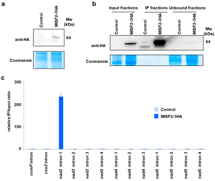
Figure 6 The MISF2 protein associates with nad2 intron 1 in vivo. (a) Immunodetection of the MISF2-3HA fusion protein in protein extracts prepared form untransformed (control) and transformed (MISF2-3HA) Arabidopsis cell cultures. (b) Immunoprecipitation assays were conducted with the anti-HA antibody and the shown immunoblot analysis attests for the strong enrichment of the MISF2-3HA fusion in the immunoprecipitated (IP) fraction derived from the Arabidopsis transgenic cell line expressing the fusion. The weak MISF2-3HA signal in the unbound fraction demonstrates the efficiency of the immunoprecipitation. Parts of the blots stained with Coomassie blue are shown to display equal loading between samples. (c) Co-immunoprecipitated RNAs were analyzed by qRT-PCR using primer pairs specific to the indicated mitochondrial introns and relative enrichment ratios (immunoprecipitation fraction/input fraction) are shown.
Figure 6 The MISF2 protein associates with nad2 intron 1 in vivo. (a) Immunodetection of the MISF2-3HA fusion protein in protein extracts prepared form untransformed (control) and transformed (MISF2-3HA) Arabidopsis cell cultures. (b) Immunoprecipitation assays were conducted with the anti-HA antibody and the shown immunoblot analysis attests for the strong enrichment of the MISF2-3HA fusion in the immunoprecipitated (IP) fraction derived from the Arabidopsis transgenic cell line expressing the fusion. The weak MISF2-3HA signal in the unbound fraction demonstrates the efficiency of the immunoprecipitation. Parts of the blots stained with Coomassie blue are shown to display equal loading between samples. (c) Co-immunoprecipitated RNAs were analyzed by qRT-PCR using primer pairs specific to the indicated mitochondrial introns and relative enrichment ratios (immunoprecipitation fraction/input fraction) are shown.CC BY · PubMed Central
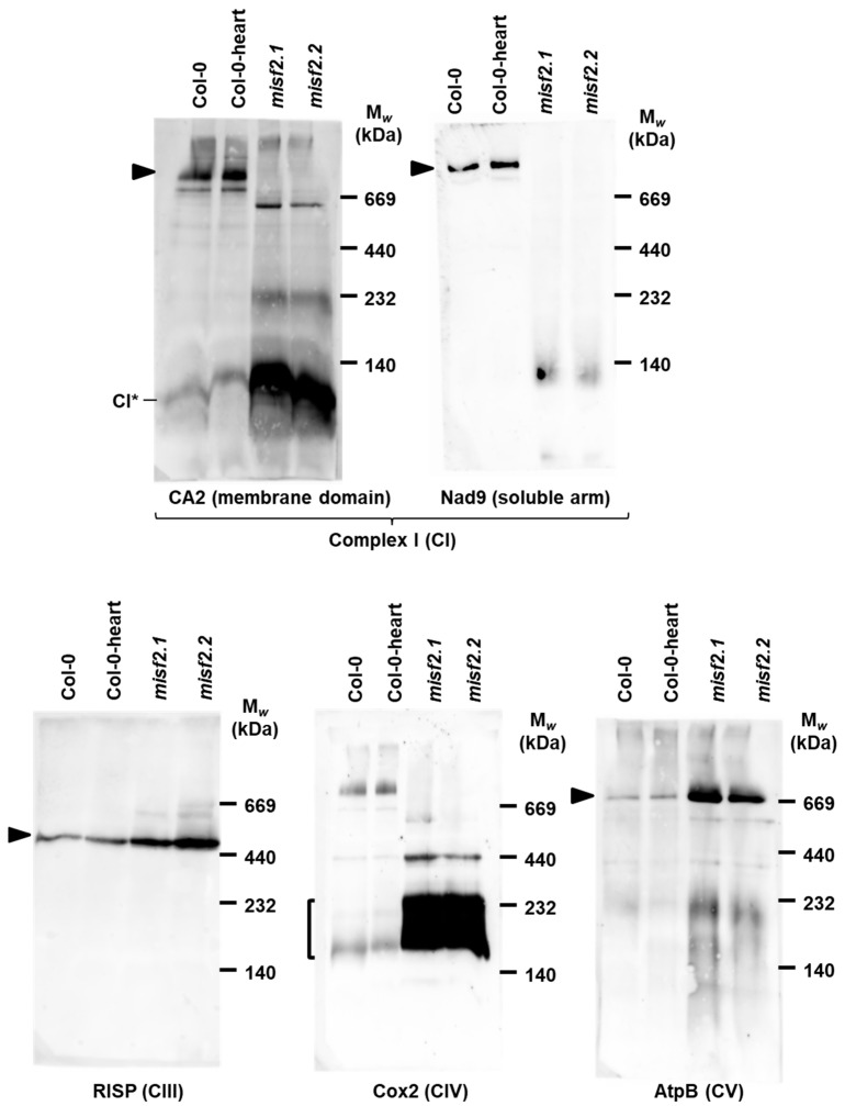
Figure 7 Holo-complex I is below detectable levels in misf2 mutants. Blue native (BN)-PAGE analysis of crude organellar fractions was performed as described by [79]. Aliquots, equivalent to 40 mg of crude organellar membrane extracts, obtained from wild-type and misf2 plants, were solubilized with digitonin and resolved by BN-PAGE. For immunodetection, the proteins were transferred onto PVDF membranes and probed with the antibodies indicated below each blot (Table S2). Arrows point toward native complexes I (~1000 kDa), CIII dimer (III2, ~500 kDa), CIV (about 200 and 220 kDa forms), and CV (~660 kDa) [69]. CI* indicates the ~85 kDa sub-CI assembly intermediate [5].
Figure 7 Holo-complex I is below detectable levels in misf2 mutants. Blue native (BN)-PAGE analysis of crude organellar fractions was performed as described by [79]. Aliquots, equivalent to 40 mg of crude organellar membrane extracts, obtained from wild-type and misf2 plants, were solubilized with digitonin and resolved by BN-PAGE. For immunodetection, the proteins were transferred onto PVDF membranes and probed with the antibodies indicated below each blot (Table S2). Arrows point toward native complexes I (~1000 kDa), CIII dimer (III2, ~500 kDa), CIV (about 200 and 220 kDa forms), and CV (~660 kDa) [69]. CI* indicates the ~85 kDa sub-CI assembly intermediate [5].CC BY · PubMed Central
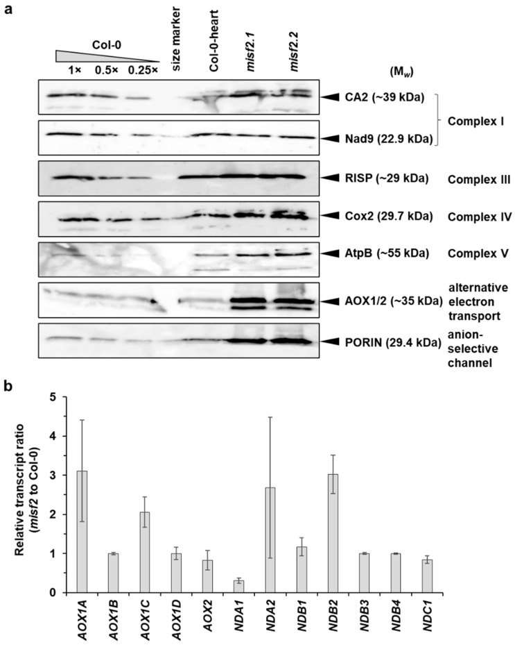
Figure 8 Relative accumulation of different mitochondrial proteins and AOX or ND transcripts in wild-type and misf2 plants. (a) Immunoblots made with crude organellar fractions (equivalent to ~40 mg FW) extracted from 3-week-old MS-grown wild-type plants, in vitro germinated wild-type embryos at the heart to torpedo stages (Col-0 torpedo), and homozygous misf2 plantlets. The blots were probed with antibodies raised against the indicated mitochondrial proteins. (b) Analysis of the steady-state levels of various alternative oxidase (AOX) and rotenone-insensitive NAD(P)H dehydrogenase (ND) mRNAs by RT-qPCR. The histogram displays relative mRNAs levels in misf2 plantlets to 3-week-old MS-grown wild-type plants.
Figure 8 Relative accumulation of different mitochondrial proteins and AOX or ND transcripts in wild-type and misf2 plants. (a) Immunoblots made with crude organellar fractions (equivalent to ~40 mg FW) extracted from 3-week-old MS-grown wild-type plants, in vitro germinated wild-type embryos at the heart to torpedo stages (Col-0 torpedo), and homozygous misf2 plantlets. The blots were probed with antibodies raised against the indicated mitochondrial proteins. (b) Analysis of the steady-state levels of various alternative oxidase (AOX) and rotenone-insensitive NAD(P)H dehydrogenase (ND) mRNAs by RT-qPCR. The histogram displays relative mRNAs levels in misf2 plantlets to 3-week-old MS-grown wild-type plants.CC BY · PubMed Central

Verification

Claims the paper makes

The claims and statistics of this paper haven’t been checked yet.

Run the check — free, no account needed. It reads the open-access text and checks the sources it cites.

A few free checks a day · papers already verified open instantly · results are public

Self-check

Percentages, sums and reported differences re-computed from the paper’s own numbers.

The self-check needs the paper’s full text.

Citations

Cites

Show all 107 references

Metadata

Authors
Nguyen, T.-T., Best, C., Shevtsov, S., Zmudjak, M., Quadrado, M., Mizrahi, R., Zer, H., Mireau, H., & Ostersetzer-Biran, O.
Affiliations
  • Centre National de la Recherche Scientifique (FR)
  • Hebrew University of Jerusalem (IL)
  • Department of Plant and Environmental Sciences, The Alexander Silberman Institute of Life Sciences, The Hebrew University of Jerusalem, Givat-Ram, Jerusalem 91904, Israel
Corresponding author
Mireau, Hakim
Venue
International Journal of Molecular Sciences
Publisher
MDPI AG
Published
2022-02-28
Volume
23
Issue
5
Pages
2670
ISSN
1422-0067 (ISSN-L)
OpenAlex
W4220848332
Cited by
11
References
107
Citations by year
2026: 1 · 2025: 3 · 2024: 3 · 2023: 1 · 2022: 3
FWCI
0.81
Type
journal article
Language
en
Open access
Locations
  • International Journal of Molecular Sciences (publishedVersion, cc-by)
  • Preprints.org (acceptedVersion, cc-by)
  • PubMed Central (publishedVersion, cc-by)
  • PubMed (publishedVersion)
  • HAL (submittedVersion, cc-by)
  • DOAJ (DOAJ: Directory of Open Access Journals) (submittedVersion, cc-by-sa)
Funders
  • Israel Science Foundation (1834/20, 3254-2020)
  • French National Centre for Scientific Research (ANR-16-CE11- 0024-01)
  • University of Paris-Saclay (ANR-17-EUR-0007)
Topics
  • Life Sciences › Biochemistry, Genetics and Molecular Biology › Molecular Biology › RNA Research and Splicing
  • Life Sciences › Biochemistry, Genetics and Molecular Biology › Molecular Biology › Mitochondrial Function and Pathology
  • Life Sciences › Biochemistry, Genetics and Molecular Biology › Molecular Biology › Photosynthetic Processes and Mechanisms
Keywords
pentatricopeptide repeat proteins, mitochondrial RNA splicing, mitochondrial gene expression, early embryonic development, embryo rescue, monocot-dicot divergence, Arabidopsis thaliana, maize
MeSH terms
Introns, Mitochondria, Mutation, Plant Proteins, RNA, RNA Splicing, RNA, Messenger, Arabidopsis, Gene Expression Regulation, Plant, Arabidopsis Proteins, Electron Transport Complex I, Embryonic Development

About these checks

Registry, retraction, Cites and statistics results are computed, not judged, with one exception: a printed reference that no index lists is read once by a model before it counts as a citation error. Claims are extracted by a model and kept only when their quote appears verbatim in the text; each cited source is then checked, with a model’s help, for whether it says what the claim relies on. Those verdicts can be wrong — read the claim’s quote and the cited source before relying on one.

Statistics and arithmetic are recomputed, not judged.

Confidence bands are shown only for fields calibrated on our annotated test set. None is calibrated yet, so no band is shown.

What counts as a claim ›How we measure confidence ›How we check this ›

Are you an author, or do you see a problem with this page? Request a review

How to cite

Cite CoChat for the checks on this page, and the paper for its own findings.

This page
CoChat. (2026). Citation check: MISF2 Encodes an Essential Mitochondrial Splicing Cofactor Required for nad2 mRNA Processing and Embryo Development in Arabidopsis thaliana. https://app.cochat.ai/papers/10.3390/ijms23052670
The paper
Nguyen, T.-T., Best, C., Shevtsov, S., Zmudjak, M., Quadrado, M., Mizrahi, R., Zer, H., Mireau, H., & Ostersetzer-Biran, O. (2022). MISF2 Encodes an Essential Mitochondrial Splicing Cofactor Required for nad2 mRNA Processing and Embryo Development in Arabidopsis thaliana. International Journal of Molecular Sciences. https://doi.org/10.3390/ijms23052670

Data sources

Bibliographic data: Semantic Scholar, OpenAlex, Crossref.

Contains information from the Semantic Scholar Academic Graph Datasets, made available under the ODC Attribution License (ODC-BY 1.0).

Semantic Scholar’s data is described in: Kinney, R., et al. (2023). The Semantic Scholar Open Data Platform. arXiv:2301.10140.