

The DOI matches its Crossref record; 229 of the 233 references it cites were found in a registry or an index.
Therapeutic Effects of Mesenchymal Stromal Cells Require Mitochondrial Transfer and Quality Control
Open accessAbstract
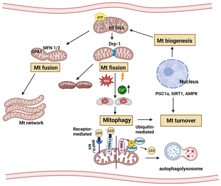
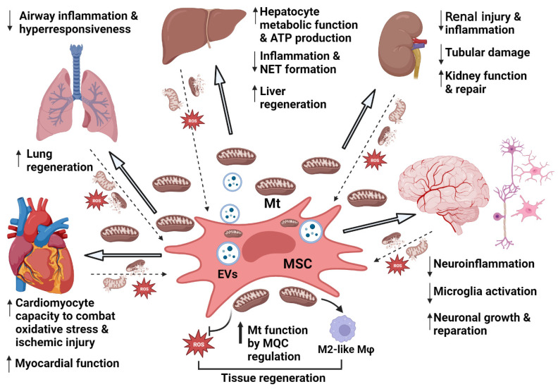
Due to their beneficial effects in an array of diseases, Mesenchymal Stromal Cells (MSCs) have been the focus of intense preclinical research and clinical implementation for decades. MSCs have multilineage differentiation capacity, support hematopoiesis, secrete pro-regenerative factors and exert immunoregulatory functions promoting homeostasis and the resolution of injury/inflammation. The main effects of MSCs include modulation of immune cells (macrophages, neutrophils, and lymphocytes), secretion of antimicrobial peptides, and transfer of mitochondria (Mt) to injured cells. These actions can be enhanced by priming (i.e., licensing) MSCs prior to exposure to deleterious microenvironments. Preclinical evidence suggests that MSCs can exert therapeutic effects in a variety of pathological states, including cardiac, respiratory, hepatic, renal, and neurological diseases. One of the key emerging beneficial actions of MSCs is the improvement of mitochondrial functions in the injured tissues by enhancing mitochondrial quality control (MQC). Recent advances in the understanding of cellular MQC, including mitochondrial biogenesis, mitophagy, fission, and fusion, helped uncover how MSCs enhance these processes. Specifically, MSCs have been suggested to regulate peroxisome proliferator-activated receptor-gamma coactivator 1 alpha (PGC1α)-dependent biogenesis, Parkin-dependent mitophagy, and Mitofusins (Mfn1/2) or Dynamin Related Protein-1 (Drp1)-mediated fission/fusion. In addition, previous studies also verified mitochondrial transfer from MSCs through tunneling nanotubes and via microvesicular transport. Combined, these effects improve mitochondrial functions, thereby contributing to the resolution of injury and inflammation. Thus, uncovering how MSCs affect MQC opens new therapeutic avenues for organ injury, and the transplantation of MSC-derived mitochondria to injured tissues might represent an attractive new therapeutic approach.
Overview
The claims and statistics of this paper haven’t been checked yet.
Run the check — free, no account needed. It reads the open-access text and checks the sources it cites.
Integrity signals
Registry
Cites
Statistics
Figures
Verification
Claims the paper makes
The claims and statistics of this paper haven’t been checked yet.
Run the check — free, no account needed. It reads the open-access text and checks the sources it cites.
Claims other papers make about this one
Each is checked against this paper’s own text: does it say what the citing paper says it does?
The cited source says what the claim attributes to it.
In the citing paper
“MSCs can engulf and degrade dysfunctional Mt released from injured cells [122].”
From Mechanisms, efficacy, and future perspectives of cellular-based therapies for liver fibrosis/cirrhosis: focusing on mesenchymal stromal cells (2025) · cites this paper as [122]
The cited source says what the claim attributes to it.
In the citing paper
“This process, in turn, boosts mitochondrial biogenesis within the MSCs, enhancing their capacity to donate functional Mt to recipient cells, thereby improving overall mitochondrial quality control (MQC) and metabolic function [122, 123].”
From Mechanisms, efficacy, and future perspectives of cellular-based therapies for liver fibrosis/cirrhosis: focusing on mesenchymal stromal cells (2025) · cites this paper as [122]
Self-check
Percentages, sums and reported differences re-computed from the paper’s own numbers.
The self-check needs the paper’s full text.
Citations
Cites
Parkin is recruited selectively to impaired mitochondria and promotes their autophagy
Narendra, D., Tanaka, A., Suen, D.-F., & Youle, R. J. (2008) · The Journal of Cell Biology · doi:10.1083/jcb.200809125 · Cited by 4,026
PINK1 Is Selectively Stabilized on Impaired Mitochondria to Activate Parkin
Narendra, D. P., Jin, S. M., Tanaka, A., Suen, D.-F., Gautier, C. A., Shen, J., Cookson, M. R., & Youle, R. J. (2010) · PLoS Biology · doi:10.1371/journal.pbio.1000298 · Cited by 2,944
Chen, H., Detmer, S. A., Ewald, A. J., Griffin, E. E., Fraser, S. E., & Chan, D. C. (2003) · The Journal of Cell Biology · doi:10.1083/jcb.200211046 · Cited by 2,601
Mitophagy and Quality Control Mechanisms in Mitochondrial Maintenance
Pickles, S., Vigié, P., & Youle, R. J. (2018) · Current Biology · doi:10.1016/j.cub.2018.01.004 · Cited by 2,081
Nanotubular Highways for Intercellular Organelle Transport
Rustom, A., Saffrich, R., Markovic, I., Walther, P., & Gerdes, H.-H. (2004) · Science · doi:10.1126/science.1093133 · Cited by 2,038
The multifaceted contributions of mitochondria to cellular metabolism
Spinelli, J. B., & Haigis, M. C. (2018) · Nature Cell Biology · doi:10.1038/s41556-018-0124-1 · Cited by 1,935
Islam, M. N., Das, S. R., Emin, M. T., Wei, M., Sun, L., Westphalen, K., Rowlands, D. J., Quadri, S. K., Bhattacharya, S., & Bhattacharya, J. (2012) · Nature Medicine · doi:10.1038/nm.2736 · Cited by 1,585
Transfer of mitochondria from astrocytes to neurons after stroke
Hayakawa, K., Esposito, E., Wang, X., Terasaki, Y., Liu, Y., Xing, C., Ji, X., & Lo, E. H. (2016) · Nature · doi:10.1038/nature18928 · Cited by 1,423
Mitochondrial dynamics: overview of molecular mechanisms
Tilokani, L., Nagashima, S., Paupe, V., & Prudent, J. (2018) · Essays in Biochemistry · doi:10.1042/ebc20170104 · Cited by 1,397
Disruption of Fusion Results in Mitochondrial Heterogeneity and Dysfunction
Chen, H., Chomyn, A., & Chan, D. C. (2005) · Journal of Biological Chemistry · doi:10.1074/jbc.m503062200 · Cited by 1,376 · cited 1×
“However, excessive fusion (hyperfusion) promotes cellular senescence [44], disrupts normal mitochondrial energy production, and interferes with mitophagy [45–47].”
Background
Research works printed but not linked: 1
- Maumus, 2018, Mesenchymal Stem Cells and Regenerative Medicine: Future Perspectives in Osteoarthritis, Med. Sci., 34, 1092
Cited by
Sorted by citations
Tissue Engineering and Regenerative Medicine: Perspectives and Challenges
Hoang, V. T., Nguyen, Q. T., Phan, T. T. K., Pham, T. H., Dinh, N. T. H., Anh, L. P. H., Dao, L. T. M., Bui, V. D., Dao, H., Le, D. S., et al. (2025) · MedComm · doi:10.1002/mco2.70192 · Cited by 67
Gao, F., Xu, T., Zang, F., Luo, Y., & Pan, D. (2024) · Drug Design Development and Therapy · doi:10.2147/dddt.s469331 · Cited by 60
Tripathi, K. K., & Ben‐Shachar, D. (2024) · Cells · doi:10.3390/cells13050410 · Cited by 47
Bodenstein, D. F., Siebiger, G., Zhao, Y., Clasky, A. J., Mukkala, A. N., Beroncal, E. L., Banh, L., Aslostovar, L., Brijbassi, S., Hogan, S. E., et al. (2024) · Stem Cell Research & Therapy · doi:10.1186/s13287-024-03771-8 · Cited by 41
Self-citationHe, X., Zhong, L., Wang, N., Zhao, B., Wang, Y., Wu, X., Zheng, C., Ruan, Y., Hou, J., Luo, Y., et al. (2024) · Advanced Science · doi:10.1002/advs.202404994 · Cited by 34
Metadata
- Authors
- Mukkala, A. N., Jerkic, M., Khan, Z., Szaszi, K., Kapus, A., & Rotstein, O.
- Affiliations
- St. Michael's Hospital (CA)
- Unity Health Toronto, The Keenan Research Centre for Biomedical Science of St. Michael’s Hospital, University of Toronto, Toronto, ON M5B 1T8, Canada
- Venue
- International Journal of Molecular Sciences
- Publisher
- MDPI AG
- Published
- 2023-10-31
- Volume
- 24
- Issue
- 21
- Pages
- 15788
- ISSN
- 1422-0067 (ISSN-L)
- PMC
- PMC10647450
- OpenAlex
- W4388048619
- Cited by
- 45
- References
- 233
- Citations by year
- 2026: 10 · 2025: 19 · 2024: 16
- FWCI
- 2.12
- Type
- journal article
- Language
- en
- Open access
- gold
- doi.org
- Licence
- Locations
- International Journal of Molecular Sciences (publishedVersion, cc-by)
- PubMed Central (publishedVersion, cc-by)
- PubMed (publishedVersion)
- TSpace (University of Toronto) (submittedVersion, cc-by)
- Funders
- Canadian Institutes of Health Research (CIHR) (#23766, PJT-180624, PJT-178152, AK2023)
- Kidney Foundation of Canada (#23766, PJT-180624, PJT-178152, AK2023)
- Topics
- Life Sciences › Biochemistry, Genetics and Molecular Biology › Cell Biology › Mesenchymal stem cell research
- Life Sciences › Biochemistry, Genetics and Molecular Biology › Molecular Biology › Mitochondrial Function and Pathology
- Life Sciences › Biochemistry, Genetics and Molecular Biology › Cell Biology › Autophagy in Disease and Therapy
- Keywords
- mesenchymal stem cells, mitochondrial transfer, mitochondrial quality control, mitophagy, mitochondrial biogenesis, mitochondrial fission and fusion, PGC-1α, Parkin, mitofusins, Drp1, tunneling nanotubes, extracellular vesicles, organ damage, inflammation resolution, immunomodulation
- MeSH terms
- Humans, Inflammation, Mitochondria, Cell Membrane Structures, Nanotubes, Mesenchymal Stem Cells
About these checks
Registry, retraction, Cites and statistics results are computed, not judged, with one exception: a printed reference that no index lists is read once by a model before it counts as a citation error. Claims are extracted by a model and kept only when their quote appears verbatim in the text; each cited source is then checked, with a model’s help, for whether it says what the claim relies on. Those verdicts can be wrong — read the claim’s quote and the cited source before relying on one.
Statistics and arithmetic are recomputed, not judged.
Confidence bands are shown only for fields calibrated on our annotated test set. None is calibrated yet, so no band is shown.
What counts as a claim ›How we measure confidence ›How we check this ›
Are you an author, or do you see a problem with this page? Request a review
