

The DOI matches its Crossref record; 6 of the 6 references it cites were found in a registry or an index.
Innovative Therapeutic Approaches for Duchenne Muscular Dystrophy
Abstract
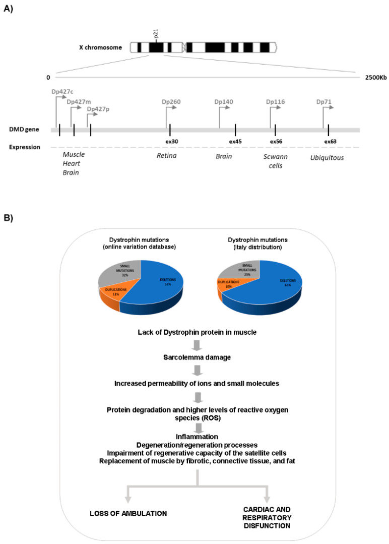
Duchenne muscular dystrophy (DMD) is the most common childhood muscular dystrophy affecting ~1:5000 live male births. Following the identification of pathogenic variations in the dystrophin gene in 1986, the underlining genotype/phenotype correlations emerged and the role of the dystrophin protein was elucidated in skeletal, smooth, and cardiac muscles, as well as in the brain. When the dystrophin protein is absent or quantitatively or qualitatively modified, the muscle cannot sustain the stress of repeated contractions. Dystrophin acts as a bridging and anchoring protein between the sarcomere and the sarcolemma, and its absence or reduction leads to severe muscle damage that eventually cannot be repaired, with its ultimate substitution by connective tissue and fat. The advances of an understanding of the molecular pathways affected in DMD have led to the development of many therapeutic strategies that tackle different aspects of disease etiopathogenesis, which have recently led to the first successful approved orphan drugs for this condition. The therapeutic advances in this field have progressed exponentially, with second-generation drugs now entering in clinical trials as gene therapy, potentially providing a further effective approach to the condition.
Overview
The claims and statistics of this paper haven’t been checked yet.
Run the check — free, no account needed. It reads the open-access text and checks the sources it cites.
Integrity signals
Registry
Cites
Statistics
Figures
Verification
Claims the paper makes
The claims and statistics of this paper haven’t been checked yet.
Run the check — free, no account needed. It reads the open-access text and checks the sources it cites.
Claims other papers make about this one
Each is checked against this paper’s own text: does it say what the citing paper says it does?
The cited source says what the claim attributes to it.
In the citing paper
“Currently, several therapeutic approaches (gene therapy, cell therapy, and exon skipping) have been investigated to restore the expression of dystrophin in DMD muscles.35,36”
From Mechanism and Applications of CRISPR/Cas-9-Mediated Genome Editing (2021) · cites this paper as [35]
In the citing paper
“Despite considerable efforts, the treatment available for DMD remains supportive rather than curative.”
From Mechanism and Applications of CRISPR/Cas-9-Mediated Genome Editing (2021) · cites this paper as [35]
Self-check
Percentages, sums and reported differences re-computed from the paper’s own numbers.
The self-check needs the paper’s full text.
Citations
Cites
CRISPR-Cas systems for editing, regulating and targeting genomes
Sander, J. D., & Joung, J. K. (2014) · Nature Biotechnology · doi:10.1038/nbt.2842 · Cited by 3,153
Improving CRISPR-Cas nuclease specificity using truncated guide RNAs
Fu, Y., Sander, J. D., Reyon, D., Cascio, V. M., & Joung, J. K. (2014) · Nature Biotechnology · doi:10.1038/nbt.2808 · Cited by 1,993
Dystrophin Immunity in Duchenne's Muscular Dystrophy
Mendell, J. R., Campbell, K., Rodino-Klapac, L., Sahenk, Z., Shilling, C., Lewis, S., Bowles, D., Gray, S., Li, C., Galloway, G., et al. (2010) · New England Journal of Medicine · doi:10.1056/nejmoa1000228 · Cited by 655
Very mild muscular dystrophy associated with the deletion of 46% of dystrophin
England, S. B., Nicholson, L. V. B., Johnson, M. A., Forrest, S. M., Love, D. R., Zubrzycka-Gaarn, E. E., Bulman, D. E., Harris, J. B., & Davies, K. E. (1990) · Nature · doi:10.1038/343180a0 · Cited by 623
Gene editing restores dystrophin expression in a canine model of Duchenne muscular dystrophy
Amoasii, L., Hildyard, J. C. W., Li, H., Sanchez-Ortiz, E., Mireault, A., Caballero, D., Harron, R., Stathopoulou, T.-R., Massey, C., Shelton, J. M., et al. (2018) · Science · doi:10.1126/science.aau1549 · Cited by 546
Long-term microdystrophin gene therapy is effective in a canine model of Duchenne muscular dystrophy
Le Guiner, C., Servais, L., Montus, M., Larcher, T., Fraysse, B., Moullec, S., Allais, M., François, V., Dutilleul, M., Malerba, A., et al. (2017) · Nature Communications · doi:10.1038/ncomms16105 · Cited by 246
Research works printed but not linked: 119
- Koenig, 1987, Complete cloning of the Duchenne muscular dystrophy (DMD) cDNA and preliminary genomic organization of the DMD gene in normal and affected individuals, Cell, 50, 509, 10.1016/0092-8674(87)90504-6 · DOI · Paper page
- Muntoni, 2003, Dystrophin and mutations: One gene, several proteins, multiple phenotypes, Lancet Neurol., 2, 731, 10.1016/S1474-4422(03)00585-4 · DOI
- Neri, 2020, The Genetic Landscape of Dystrophin Mutations in Italy: A Nationwide Study, Front. Genet., 11, 131, 10.3389/fgene.2020.00131 · DOI
- Falzarano, 2015, Duchenne Muscular Dystrophy: From Diagnosis to Therapy, Molecules, 20, 18168, 10.3390/molecules201018168 · DOI
- Barseghyan, 2017, Next-generation mapping: A novel approach for detection of pathogenic structural variants with a potential utility in clinical diagnosis, Genome Med., 9, 90, 10.1186/s13073-017-0479-0 · DOI
- Parsons, 2002, Newborn screening for Duchenne muscular dystrophy: A psychosocial study, Arch. Dis. Child. Fetal Neonatal Ed., 86, 91F, 10.1136/fn.86.2.F91 · DOI
- Fraser, 2018, Experiences of Women Who Have Had Carrier Testing for Duchenne Muscular Dystrophy and Becker Muscular Dystrophy During Adolescence, J. Genet. Couns., 27, 1349, 10.1007/s10897-018-0266-0 · DOI
- Breveglieri, 2019, Non-invasive Prenatal Testing Using Fetal DNA, Mol. Diagn. Ther., 23, 291, 10.1007/s40291-019-00385-2 · DOI
- Fokkema, 2006, Entries in the Leiden Duchenne muscular dystrophy mutation database: An overview of mutation types and paradoxical cases that confirm the reading-frame rule, Muscle Nerve, 34, 135, 10.1002/mus.20586 · DOI
- White (2006). S.J.; den Dunnen, J.T. Copy number variation in the genome; the human DMD gene as an example. Cytogenet. Genome Res., 115, 240–246. · DOI
- Batchelor, 2006, Sparks, signals and shock absorbers: How dystrophin loss causes muscular dystrophy, Trends Cell Biol., 16, 198, 10.1016/j.tcb.2006.02.001 · DOI
- Naidoo, 2019, Dystrophin Dp71 and the Neuropathophysiology of Duchenne Muscular Dystrophy, Mol. Neurobiol., 57, 1748, 10.1007/s12035-019-01845-w · DOI
- McElhanon, 2018, Altered membrane integrity in the progression of muscle diseases, Life Sci., 192, 166, 10.1016/j.lfs.2017.11.035 · DOI
- Ding, 2018, An Overview of Muscle Atrophy, Adv. Exp. Med. Biol., 1088, 3, 10.1007/978-981-13-1435-3_1 · DOI
- Boldrin, 2015, Satellite cells from dystrophic muscle retain regenerative capacity, Stem Cell Res., 14, 20, 10.1016/j.scr.2014.10.007 · DOI
- Dumont, 2015, Dystrophin expression in muscle stem cells regulates their polarity and asymmetric division, Nat. Med., 21, 1455, 10.1038/nm.3990 · DOI
- Mukherjee, 2015, Cancer Stem Cell Division: When the Rules of Asymmetry Are Broken, Stem Cells Dev., 24, 405, 10.1089/scd.2014.0442 · DOI
- Wang, 2014, Dystrophin is a tumor suppressor in human cancers with myogenic programs, Nat. Genet., 46, 601, 10.1038/ng.2974 · DOI
- Ruggieri, S., De Giorgis, M., Annese, T., Tamma, R., Notarangelo, A., Marzullo, A., Senetta, R., Cassoni, P., Notarangelo, M., and Ribatti, D. (2019). Dp71 Expression in Human Glioblastoma. Int. J. Mol. Sci., 20. · DOI
- Gavillet, 2006, Cardiac Sodium Channel Na v 1.5 Is Regulated by a Multiprotein Complex Composed of Syntrophins and Dystrophin, Circ. Res., 99, 407, 10.1161/01.RES.0000237466.13252.5e · DOI
- Sadeghi, 2002, Regulation of the cardiac L-type Ca2+ channel by the actin-binding proteins α-actinin and dystrophin, Am. J. Physiol. Cell Physiol., 282, C1502, 10.1152/ajpcell.00435.2001 · DOI
- Koenig, X., Dysek, S., Kimbacher, S., Mike, A.K., Cervenka, R., Lukacs, P., Nagl, K., Dang, X.B., Todt, H., and Bittner, R.E. (2011). Voltage-Gated Ion Channel Dysfunction Precedes Cardiomyopathy Development in the Dystrophic Heart. PLoS ONE, 6. · DOI
- Garbincius, 2015, Dystrophin-glycoprotein complex regulates muscle nitric oxide production through mechanoregulation of AMPK signaling, Proc. Natl. Acad. Sci. USA, 112, 13663, 10.1073/pnas.1512991112 · DOI
- Tidball, 2014, Nitric oxide synthase deficiency and the pathophysiology of muscular dystrophy, J. Physiol., 592, 4627, 10.1113/jphysiol.2014.274878 · DOI
- Lee, 2018, Descriptive Phenotype of Obsessive-Compulsive Symptoms in Males with Duchenne Muscular Dystrophy, J. Child Neurol., 33, 572, 10.1177/0883073818774439 · DOI
- Ricotti, 2016, Neurodevelopmental, emotional, and behavioural problems in Duchenne muscular dystrophy in relation to underlying dystrophin gene mutations, Dev. Med. Child Neurol., 58, 77, 10.1111/dmcn.12922 · DOI
- Doorenweerd, 2017, Timing and localization of human dystrophin isoform expression provide insights into the cognitive phenotype of Duchenne muscular dystrophy, Sci. Rep., 7, 12575, 10.1038/s41598-017-12981-5 · DOI
- Fritschy, 2012, Molecular and functional heterogeneity of GABAergic synapses, Cell. Mol. Life Sci., 69, 2485, 10.1007/s00018-012-0926-4 · DOI
- Daoud, F., Candelario-Martínez, A., Billard, J.-M., Avital, A., Khelfaoui, M., Rozenvald, Y., Guegan, M., Mornet, D., Jaillard, D., and Nudel, U. (2009). Role of Mental Retardation-Associated Dystrophin-Gene Product Dp71 in Excitatory Synapse Organization, Synaptic Plasticity and Behavioral Functions. PLoS ONE, 4. · DOI
- Taylor, P.J., Betts, G.A., Maroulis, S., Gilissen, C., Pedersen, R.L., Mowat, D.R., Johnston, H.M., and Buckley, M.F. (2010). Dystrophin Gene Mutation Location and the Risk of Cognitive Impairment in Duchenne Muscular Dystrophy. PLoS ONE, 5. · DOI
- Lorusso, 2011, Neurocognitive Profiles in Duchenne Muscular Dystrophy and Gene Mutation Site, Pediatr. Neurol., 45, 292, 10.1016/j.pediatrneurol.2011.08.003 · DOI
- Shieh, 2018, Emerging Strategies in the Treatment of Duchenne Muscular Dystrophy, Neurotherapeutics, 15, 840, 10.1007/s13311-018-00687-z · DOI
- Takeshima, 1995, Modulation of in vitro splicing of the upstream intron by modifying an intra-exon sequence which is deleted from the dystrophin gene in dystrophin Kobe, J. Clin. Investig., 95, 515, 10.1172/JCI117693 · DOI
- Pramono, 1996, Induction of exon skipping of the dystrophin transcript in lymphoblastoid cells by transfecting an antisense oligodeoxynucleotide complementary to an exon recognition sequence, Biochem. Biophys. Res. Commun., 226, 445, 10.1006/bbrc.1996.1375 · DOI
- Matsuo, 1991, Exon skipping during splicing of dystrophin mRNA precursor due to an intraexon deletion in the dystrophin gene of Duchenne muscular dystrophy kobe, J. Clin. Investig., 87, 2127, 10.1172/JCI115244 · DOI
- Matsuo, 1990, A very small frame-shifting deletion within exon 19 of the Duchenne muscular dystrophy gene, Biochem. Biophys. Res. Commun., 170, 963, 10.1016/0006-291X(90)92185-3 · DOI
- Nicholson, 1993, The “rescue” of dystrophin synthesis in boys with Duchenne muscular dystrophy, Neuromuscul. Disord., 3, 525, 10.1016/0960-8966(93)90109-W · DOI
- Sherratt, 1993, Exon skipping and translation in patients with frameshift deletions in the dystrophin gene, Am. J. Hum. Genet., 53, 1007
- Kinali, 2010, Revertant fibres and dystrophin traces in Duchenne muscular dystrophy: Implication for clinical trials, Neuromuscul. Disord., 20, 295, 10.1016/j.nmd.2010.03.007 · DOI
- Hoffman, 2011, Restoring dystrophin expression in duchenne muscular dystrophy muscle progress in exon skipping and stop codon read through, Am. J. Pathol., 179, 12, 10.1016/j.ajpath.2011.03.050 · DOI
- Gao, 2015, The Dystrophin Complex: Structure, Function, and Implications for Therapy, Compr. Physiol., 5, 1223, 10.1002/cphy.c140048 · DOI
- Fokkema, 2009, Theoretic applicability of antisense-mediated exon skipping for Duchenne muscular dystrophy mutations, Hum. Mutat., 30, 293, 10.1002/humu.20918 · DOI
- Sun, C., Shen, L., Zhang, Z., and Xie, X. (2020). Therapeutic Strategies for Duchenne Muscular Dystrophy: An Update. Genes, 11. · DOI
- Kuntz, 2020, Casimersen treatment in eligible patients with Duchenne muscular dystrophy: Safety, tolerability, and pharmacokinetics over 144 weeks of treatment, Neuromuscul. Disord., 30, 130, 10.1016/j.nmd.2020.08.285 · DOI
- Frank, 2020, Increased dystrophin production with golodirsen in patients with Duchenne muscular dystrophy, Neurology, 94, e2270, 10.1212/WNL.0000000000009233 · DOI
- Komaki, 2020, Viltolarsen in Japanese Duchenne muscular dystrophy patients: A phase 1/2 study, Ann. Clin. Transl. Neurol., 7, 2393, 10.1002/acn3.51235 · DOI
- Wang, 2018, Saponins as Natural Adjuvant for Antisense Morpholino Oligonucleotides Delivery in vitro and in mdx Mice, Mol. Ther. Nucleic Acids, 11, 192, 10.1016/j.omtn.2018.02.004 · DOI
- Novak, 2018, Author Correction: Myoblasts and macrophages are required for therapeutic morpholino antisense oligonucleotide delivery to dystrophic muscle, Nat. Commun., 9, 1256, 10.1038/s41467-018-03709-8 · DOI
- Tsoumpra, 2019, Peptide-conjugate antisense based splice-correction for Duchenne muscular dystrophy and other neuromuscular diseases, EBioMedicine, 45, 630, 10.1016/j.ebiom.2019.06.036 · DOI
- Sardone, V., Zhou, H., Muntoni, F., Ferlini, A., and Falzarano, M.S. (2017). Antisense Oligonucleotide-Based Therapy for Neuromuscular Disease. Molecules, 22. · DOI
- Aupy, 2019, Evaluating the Impact of Variable Phosphorothioate Content in Tricyclo-DNA Antisense Oligonucleotides in a Duchenne Muscular Dystrophy Mouse Model, Nucleic Acid Ther., 29, 148, 10.1089/nat.2018.0773 · DOI
- McDonald, 2018, Placebo-controlled Phase 2 Trial of Drisapersen for Duchenne Muscular Dystrophy, Ann. Clin. Transl. Neurol., 5, 913, 10.1002/acn3.579 · DOI
- Goemans, 2018, A randomized placebo-controlled phase 3 trial of an antisense oligonucleotide, drisapersen, in Duchenne muscular dystrophy, Neuromuscul. Disord., 28, 4, 10.1016/j.nmd.2017.10.004 · DOI
- Schneider, 2020, Developments in reading frame restoring therapy approaches for Duchenne muscular dystrophy, Expert Opin. Biol. Ther., 19, 1
- Wan, 2014, Synthesis, biophysical properties and biological activity of second generation antisense oligonucleotides containing chiral phosphorothioate linkages, Nucleic Acids Res., 42, 13456, 10.1093/nar/gku1115 · DOI
- Iwamoto, 2017, Control of phosphorothioate stereochemistry substantially increases the efficacy of antisense oligonucleotides, Nat. Biotechnol., 35, 845, 10.1038/nbt.3948 · DOI
- Campbell, 2020, Meta-analyses of ataluren randomized controlled trials in nonsense mutation Duchenne muscular dystrophy, J. Comp. Eff. Res., 9, 973, 10.2217/cer-2020-0095 · DOI
- Berger, 2020, Effect of Ataluren on dystrophin mutations, J. Cell. Mol. Med., 24, 6680, 10.1111/jcmm.15319 · DOI
- Wein, 2018, Personalized gene and cell therapy for Duchenne Muscular Dystrophy, Neuromuscul. Disord., 28, 803, 10.1016/j.nmd.2018.06.009 · DOI
- Galibert, 2011, Latest developments in the large-scale production of adeno-associated virus vectors in insect cells toward the treatment of neuromuscular diseases, J. Invertebr. Pathol., 107, S80, 10.1016/j.jip.2011.05.008 · DOI
- Dulak, 2020, Muscle and cardiac therapeutic strategies for Duchenne muscular dystrophy: Past, present, and future, Pharmacol. Rep., 72, 1227, 10.1007/s43440-020-00134-x · DOI
- Crudele, 2019, AAV-based gene therapies for the muscular dystrophies, Hum. Mol. Genet., 28, R102, 10.1093/hmg/ddz128 · DOI
- Chiu, W., Hsun, Y.-H., Chang, K.-J., Yarmishyn, A.A., Hsiao, Y.-J., Chien, Y., Chien, C.S., Ma, C., Yang, Y.-P., and Tsai, P.-H. (2020). Current Genetic Survey and Potential Gene-Targeting Therapeutics for Neuromuscular Diseases. Int. J. Mol. Sci., 21. · DOI
- Amjad, 2020, Novel genetic therapeutic approaches for modulating the severity of β-thalassemia, Biomed. Rep., 13, 48, 10.3892/br.2020.1355 · DOI
- Ramos, 2019, Development of Novel Mi-cro-dystrophins with Enhanced Functionality, Mol. Ther., 27, 623, 10.1016/j.ymthe.2019.01.002 · DOI
- Watchko, 2002, Adeno-associated virus vector-mediated mini-dystrophin gene therapy improves dystrophic muscle contractile function in mdx mice, Hum. Gene Ther., 13, 1451, 10.1089/10430340260185085 · DOI
- Kornegay, 2010, Widespread Muscle Expression of an AAV9 Human Mini-dystrophin Vector After Intravenous Injection in Neonatal Dystrophin-deficient Dogs, Mol. Ther., 18, 1501, 10.1038/mt.2010.94 · DOI
- Martino, 2011, The genome of self-complementary adeno-associated viral vectors increases Toll-like receptor 9–dependent innate immune responses in the liver, Blood, 117, 6459, 10.1182/blood-2010-10-314518 · DOI · Paper page
- Song, 2019, Non-immunogenic utrophin gene therapy for the treatment of muscular dystrophy animal models, Nat. Med., 25, 1505, 10.1038/s41591-019-0594-0 · DOI
- Thomas, 2016, B4GALNT2 (GALGT2) Gene Therapy Reduces Skeletal Muscle Pathology in the FKRP P448L Mouse Model of Limb Girdle Muscular Dystrophy 2I, Am. J. Pathol., 186, 2429, 10.1016/j.ajpath.2016.05.021 · DOI
- Martin, 2009, Overexpression of Galgt2 in skeletal muscle prevents injury resulting from eccentric contractions in both mdx and wild-type mice, Am. J. Physiol. Physiol., 296, C476, 10.1152/ajpcell.00456.2008 · DOI
- Chicoine, 2014, Vascular delivery of rAAVrh74.MCK.GALGT2 to the gastrocnemius muscle of the rhesus macaque stimulates the expression of dystrophin and laminin α2 surrogates, Mol. Ther., 22, 713, 10.1038/mt.2013.246 · DOI
- Xu, 2019, rAAVrh74.MCK.GALGT2 Protects against Loss of Hemodynamic Function in the Aging mdx Mouse Heart, Mol. Ther., 27, 636, 10.1016/j.ymthe.2019.01.005 · DOI
- Gaj, 2013, ZFN, TALEN, and CRISPR/Cas-based methods for genome engineering, Trends Biotechnol., 31, 397, 10.1016/j.tibtech.2013.04.004 · DOI · Paper page
- Cox, 2015, Therapeutic genome editing: Prospects and challenges, Nat. Med., 21, 121, 10.1038/nm.3793 · DOI · Paper page
- Epinat, 2003, A novel engineered meganuclease induces homologous recombination in yeast and mammalian cells, Nucleic Acids Res., 31, 2952, 10.1093/nar/gkg375 · DOI
- Urnov, 2010, Genome editing with engineered zinc finger nucleases, Nat. Rev. Genet., 11, 636, 10.1038/nrg2842 · DOI · Paper page
- Miller, 2011, A TALE nuclease architecture for efficient genome editing, Nat. Biotechnol., 29, 143, 10.1038/nbt.1755 · DOI · Paper page
- Jinek, 2012, A Programmable Dual-RNA-Guided DNA Endonuclease in Adaptive Bacterial Immunity, Science, 337, 816, 10.1126/science.1225829 · DOI · Paper page
- Ye, 2014, Seamless modi-fication of wild-type induced pluripotent stem cells to the natural CCR5Δ32 mutation confers resistance to HIV infection, Proc. Natl. Acad. Sci. USA, 111, 9591, 10.1073/pnas.1407473111 · DOI · Paper page
- Long, 2016, Postnatal genome editing partially restores dystrophin expression in a mouse model of muscular dystrophy, Science, 351, 400, 10.1126/science.aad5725 · DOI
- Amoasii, 2017, Single-cut genome editing restores dystrophin expression in a new mouse model of muscular dystrophy, Sci. Transl. Med., 9, eaan8081, 10.1126/scitranslmed.aan8081 · DOI · Paper page
- Sicinski, 1989, The molecular basis of muscular dystrophy in the mdx mouse: A point mutation, Science, 244, 1578, 10.1126/science.2662404 · DOI · Paper page
- Nelson, 2015, In vivo genome editing improves muscle function in a mouse model of Duchenne muscular dystrophy, Science, 351, 403, 10.1126/science.aad5143 · DOI · Paper page
- Tabebordbar, 2015, In vivo gene editing in dystrophic mouse muscle and muscle stem cells, Science, 351, 407, 10.1126/science.aad5177 · DOI · Paper page
- Xu, 2017, In Vivo Genome Editing Restores Dystrophin Expression and Cardiac Function in Dystrophic Mice, Circ. Res., 121, 923, 10.1161/CIRCRESAHA.117.310996 · DOI
- Min, 2019, CRISPR-Cas9 corrects Duchenne muscular dystrophy exon 44 deletion mutations in mice and human cells, Sci. Adv., 5, eaav4324, 10.1126/sciadv.aav4324 · DOI
- Chemello, 2020, Correction of muscular dystrophies by CRISPR gene editing, J. Clin. Investig., 130, 2766, 10.1172/JCI136873 · DOI
- Hakim, C.H., Wasala, N.B., Nelson, C.E., Wasala, L.P., Yue, Y., Louderman, J.A., Lessa, T.B., Dai, A., Zhang, K., and Jenkins, G.J. (2018). AAV CRISPR editing rescues cardiac and muscle function for 18 months in dystrophic mice. JCI Insight, 3. · DOI
- Nelson, 2019, Long-term evaluation of AAV-CRISPR genome editing for Duchenne muscular dystrophy, Nat. Med., 25, 427, 10.1038/s41591-019-0344-3 · DOI
- Chew, 2016, A multi-functional AAV-CRISPR-Cas9 and its host response, Nat. Methods, 13, 868, 10.1038/nmeth.3993 · DOI · Paper page
- Cho, 2014, Analysis of off-target effects of CRISPR/Cas-derived RNA-guided endonucleases and nickases, Genome Res., 24, 132, 10.1101/gr.162339.113 · DOI · Paper page
- Matre, 2019, CRISPR/Cas9-Based Dystrophin Restoration Reveals a Novel Role for Dystrophin in Bioenergetics and Stress Resistance of Muscle Progenitors, Stem Cells, 37, 1615, 10.1002/stem.3094 · DOI
- Chang, 2018, The Dystrophin Glycoprotein Complex Regulates the Epigenetic Activation of Muscle Stem Cell Commitment, Cell Stem Cell, 22, 755, 10.1016/j.stem.2018.03.022 · DOI
- Sun, 2020, Stem cell-based therapies for Duchenne muscular dystrophy, Exp. Neurol., 323, 113086, 10.1016/j.expneurol.2019.113086 · DOI
- Fukada, 2018, The roles of muscle stem cells in muscle injury, atrophy and hypertrophy, J. Biochem., 163, 353, 10.1093/jb/mvy019 · DOI
- Huard, 2019, Stem cells, blood vessels, and angiogenesis as major determinants for musculoskeletal tissue repair, J. Orthop. Res., 37, 1212, 10.1002/jor.24058 · DOI
- Cossu, 2015, Intra-arterial transplantation of HLA-matched donor mesoangioblasts in Duchenne muscular dystrophy, EMBO Mol. Med., 7, 1513, 10.15252/emmm.201505636 · DOI
- Angelis, 1999, Skeletal Myogenic Progenitors Originating from Embryonic Dorsal Aorta Coexpress Endothelial and Myogenic Markers and Contribute to Postnatal Muscle Growth and Regeneration, J. Cell Biol., 147, 869, 10.1083/jcb.147.4.869 · DOI
- Minasi, 2002, The meso-angioblast: A multipotent, self-renewing cell that originates from the dorsal aorta and differentiates into most mesodermal tissues, Development, 129, 2773, 10.1242/dev.129.11.2773 · DOI · Paper page
- Dellavalle, 2011, Pericytes resident in postnatal skeletal muscle differentiate into muscle fibres and generate satellite cells, Nat. Commun., 2, 499, 10.1038/ncomms1508 · DOI
- Dellavalle, 2007, Pericytes of human skeletal muscle are myogenic precursors distinct from satellite cells, Nat. Cell Biol., 9, 255, 10.1038/ncb1542 · DOI
- Sampaolesi, 2003, Cell Therapy of -Sarcoglycan Null Dystrophic Mice Through Intra-Arterial Delivery of Mesoangioblasts, Science, 301, 487, 10.1126/science.1082254 · DOI
- Sampaolesi, 2006, Mesoangioblast stem cells ameliorate muscle function in dystrophic dogs, Nat. Cell Biol., 444, 574
- Taylor, 2019, Cardiac and skeletal muscle effects in the randomized HOPE-Duchenne trial, Neurology, 92, e866, 10.1212/WNL.0000000000006950 · DOI
- Takeda, 2017, Skeletal muscle generated from induced pluripotent stem cells—Induction and application, World J. Stem Cells, 9, 89
- Abujarour, 2014, Myogenic differentiation of muscular dystrophy-specific induced pluripotent stem cells for use in drug discovery, Stem Cells Transl. Med., 3, 149, 10.5966/sctm.2013-0095 · DOI
- Shoji, 2015, Early pathogenesis of Duchenne muscular dystrophy modelled in patient-derived human induced pluripotent stem cells, Sci. Rep., 5, 12831, 10.1038/srep12831 · DOI
- Choi, 2016, Concordant but Varied Phenotypes among Duchenne Muscular Dystrophy Patient-Specific Myoblasts Derived using a Human iPSC-Based Model, Cell Rep., 15, 2301, 10.1016/j.celrep.2016.05.016 · DOI
- Young, 2016, A Single CRISPR-Cas9 Deletion Strategy that Targets the Majority of DMD Patients Restores Dystrophin Function in hiPSC-Derived Muscle Cells, Cell Stem Cell, 18, 533, 10.1016/j.stem.2016.01.021 · DOI
- Uchimura, 2017, A human iPS cell myogenic differentiation system permitting high-throughput drug screening, Stem Cell Res., 25, 98, 10.1016/j.scr.2017.10.023 · DOI
- Maffioletti, 2018, Three-Dimensional Human iPSC-Derived Artificial Skeletal Muscles Model Muscular Dystrophies and Enable Multilineage Tissue Engineering, Cell Rep., 23, 899, 10.1016/j.celrep.2018.03.091 · DOI
- Perlingeiro, 2018, Recapitulating muscle disease phenotypes with myotonic dystrophy 1 induced pluripotent stem cells: A tool for disease modeling and drug discovery, Dis. Model. Mech., 11, dmm034728, 10.1242/dmm.034728 · DOI
- Cordova, 2018, Combined Therapies for Duchenne Muscular Dystrophy to Optimize Treatment Efficacy, Front. Genet., 9, 114, 10.3389/fgene.2018.00114 · DOI
- Tinsley, 1998, Expression of full-length utrophin prevents muscular dystrophy in mdx mice, Nat. Med., 4, 1441, 10.1038/4033 · DOI
- Guiraud, 2015, Second-generation compound for the modulation of utrophin in the therapy of DMD, Hum. Mol. Genet., 24, 4212, 10.1093/hmg/ddv154 · DOI
- Moorwood, C., Lozynska, O., Suri, N., Napper, A.D., Diamond, S.L., and Khurana, T.S. (2011). Drug Discovery for Duchenne Muscular Dystrophy via Utrophin Promoter Activation Screening. PLoS ONE, 6. · DOI
- Spinazzola, 2016, Pharmacological therapeutics targeting the secondary defects and downstream pathology of Duchenne muscular dystrophy, Expert Opin. Orphan Drugs, 4, 1179, 10.1080/21678707.2016.1240613 · DOI
- Guiraud, 2017, Pharmacological advances for treatment in Duchenne muscular dystrophy, Curr. Opin. Pharmacol., 34, 36, 10.1016/j.coph.2017.04.002 · DOI
Cited by
Sorted by citations
Mechanism and Applications of CRISPR/Cas-9-Mediated Genome Editing
Mengstie, M. A., & Wondimu, B. Z. (2021) · Biologics: Targets and Therapy · doi:10.2147/btt.s326422 · Cited by 478
Metadata
- Authors
- Fortunato, F., Rossi, R., Falzarano, M. S., & Ferlini, A.
- Affiliations
- Department of Medical Sciences, University of Ferrara, 44121 Ferrara, Italy
- Venue
- Journal of Clinical Medicine
- Publisher
- MDPI AG
- Published
- 2021-02-17
- Volume
- 10
- Issue
- 4
- Pages
- 820
- ISSN
- 2077-0383
- PMC
- PMC7922390
- Cited by
- 64
- References
- 6
- Language
- en
About these checks
Registry, retraction, Cites and statistics results are computed, not judged, with one exception: a printed reference that no index lists is read once by a model before it counts as a citation error. Claims are extracted by a model and kept only when their quote appears verbatim in the text; each cited source is then checked, with a model’s help, for whether it says what the claim relies on. Those verdicts can be wrong — read the claim’s quote and the cited source before relying on one.
Statistics and arithmetic are recomputed, not judged.
Confidence bands are shown only for fields calibrated on our annotated test set. None is calibrated yet, so no band is shown.
What counts as a claim ›How we measure confidence ›How we check this ›
Are you an author, or do you see a problem with this page? Request a review
How to cite
Cite CoChat for the checks on this page, and the paper for its own findings.
- This page
- CoChat. (2026). Citation check: Innovative Therapeutic Approaches for Duchenne Muscular Dystrophy. https://app.cochat.ai/papers/10.3390/jcm10040820
- The paper
- Fortunato, F., Rossi, R., Falzarano, M. S., & Ferlini, A. (2021). Innovative Therapeutic Approaches for Duchenne Muscular Dystrophy. Journal of Clinical Medicine. https://doi.org/10.3390/jcm10040820
