

The DOI matches its Crossref record; 89 of the 89 references it cites were found in a registry or an index.
Subretinal Injection Techniques for Retinal Disease: A Review
Open accessAbstract
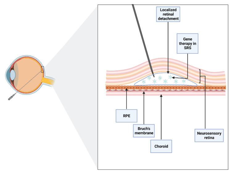
Inherited retinal dystrophies (IRDs) affect an estimated 1 in every 2000 people, this corresponding to nearly 2 million cases worldwide. Currently, 270 genes have been associated with IRDs, most of them altering the function of photoreceptors and retinal pigment epithelium. Gene therapy has been proposed as a potential tool for improving visual function in these patients. Clinical trials in animal models and humans have been successful in various types of IRDs. Recently, voretigene neparvovec (Luxturna®) has been approved by the US Food and Drug Administration for the treatment of biallelic mutations in the RPE65 gene. The current state of the art in gene therapy involves the delivery of various types of viral vectors into the subretinal space to effectively transduce diseased photoreceptors and retinal pigment epithelium. For this, subretinal injection is becoming increasingly popular among researchers and clinicians. To date, several approaches for subretinal injection have been described in the scientific literature, all of them effective in accessing the subretinal space. The growth and development of gene therapy give rise to the need for a standardized procedure for subretinal injection that ensures the efficacy and safety of this new approach to drug delivery. The goal of this review is to offer an insight into the current subretinal injection techniques and understand the key factors in the success of this procedure.
Overview
The claims and statistics of this paper haven’t been checked yet.
Run the check — free, no account needed. It reads the open-access text and checks the sources it cites.
Integrity signals
Registry
Cites
Statistics
Figures
Verification
Claims the paper makes
The claims and statistics of this paper haven’t been checked yet.
Run the check — free, no account needed. It reads the open-access text and checks the sources it cites.
Claims other papers make about this one
Each is checked against this paper’s own text: does it say what the citing paper says it does?
In the citing paper
“The subretinal space is thought to be anatomically more closed and immune-privileged than the intravitreal space (Irigoyen et al., 2022).”
From AAV2 and AAV9 tropism and transgene expression in the mouse eye and major tissues after intravitreal and subretinal delivery (2023) · cites this paper as Irigoyen et al., 2022
In the citing paper
“IVT injection potentially exposes the entire retina to gene therapy (Koponen et al., 2021; Puranen et al., 2022), whereas delivery to subretinal space allows direct contact of the vector with photoreceptors and the RPE layers (Irigoyen et al., 2022).”
From AAV2 and AAV9 tropism and transgene expression in the mouse eye and major tissues after intravitreal and subretinal delivery (2023) · cites this paper as Irigoyen et al., 2022
Self-check
Percentages, sums and reported differences re-computed from the paper’s own numbers.
The self-check needs the paper’s full text.
Citations
Cites
Russell, S., Bennett, J., Wellman, J. A., Chung, D. C., Yu, Z.-F., Tillman, A., Wittes, J., Pappas, J., Elci, O., McCague, S., et al. (2017) · The Lancet · doi:10.1016/s0140-6736(17)31868-8 · Cited by 1,952
Effect of gene therapy on visual function in Leber’s congenital amaurosis
Bainbridge, J. W. B., Smith, A. J., Barker, S. S., Robbie, S., Henderson, R., Balaggan, K., Viswanathan, A., Holder, G. E., Stockman, A., Tyler, N., et al. (2008) · New England Journal of Medicine · doi:10.1056/nejmoa0802268 · Cited by 1,924
Schwartz, S. D., Regillo, C. D., Lam, B. L., Eliott, D., Rosenfeld, P. J., Gregori, N. Z., Hubschman, J.-P., Davis, J. L., Heilwell, G., Spirn, M., et al. (2015) · The Lancet · doi:10.1016/s0140-6736(14)61376-3 · Cited by 1,327
Gene therapy restores vision in a canine model of childhood blindness
Acland, G. M., Aguirre, G. D., Ray, J., Zhang, Q., Aleman, T. S., Cideciyan, A. V., Pearce-Kelling, S. E., Anand, V., Zeng, Y., Maguire, A. M., et al. (2001) · Nature Genetics · doi:10.1038/ng0501-92 · Cited by 1,202 · cited 1×
“The vector was injected using a 30G anterior chamber cannula through a pars plana approach [35].”
MethodMaguire, A. M., High, K. A., Auricchio, A., Wright, J. F., Pierce, E. A., Testa, F., Mingozzi, F., Bennicelli, J. L., Ying, G.-S., Rossi, S., et al. (2009) · The Lancet · doi:10.1016/s0140-6736(09)61836-5 · Cited by 865
MacLaren, R. E., Groppe, M., Barnard, A. R., Cottriall, C. L., Tolmachova, T., Seymour, L., Clark, K. R., During, M. J., Cremers, F. P. M., Black, G. C. M., et al. (2014) · The Lancet · doi:10.1016/s0140-6736(13)62117-0 · Cited by 796
Pharmacokinetic aspects of retinal drug delivery
del Amo, E. M., Rimpelä, A.-K., Heikkinen, E., Kari, O. K., Ramsay, E., Lajunen, T., Schmitt, M., Pelkonen, L., Bhattacharya, M., Richardson, D., et al. (2017) · Progress in Retinal and Eye Research · doi:10.1016/j.preteyeres.2016.12.001 · Cited by 693
The molecular basis of human retinal and vitreoretinal diseases
Berger, W., Kloeckener‐Gruissem, B., & Neidhardt, J. (2010) · Progress in Retinal and Eye Research · doi:10.1016/j.preteyeres.2010.03.004 · Cited by 609
Stable and efficient gene transfer into the retina using an HIV-based lentiviral vector
Miyoshi, H., Takahashi, M., Gage, F. H., & Verma, I. M. (1997) · Proceedings of the National Academy of Sciences · doi:10.1073/pnas.94.19.10319 · Cited by 497
Varela‐Fernández, R., Díaz-Tomé, V., Luaces-Rodríguez, A., Conde-Penedo, A., García‐Otero, X., Álvarez, A. L., Fernández‐Ferreiro, A., & Otero-Espinar, F. J. (2020) · Pharmaceutics · doi:10.3390/pharmaceutics12030269 · Cited by 374
Cited by
Sorted by citations
Overview of Recent Advances in Nano-Based Ocular Drug Delivery
Liu, L.-C., Chen, Y., & Lu, D. (2023) · International Journal of Molecular Sciences · doi:10.3390/ijms242015352 · Cited by 152
Near-infrared spatiotemporal color vision in humans enabled by upconversion contact lenses
Ma, Y., Chen, Y., Wang, S., Chen, Z., Zhang, Y., Huang, L. F., Zhang, X., Yin, F., Wang, Y., Yang, M., et al. (2025) · Cell · doi:10.1016/j.cell.2025.04.019 · Cited by 55
Next generation therapeutics for retinal neurodegenerative diseases
Appell, M. B., Pejavar, J., Pasupathy, A., Rompicharla, S. V. K., Abbasi, S., Malmberg, K., Kolodziejski, P., & Ensign, L. M. (2024) · Journal of Controlled Release · doi:10.1016/j.jconrel.2024.01.063 · Cited by 33
Tripepi, D., Jalil, A., Ally, N., Buzzi, M., Moussa, G., Rothschild, P.-R., Rossi, T., Ferrara, M., & Romano, M. R. (2023) · International Journal of Molecular Sciences · doi:10.3390/ijms241310535 · Cited by 31
Biomechanical considerations for optimising subretinal injections
L’Abbate, D., Prescott, K., Geraghty, B., Kearns, V., & STEEL, D. H. (2024) · Survey of Ophthalmology · doi:10.1016/j.survophthal.2024.05.004 · Cited by 26
Beyond the injection: delivery systems reshaping retinal disease management
Rowe, L. W., Akotoye, C., Harris, A., & Ciulla, T. A. (2025) · Expert Opinion on Pharmacotherapy · doi:10.1080/14656566.2025.2496424 · Cited by 22
Chen, K.-S., Chan, H.-C., & Chan, C.-M. (2025) · Drugs · doi:10.1007/s40265-025-02237-2 · Cited by 22
Koponen, S., Kokki, E., Tamminen, T., & Ylä-Herttuala, S. (2023) · Frontiers in Drug Delivery · doi:10.3389/fddev.2023.1148795 · Cited by 21
Seeing the Future: A Review of Ocular Therapy
Whalen, M., Akula, M., McNamee, S. M., DeAngelis, M. M., & Haider, N. B. (2024) · Bioengineering · doi:10.3390/bioengineering11020179 · Cited by 21
Malvasi, M., Casillo, L., Avogaro, F., Abbouda, A., & Vingolo, E. M. (2023) · International Journal of Molecular Sciences · doi:10.3390/ijms241813756 · Cited by 21
Novel Approaches in the Drug Development and Delivery Systems for Age-Related Macular Degeneration
Paliwal, H., Prajapati, B. G., Srichana, T., Singh, S. K., & Patel, R. J. (2023) · Life · doi:10.3390/life13020568 · Cited by 21
Choi, S. W., Seo, S., Hong, H. K., Yoon, S. J., Kim, M., Moon, S., Lee, J. Y., Lim, J., Lee, J. B., & Woo, S. J. (2023) · Tissue Engineering and Regenerative Medicine · doi:10.1007/s13770-023-00555-8 · Cited by 18
Xiao, K., Jiang, H., Li, Q., Luo, S., Qin, Y., Li, J., Xie, Q., & Zheng, Q. (2023) · Molecular Therapy · doi:10.1016/j.ymthe.2023.09.019 · Cited by 17
MacLaren, R. E., Audo, I., Fischer, M. D., Huckfeldt, R. M., Lam, B. L., Pennesi, M. E., Sisk, R., Gow, J. A., Li, J., Zhu, K., et al. (2024) · Human Gene Therapy · doi:10.1089/hum.2024.017 · Cited by 15
A Comparative Analysis of Models for AAV-Mediated Gene Therapy for Inherited Retinal Diseases
Alsalloum, A., Gornostal, E. A., Mingaleva, N., Pavlov, R. E., Kuznetsova, E., Antonova, E., Nadzhafova, A., Колотова, Д. Е., Кадышев, В. В., Mityaeva, O. N., et al. (2024) · Cells · doi:10.3390/cells13201706 · Cited by 17
Real-Time Deformation-Aware Control for Autonomous Robotic Subretinal Injection Under iOCT Guidance
Arikan, D., Zhang, P., Sommersperger, M., Dehghani, S., Esfandiari, M., Taylor, R. H., Nasseri, M. A., Gehlbach, P. L., Navab, N., & Iordachita, I. I. (2025) · Proceedings - IEEE International Conference on Robotics and Automation/Proceedings · doi:10.1109/icra55743.2025.11128595 · Cited by 14
Pharmacotherapy and Nutritional Supplements for Neovascular Eye Diseases
Rusciano, D., & Bagnoli, P. (2023) · Medicina · doi:10.3390/medicina59071334 · Cited by 14
Cao, Y., Yin, X., Wu, L., Huang, D., Wang, Z., Wu, F., Jiang, J., Chen, G., & Wang, Q. (2025) · Advanced Materials · doi:10.1002/adma.202500623 · Cited by 12
Recent advances and future challenges in nanosystems for ocular drug delivery
Shahror, R. A., & Fouda, A. Y. (2025) · Journal of Pharmacology and Experimental Therapeutics · doi:10.1016/j.jpet.2025.103738 · Cited by 12
Advances and Challenges in Gene Therapy for Inherited Retinal Dystrophies: A Comprehensive Review
Jain, R., & Daigavane, S. V. (2024) · Cureus · doi:10.7759/cureus.69895 · Cited by 14
Head-mounted surgical robots are an enabling technology for subretinal injections
Posselli, N. R., Hwang, E. S., Olson, Z. J., Nagiel, A., Bernstein, P. S., & Abbott, J. J. (2025) · Science Robotics · doi:10.1126/scirobotics.adp7700 · Cited by 12
Chronic In Vivo CRISPR-Cas Genome Editing: Challenges, Long-Term Safety, and Outlook
Bao, C., Channell, C. I., Tseng, Y.-H., Bailey, J., Sbaiti, N., Demirkol, A., & Tsang, S. H. (2026) · Cells · doi:10.3390/cells15020156 · Cited by 14
New Directions for Ophthalmic OCT – Handhelds, Surgery, and Robotics
Foust, J. S., McCloud, M., Narawane, A. M., Trout, R. M., Chen, X., Dhalla, A.-H. Z., Li, J. D., Viehland, C., Draelos, M., Vajzovic, L. M., et al. (2025) · Translational Vision Science & Technology · doi:10.1167/tvst.14.1.14 · Cited by 11
Swirski, S., May, O., Ahlers, M. T., Wissinger, B., Greschner, M., Jüschke, C., & Neidhardt, J. (2023) · Cells · doi:10.3390/cells12060955 · Cited by 10
Sadeghi, E., Mohan, S., Iannetta, D., & Chhablani, J. K. (2023) · Expert Review of Medical Devices · doi:10.1080/17434440.2023.2231841 · Cited by 10
Subretinal Gene Therapy for Treatment of Retinal and Choroidal Vascular Diseases
Khanani, A. M., Aziz, A. A., Khanani, Z. A., Khan, H., Mojumder, O., Sulahria, H., Khanani, I., Khan, H., Gahn, G. M., & Mishra, K. (2024) · American Journal of Ophthalmology · doi:10.1016/j.ajo.2024.12.002 · Cited by 9
Co-delivery of antioxidants and siRNA-VEGF: promising treatment for age-related macular degeneration
Dias, M. F., da Cruz-Cazarim, E. L. C., Pittella, F., Baião, A., Pacheco, C., Sarmento, B. F. C. C., & Fialho, S. L. (2025) · Drug Delivery and Translational Research · doi:10.1007/s13346-024-01772-x · Cited by 9
Towards Motion Compensation in Autonomous Robotic Subretinal Injections
Arikan, D., Esfandiari, M., Zhang, P., Sommersperger, M., Dehghani, S., Taylor, R. H., Nasseri, M. A., Gehlbach, P. L., Navab, N., & Iordachita, I. I. (2025) · International Symposium on Medical Robotics · doi:10.1109/ismr67322.2025.11025990 · Cited by 9
Im, J., Park, S., & Song, C. O. (2023) · International Journal of Optomechatronics · doi:10.1080/15599612.2023.2299023 · Cited by 9
Zhao, X., Zhao, Q., Wang, E., Li, N., Meng, L., Zhang, W., Wang, T., Chen, Y., & Min, H. (2023) · Eye and Vision · doi:10.1186/s40662-023-00326-5 · Cited by 8
Optical coherence tomography for noninvasive monitoring of drug delivery
Aglyamov, S. R., & Larin, K. V. (2025) · Advanced Drug Delivery Reviews · doi:10.1016/j.addr.2025.115571 · Cited by 8
Feasibility of direct vitrectomy-sparing subretinal injection for gene delivery in large animals
Stranak, Z., Ardan, T., Nemesh, Y., Toms, M., Toualbi, L., Harbottle, R., Ellederova, Z., Lytvynchuk, L., Petrovski, G., Motlik, J., et al. (2024) · Current Eye Research · doi:10.1080/02713683.2024.2343335 · Cited by 7
Marie, M., Churet, L., Gautron, A.-S., Farjo, R. A., Mizuyoshi, K., Stevenson, V. A., Khabou, H., Léveillard, T., Sahel, J., & Lorget, F. (2025) · Gene Therapy · doi:10.1038/s41434-025-00556-3 · Cited by 7
A soft micron accuracy robot design and clinical validation for retinal surgery
Chen, Y., Guo, X., Ye, X., Jiang, T., Chen, H., Yu, J., Yang, G., Knoll, A., Cui, D., Zhou, M., et al. (2025) · Microsystems & Nanoengineering · doi:10.1038/s41378-025-01002-5 · Cited by 7
Design and analyses of tendon-driven continuum robots for minimally invasive surgery
Varga, M., Sinčák, P. J., Merva, T., Marcinko, P., Miková, Ľ., Prada, E., Schnitzer, M., & Virgala, I. (2024) · IFAC-PapersOnLine · doi:10.1016/j.ifacol.2024.07.366 · Cited by 7
Development of a novel SupraChoroidal-to-Optic-NervE (SCONE) drug delivery system
Chiang, B., Heng, K., Jang, K., Dalal, R., Liao, Y. J., Myung, D. J., & Goldberg, J. L. (2024) · Drug Delivery · doi:10.1080/10717544.2024.2379369 · Cited by 6
Wang, Q. K., Xu, X., Chen, S., Lu, R., Li, L., Lo, C., Liu, Z., Ning, K., Li, T., Kowal, T. J., et al. (2024) · Science Advances · doi:10.1126/sciadv.adn7540 · Cited by 6
“Blindness” is not a contraindication for voretigene neparvovec-rzyl treatment: a review of 9 cases
Parameswarappa, D. C., Stephenson, K. A. J., Seamone, M. E., Qian, C. X., Muni, R. H., Kertes, P. J., Vincent, A., & Héon, E. (2025) · Canadian Journal of Ophthalmology · doi:10.1016/j.jcjo.2024.12.013 · Cited by 6
Latest trends & strategies in ocular drug delivery
Kulkarni, N. S., Josowitz, A. D., James, R., Liu, Y., Rayaprolu, B. M., Sagdullaev, B. T., Bhalla, A. S., & Shameem, M. (2025) · Methods · doi:10.1016/j.ymeth.2025.02.003 · Cited by 7
Parsons, N., Annamalai, B., & Rohrer, B. (2023) · Translational Vision Science & Technology · doi:10.1167/tvst.12.7.17 · Cited by 6
Cai, Y., Gu, Y., Zhang, J., Zhu, Y. Y., Ma, Z., He, Q., Sun, Y. J., Yuan, M., Li, X., Zhu, K., et al. (2025) · Research · doi:10.34133/research.0717 · Cited by 5
Jiang, Y. Z., Tang, Y., Liu, Z., Yang, X., Zhou, M.-Y., Liang, Y., Ding, J., & Liao, Y. (2025) · Inflammopharmacology · doi:10.1007/s10787-025-02022-3 · Cited by 5
Wu, K. Y., Osman, R. M., Esomchukwu, O., Marchand, M., Nguyen, B. H., & Tran, S. D. (2025) · Advances in experimental medicine and biology · doi:10.1007/5584_2025_854 · Cited by 5
Nardella, M., Pellegrini, M., Yu, A. C., Adamo, G., Mura, M., & Busin, M. (2025) · Biomolecules · doi:10.3390/biom15111592 · Cited by 4
Butt, F. R., Dhivagaran, T., Li, B., Ashamalla, M., Tao, B. K.-L., Balas, M., Pereira, A., Yan, P., & Arjmand, P. (2025) · Journal of Personalized Medicine · doi:10.3390/jpm15120619 · Cited by 6
Techniques for subretinal injections in animals
Boyd, R. F., & Petersen‐Jones, S. M. (2024) · Veterinary Ophthalmology · doi:10.1111/vop.13219 · Cited by 4
Metabolic plasticity in a Pde6bSTOP/STOP retinitis pigmentosa mouse model following rescue
Ayten, M., Díaz‐Lezama, N., Ghanawi, H., Haffelder, F. C., Kajtna, J., Straub, T., Borsò, M., Imhof, A., Hauck, S. M., & Koch, S. (2024) · Molecular Metabolism · doi:10.1016/j.molmet.2024.101994 · Cited by 4
How Advanced are Nanocarriers for Effective Subretinal Injection?
Guan, J.-X., Wang, Y.-L., & Wang, J. (2024) · International Journal of Nanomedicine · doi:10.2147/ijn.s479327 · Cited by 4
Tayyib, A., Parameswarappa, D. C., Kertes, P. J., Muni, R. H., Tumber, A. K., Costain, G., Schramm, A., MacDonald, H. R., Klatt, R., Vincent, A., et al. (2025) · Canadian Journal of Ophthalmology · doi:10.1016/j.jcjo.2024.12.006 · Cited by 4
Kwok, J., Gray, A. P., Sato, Y., Savina, S. V., Dolgova, N., Sudharsan, R., Boye, S. L., Boye, S. L., Reichenbacker, S., Meyer, T., et al. (2025) · Drug Delivery and Translational Research · doi:10.1007/s13346-025-01929-2 · Cited by 3
Robotic augmentation of surgical skills in subretinal injection: a novel semiautomated system
Lian, Z., Xu, A., Li, Y., Liu, J., Song, R., Liang, H., Wang, R., Yang, Y., Wei, X., Zhang, Z., et al. (2025) · International Journal of Surgery · doi:10.1097/js9.0000000000004501 · Cited by 3
Djaksigulova, D., Kaad, S. G., Korecki, A. J., Lam, S. L., Lengyell, T. C., & Simpson, E. M. (2026) · Gene Therapy · doi:10.1038/s41434-026-00605-5 · Cited by 3
Paoletti, L., Clerck, K. D., Coster, E. D., Matricardi, P., Remaut, K., & Peynshaert, K. (2025) · Drug Delivery and Translational Research · doi:10.1007/s13346-025-01921-w · Cited by 3
Sub-Retinal Delivery of Human Embryonic Stem Cell Derived Photoreceptor Progenitors in rd10 Mice
Tun, S. B. B., Shepherdson, E., Tay, H. G., & Barathi, V. A. (2023) · Journal of Visualized Experiments · doi:10.3791/65848 · Cited by 3
Stem cell therapy in retinal diseases: current evidence
Rubinato, L., Zan, E. R. D., Sarao, V., Veritti, D., & Lanzetta, P. (2023) · Expert Review of Ophthalmology · doi:10.1080/17469899.2023.2296895 · Cited by 3
CLINICAL FEATURES AND OUTCOMES OF LATE RECURRENT RHEGMATOGENOUS RETINAL DETACHMENT
Ghalibafan, S., Volante, V. A. S., Kim, C. K., Rohowetz, L. J., Flynn, H. W., & Yannuzzi, N. A. (2025) · Retina · doi:10.1097/iae.0000000000004472 · Cited by 2
Advances in technical methods and applications of subretinal injections in experimental animals
Song, C., Ji, Y., Wang, Y., & Yang, W. (2025) · Frontiers in Veterinary Science · doi:10.3389/fvets.2025.1574519 · Cited by 2
Balogh, B., Zille, M., Szarka, G., Péntek, L., Futácsi, A., Völgyi, B., & Kovács‐Öller, T. (2025) · Scientific Reports · doi:10.1038/s41598-025-09007-w · Cited by 2
Tropism and Retinal Transduction Efficiency of Adeno-Associated Virus Serotypes in Mice
Wu, W., McRae, J., Brown, A., & Agbaga, M. (2025) · Investigative Ophthalmology & Visual Science · doi:10.1167/iovs.66.12.18 · Cited by 2
Lipid nanoparticle mediated delivery of dsDNA to the murine retina
Fehrman, R. L., Sahel, D. K., Sahay, G., Lipinski, D. M., & Ryals, R. C. (2025) · Journal of Controlled Release · doi:10.1016/j.jconrel.2025.114194 · Cited by 2
Fehrman, R. L., & Lipinski, D. M. (2025) · Translational Vision Science & Technology · doi:10.1167/tvst.14.11.38 · Cited by 2
mRNA therapy: A novel approach for retinal neurodegenerative diseases
Zhang, B., Tang, J., Zhang, Q., Qiu, Y., Cheng, Y., Yu, S., Liu, L., Xue, J., Liu, X., He, Q., et al. (2026) · Neural Regeneration Research · doi:10.4103/nrr.nrr-d-25-01306 · Cited by 2
Extracellular vesicles in ophthalmology: innovations in diagnosis, therapy, and drug delivery
Aziziyan, F., Mahdipour, M., Hadi, B., Asl, S. S., Afkhami, H., & Sheykhhasan, M. (2026) · Molecular Biology Reports · doi:10.1007/s11033-026-11575-7 · Cited by 2
mRNA delivery to the retina restores REP1 function in choroideremia
de Lemos, L., Antas, P., Carvalho, C., Castro, M., Bruno, D., Fonseca, A. F., Rakshit, S., & Seabra, M. C. (2026) · Molecular Therapy Advances · doi:10.1016/j.omta.2026.201768 · Cited by 2
Giansanti, F., Nicolosi, C., Giorgio, D., Sodi, A., Mucciolo, D. P., Pavese, L., Pollazzi, L., Virgili, G., Vicini, G., Passerini, I., et al. (2024) · Genes · doi:10.3390/genes15070879 · Cited by 2
Gene Therapy in Age-related Macular Degeneration
Zhou, H. W., & Kim, L. A. (2025) · International Ophthalmology Clinics · doi:10.1097/iio.0000000000000567 · Cited by 2
Waldock, W. J., Taylor, L. J., Sperring, S., Staurenghi, F., de la Cámara, C. M.-F., Whitfield, J., Clouston, P., Yusuf, I. H., & MacLaren, R. E. (2024) · Ophthalmic Genetics · doi:10.1080/13816810.2023.2270554 · Cited by 2
Fiber-Based OCT Guided Subretinal Injection in Ex-Vivo Mice Eyes
Davani, S., Larrivée, B., & Boutopoulos, C. (2025) · Photonics North (PN) · doi:10.1109/pn66844.2025.11097167 · Cited by 1
Ocular Pharmacokinetic Studies: Challenges and Best Practices
Bacellar‐Galdino, M., Jain, S. K., & Kaja, S. (2025) · Pharmaceutical science. · doi:10.5772/intechopen.1011599 · Cited by 1
Xu, P., Ourak, M., Borghesan, G., Clerck, I. D., Costa, D. R. D., Haegemans, J., Stalmans, P., & Poorten, E. V. (2025) · IEEE Robotics and Automation Letters · doi:10.1109/lra.2025.3641814 · Cited by 1
Zong, Y., Liu, J., Yang, M., Zhang, J., Zou, Y., Ye, Z., Deng, J., Gu, W., Du, J., Ohno‐Matsui, K., et al. (2026) · Biomedical Microdevices · doi:10.1007/s10544-025-00786-7 · Cited by 1
Gene therapy outcomes in young patients with RPE65-retinal degeneration
STEPHENSON, K., Alsalamah, A. K., Tumber, A. K., Mercer, G., Ballios, B. G., Muni, R. H., Tavares, E. S., Kertes, P. J., Vincent, A., & Héon, E. (2026) · Canadian Journal of Ophthalmology · doi:10.1016/j.jcjo.2026.01.016 · Cited by 1
Kim, H. J., Kwak, J. H., Choi, J., Kim, J., Moon, S. Y., Lee, S. H. S., Lee, H., Park, K., Lee, J. Y., & Won, S.-Y. (2025) · Molecular vision · doi:10.63500/mv_v31_127 · Cited by 1
Kaad, S. G., Lengyell, T. C., Mohanna, S. Z. M., Djaksigulova, D., Korecki, A. J., & Simpson, E. M. (2026) · Molecular Therapy Advances · doi:10.1016/j.omta.2026.201786 · Cited by 1
Cui, M., Liu, H., Cai, L., Zhang, Q., Yuan, L., Gao, C., Li, C., Xi, J., Li, Y., Wu, C., et al. (2026) · Molecular Therapy Advances · doi:10.1016/j.omta.2026.201806 · Cited by 1
Fiber probe design for needle tip depth sensing in subretinal injection surgery based on SD-OCT
Zheng, Y., Huang, Y., Liu, Y., Li, X., Qiao, Z., & Hao, Q. (2024) · Applied Optics and Photonics China: Biomedical Optics (AOPC) · doi:10.1117/12.3046414 · Cited by 1
Recent advances on modeling retinal disease: Towards efficient gene/drug therapy
Dolatabadi, E. N., Zaky, M. R. A., Abbas, F. H., Milani, A. E., André, H., & Alizadeh, E. (2025) · Experimental Eye Research · doi:10.1016/j.exer.2025.110416 · Cited by 1
Enzyme-functionalized microparticles to open the vitreoretinal interfacePreprint
Motyčková, L., Peter, F., Geisweid, L., Junker, N., Pérez-Serrano, M. I. R. D. A., Tabarova, M., Curticean, R., Wacker, I. U., Schröder, R. R., Hammer, M., et al. (2026) · bioRxiv (Cold Spring Harbor Laboratory) · doi:10.64898/2026.01.12.699029 · Cited by 1
CHEN, Z.-P., wan, Q., Zou, Y., Wu, X., LIU, R.-B., & You, Z. (2023) · Research Square · doi:10.21203/rs.3.rs-3551387/v1
SpecstatOR: speckle statistics-based iOCT segmentation network for ophthalmic surgery
Mach, K., Roodaki, H., Sommersperger, M., & Navab, N. (2025) · Biomedical Optics Express · doi:10.1364/boe.543611
Indications and Surgical Treatments for Inherited Retinal Dystrophies
Ríos, P., & Irigoyen, C. (2025) · IntechOpen eBooks · doi:10.5772/intechopen.1009100
Self-citationA Rectangular Toroidal Current-Based Approach for Lung Biopsy Needle Tracking
Pei, H., Fan, Q., Duan, Y., & Xiao, L. (2025) · Applied Sciences · doi:10.3390/app15094613
Wang, S. (2025) · BIO Web of Conferences · doi:10.1051/bioconf/202517401002
When less is more: the role of non-vitrectomized vitreous surgery in retinal diseases
Brown, A. D., Shakarchi, A. F., Chauhan, M. Z., Chai-Chang, L., Alapat, D. V., Badawi, A. H., & Sallam, A. B. (2025) · International Journal of Retina and Vitreous · doi:10.1186/s40942-025-00686-1
Urbina, F., Greenwald, S. H., Vignaux, P. A., Lane, T. R., Harris, J. S., Attar, M., Luhrs, K. A., & Ekins, S. (2025) · Artificial Intelligence in the Life Sciences · doi:10.1016/j.ailsci.2025.100143
Xu, K., & Zhang, X. (2025) · International Ophthalmology · doi:10.1007/s10792-025-03747-4
Rashwan, R., Fuller-Carter, P. I., Lim, X. R., Brunet, A. A., Miller, A. L., Bhatt, Y., James, R., Anderson, D., Voigt, V., Paulo, J. A., et al. (2025) · bioRxiv (Cold Spring Harbor Laboratory) · doi:10.64898/2025.12.09.692426
Xu, H., Hu, Y., Xu, Y., Yan, P., Zhong, B., & Liu, W. (2025) · Research Square · doi:10.21203/rs.3.rs-8029636/v1
Ultrasound-Enhanced Retinal Delivery of Engineered Viral Vectors
Link, S. S., Fan, K., Adame, J., Keehn, C. C., Shen, G., Frankfort, B. J., & Szablowski, J. (2026) · Investigative Ophthalmology & Visual Science · doi:10.64898/2026.01.04.697446
Research and Realization of an OCT-Guided Robotic System for Subretinal Injections
Li, Y., Wu, S., & Shi, G. (2026) · Actuators · doi:10.3390/act15010053
Deep learning-based autonomous robotic retinal surgery
Arikan, D., Esfandiari, M., Gehlbach, P. L., & Nasseri, M. A. (2026) · Elsevier eBooks · doi:10.1016/b978-0-443-36556-0.00009-1
Gene therapy in ophthalmology: new prospects
Kharisova, C. B., Kitaeva, K. V., Solovyeva, V. V., Фаисович, А. Р., & Rizvanov, A. A. (2026) · Kazan medical journal · doi:10.17816/kmj677044
Fabrication and use of a low-cost simplified device for subretinal injection and aspiration
Chen, X., He, J., & Liu, G. (2026) · Asian Journal of Surgery · doi:10.1016/j.asjsur.2026.02.024
A theoretical and experimental model of flow characteristics in subretinal injections
Ladha, R., Merveille, B., Wyns, R., Scheid, B., Willermain, F., & de Smet, M. D. (2026) · PLoS ONE · doi:10.1371/journal.pone.0344836
Basics of Intraocular Pharmacotherapy
Melo, G. B., & Badaró, E. (2026) · Springer eBooks · doi:10.1007/978-3-031-29358-0_17-1
Huang, L., Wei, J., Wang, X., Peng, S., Deng, Z., Xu, Y., Wang, Y., & Zhang, Q. (2026) · Research Square · doi:10.21203/rs.3.rs-9000454/v1
In vivo elastography of the human retina using light-evoked intrinsic actuationPreprint
Liu, T., Li, H., Pandiyan, V. P., Chen, K. C., Bharadwaj, P., Wendel, B. J., Mustafi, D., Chao, J. R., Ling, T., & Sabesan, R. (2026) · bioRxiv (Cold Spring Harbor Laboratory) · doi:10.64898/2026.05.01.722017
Physics-Based iOCT Sonification for Real-Time Interaction Awareness in Subretinal Injection
Vargas, L. D. R., Ruozzi, V., Ross, A. K. M., Dehghani, S., Sommersperger, M., Faridpooya, K., Nasseri, M. A., Fairhurst, M., Navab, N., & Matinfar, S. (2026) · Lecture notes in computer science · doi:10.1007/978-3-032-38236-8_31
Zhang, Y., Luan, F., Feng, J., Sun, W., & Tao, Y. (2026) · Journal of Controlled Release · doi:10.1016/j.jconrel.2026.115289
The subretinal space: from surgical target to therapeutic compartment — A narrative review
Grieco, G., Carlà, M. M., Stefano, G. D., & Tamburrelli, C. (2026) · Survey of Ophthalmology · doi:10.1016/j.survophthal.2026.08.013
Metadata
- Authors
- Irigoyen, C., Amenabar Alonso, A., Sanchez-Molina, J., Rodríguez-Hidalgo, M., Lara-López, A., & Ruiz-Ederra, J.
- Affiliations
- University of the Basque Country (ES)
- Department of Ophthalmology, Donostia University Hospital (HUD), 20014 Donostia San-Sebastián, Spain
- Biogipuzkoa Health Research Institute (ES)
- Miramoon Pharma S.L., Avenida de Tolosa, 72, 20018 San Sebastián, Spain
- Venue
- Journal of Clinical Medicine
- Publisher
- MDPI AG
- Published
- 2022-08-12
- Volume
- 11
- Issue
- 16
- Pages
- 4717
- ISSN
- 2077-0383 (ISSN-L)
- PMC
- PMC9409835
- OpenAlex
- W4291465575
- Cited by
- 106
- References
- 88
- Citations by year
- 2026: 28 · 2025: 45 · 2024: 17 · 2023: 16
- FWCI
- 2.03
- Type
- journal article
- Language
- en
- Open access
- gold
- doi.org
- Licence
- Locations
- Journal of Clinical Medicine (publishedVersion, cc-by)
- PubMed Central (publishedVersion, cc-by)
- PubMed (publishedVersion)
- DOAJ (DOAJ: Directory of Open Access Journals) (submittedVersion, cc-by-sa)
- Funders
- National Institute of Health Carlos III (PI20/01186, PTQ2019-010412, DIN2020-011302)
- European Regional Development Fund (Institute of Health Carlos III/ISCIII) (PI20/01186, PTQ2019-010412, DIN2020-011302)
- Retinitis Pigmentosa Patients of Gipuzkoa Foundation (BEGISARE) (PI20/01186, PTQ2019-010412, DIN2020-011302)
- Spanish Ministry of Science and Innovation (PI20/01186, PTQ2019-010412, DIN2020-011302)
- Topics
- Health Sciences › Medicine › Ophthalmology › Retinal Development and Disorders
- Health Sciences › Medicine › Ophthalmology › Retinal Diseases and Treatments
- Life Sciences › Pharmacology, Toxicology and Pharmaceutics › Pharmaceutical Science › Advanced Drug Delivery Systems
- Keywords
- subretinal injection, retinal gene therapy, inherited retinal disease, RPE65, voretigene neparvovec, viral vectors, photoreceptors, retinal pigment epithelium
About these checks
Registry, retraction, Cites and statistics results are computed, not judged, with one exception: a printed reference that no index lists is read once by a model before it counts as a citation error. Claims are extracted by a model and kept only when their quote appears verbatim in the text; each cited source is then checked, with a model’s help, for whether it says what the claim relies on. Those verdicts can be wrong — read the claim’s quote and the cited source before relying on one.
Statistics and arithmetic are recomputed, not judged.
Confidence bands are shown only for fields calibrated on our annotated test set. None is calibrated yet, so no band is shown.
What counts as a claim ›How we measure confidence ›How we check this ›
Are you an author, or do you see a problem with this page? Request a review
