

The DOI matches its Crossref record; 46 of the 46 references it cites were found in a registry or an index.
Foetal Sonographic Anogenital Distance Is Longer in Polycystic Ovary Syndrome Mothers
Open accessAbstract
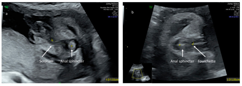
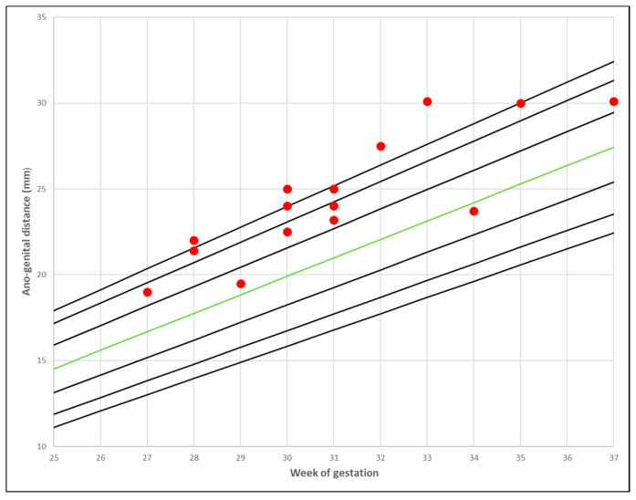
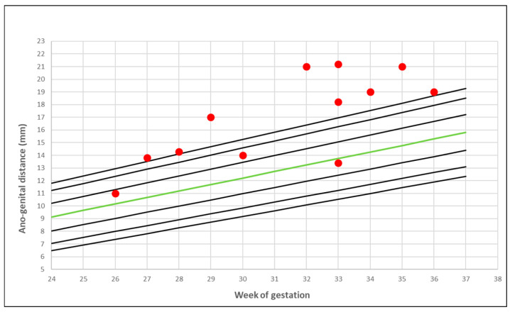
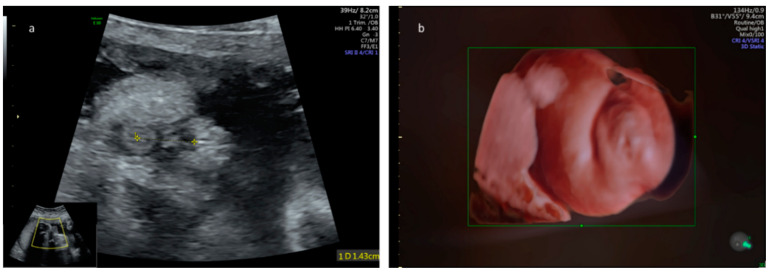
Anogenital distance (AGD) is a biomarker for the prenatal hormonal environment. Androgen excess is a key element in polycystic ovary syndrome (PCOS). The aim of this study was to assess the sonographic foetal AGD in a population of PCOS mothers in comparison to the general population. Foetal AGD was measured prospectively by 2D ultrasound in PCOS mothers and compared to prenatal AGD nomograms. The results were interpreted regarding maternal and foetal characteristics. The mean sonographic foetal AGD centile measurement in PCOS mothers was significantly longer in comparison to the general population (86.04% ± 18.22; p < 0.001). Estimated foetal weight and birthweight were appropriate for gestational age and did not correlate with AGD. Sonographic foetal AGD was significantly longer in PCOS diabetic mothers and in those who conceived following assisted reproduction treatments when compared to the general population (p < 0.001). Our results support the role of AGD as a biomarker of the prenatal hormonal environment and provide evidence for the hyperandrogenic effect in PCOS pregnancies on foetal androgenic status and genitalia development.
Overview
The claims and statistics of this paper haven’t been checked yet.
Run the check — free, no account needed. It reads the open-access text and checks the sources it cites.
Integrity signals
Registry
Cites
Statistics
Figures
Verification
Claims the paper makes
The claims and statistics of this paper haven’t been checked yet.
Run the check — free, no account needed. It reads the open-access text and checks the sources it cites.
Claims other papers make about this one
Each is checked against this paper’s own text: does it say what the citing paper says it does?
The cited source says what the claim attributes to it.
In the citing paper
“Daughters of mothers with PCOS also demonstrate evidence of longer AGDs than daughters of unaffected women (Barrett et al., 2018; Perlman et al., 2020, but see Glintborg et al., 2019), in association with high serum testosterone in affected mothers during gestation.”
From Endometriosis and polycystic ovary syndrome are diametric disorders (2021) · cites this paper as Perlman et al., 2020
Self-check
Percentages, sums and reported differences re-computed from the paper’s own numbers.
The self-check needs the paper’s full text.
Citations
Cites
(2004) · Fertility and Sterility · doi:10.1016/j.fertnstert.2003.10.004 · Cited by 9,050
Decrease in anogenital distance among male infants with prenatal phthalate exposure
Swan, S. H., Main, K. M., Liu, F., Stewart, S. L., Kruse, R. L., Calafat, A. M., Mao, C. S., Redmon, J. B., Ternand, C. L., Sullivan, S., et al. (2005) · Environmental Health Perspectives · doi:10.1289/ehp.8100 · Cited by 1,586
Swan, S. H. (2008) · Environmental Research · doi:10.1016/j.envres.2008.08.007 · Cited by 879
Obesity as disruptor of the female fertility
Silvestris, E., Pergola, G. D., Rosania, R., & Loverro, G. (2018) · Reproductive Biology and Endocrinology · doi:10.1186/s12958-018-0336-z · Cited by 737
Birth weight standards in the live-born population in Israel.
Dollberg, S., Haklai, Z., Mimouni, F. B., Gorfein, I., & Gordon, E. (2005) · PubMed · Cited by 563
Abbott, D. H., Barnett, D. K., Bruns, C. M., & Dumesic, D. A. (2005) · Human Reproduction Update · doi:10.1093/humupd/dmi013 · Cited by 555 · cited 1×
“Although the aetiology of PCOS is unclear, prenatal exposure to androgen hormones is considered an important factor in the development of this metabolic reproductive endocrine disorder [34,35].”
BackgroundGenetic, hormonal and metabolic aspects of PCOS: an update
De Leo, V., Musacchio, M. C., Cappelli, V., Massaro, M. G., Morgante, G., & Petraglia, F. (2016) · Reproductive Biology and Endocrinology · doi:10.1186/s12958-016-0173-x · Cited by 484
Sir‐Petermann, T. (2002) · Human Reproduction · doi:10.1093/humrep/17.10.2573 · Cited by 418
Association between prenatal exposure to phthalates and the health of newborns☆
Huang, P., KUO, P., CHOU, Y., LIN, S., & Lee, C.-L. (2008) · Environment International · doi:10.1016/j.envint.2008.05.012 · Cited by 346
Fetal Testosterone Influences Sexually Dimorphic Gray Matter in the Human Brain
Lombardo, M. V., Ashwin, E., Auyeung, B., Chakrabarti, B., Taylor, K., Hackett, G., Bullmore, E. T., & Baron-Cohen, S. (2012) · The Journal of Neuroscience · doi:10.1523/jneurosci.4389-11.2012 · Cited by 302
Cited by
Sorted by citations
Harada, M. (2022) · Reproductive Medicine and Biology · doi:10.1002/rmb2.12487 · Cited by 150
Endometriosis and polycystic ovary syndrome are diametric disorders
Dinsdale, N. L., & Crespi, B. J. (2021) · Evolutionary Applications · doi:10.1111/eva.13244 · Cited by 90
“Daughters of mothers with PCOS also demonstrate evidence of longer AGDs than daughters of unaffected women (Barrett et al., 2018; Perlman et al., 2020, but see Glintborg et al., 2019), in association with high serum testosterone in affected mothers during gestation.”
BackgroundDevelopmental Programming of Fertility in Cattle—Is It a Cause for Concern?
Wathes, D. C. (2022) · Animals · doi:10.3390/ani12192654 · Cited by 47
Pan, Z., Zhu, F., & Zhou, K. (2021) · Frontiers in Endocrinology · doi:10.3389/fendo.2021.696879 · Cited by 34
Variation among human populations in endometriosis and PCOS A test of the inverse comorbidity model
Crespi, B. J. (2021) · Evolution Medicine and Public Health · doi:10.1093/emph/eoab029 · Cited by 25
Metadata
- Authors
- Perlman, S., Toledano, Y., Kivilevitch, Z., Halevy, N., Rubin, E., & Gilboa, Y.
- Affiliations
- Tel Aviv University (IL)
- Rabin Medical Center (IL)
- Corresponding author
- Perlman, Sharon
- Venue
- Journal of Clinical Medicine
- Publisher
- MDPI AG
- Published
- 2020-09-04
- Volume
- 9
- Issue
- 9
- Pages
- 2863
- ISSN
- 2077-0383 (ISSN-L)
- PMC
- PMC7563834
- OpenAlex
- W3083354441
- Cited by
- 20
- References
- 46
- Citations by year
- 2026: 2 · 2025: 1 · 2024: 6 · 2023: 3 · 2022: 4 · 2021: 4
- FWCI
- 2.43
- Type
- journal article
- Language
- en
- Open access
- gold
- doi.org
- Licence
- Locations
- Journal of Clinical Medicine (publishedVersion, cc-by)
- PubMed Central (publishedVersion, cc-by)
- PubMed (publishedVersion)
- DOAJ (DOAJ: Directory of Open Access Journals) (submittedVersion, cc-by-sa)
- Topics
- Health Sciences › Medicine › Reproductive Medicine › Ovarian function and disorders
- Health Sciences › Medicine › Endocrinology, Diabetes and Metabolism › Sexual Differentiation and Disorders
- Physical Sciences › Environmental Science › Health, Toxicology and Mutagenesis › Effects and risks of endocrine disrupting chemicals
- Keywords
- polycystic ovary syndrome, anogenital distance, hyperandrogenism, prenatal biomarkers, assisted reproduction, maternal diabetes, birth weight
About these checks
Registry, retraction, Cites and statistics results are computed, not judged, with one exception: a printed reference that no index lists is read once by a model before it counts as a citation error. Claims are extracted by a model and kept only when their quote appears verbatim in the text; each cited source is then checked, with a model’s help, for whether it says what the claim relies on. Those verdicts can be wrong — read the claim’s quote and the cited source before relying on one.
Statistics and arithmetic are recomputed, not judged.
Confidence bands are shown only for fields calibrated on our annotated test set. None is calibrated yet, so no band is shown.
What counts as a claim ›How we measure confidence ›How we check this ›
Are you an author, or do you see a problem with this page? Request a review
How to cite
Cite CoChat for the checks on this page, and the paper for its own findings.
- This page
- CoChat. (2026). Citation check: Foetal Sonographic Anogenital Distance Is Longer in Polycystic Ovary Syndrome Mothers. https://app.cochat.ai/papers/10.3390/jcm9092863
- The paper
- Perlman, S., Toledano, Y., Kivilevitch, Z., Halevy, N., Rubin, E., & Gilboa, Y. (2020). Foetal Sonographic Anogenital Distance Is Longer in Polycystic Ovary Syndrome Mothers. Journal of Clinical Medicine. https://doi.org/10.3390/jcm9092863
