

Explainable Deep Learning Models in Medical Image Analysis
Open accessThe DOI matches its Crossref record; 76 of the 77 references it cites were found in a registry or an index, and 1 are not checked yet.
Abstract
Deep learning methods have been very effective for a variety of medical diagnostic tasks and have even outperformed human experts on some of those. However, the black-box nature of the algorithms has restricted their clinical use. Recent explainability studies aim to show the features that influence the decision of a model the most. The majority of literature reviews of this area have focused on taxonomy, ethics, and the need for explanations. A review of the current applications of explainable deep learning for different medical imaging tasks is presented here. The various approaches, challenges for clinical deployment, and the areas requiring further research are discussed here from a practical standpoint of a deep learning researcher designing a system for the clinical end-users.
Overview
The claims and statistics of this paper haven’t been checked yet.
Free: it reads the open-access text and checks the first 5 claims that cite a source we can check. Create a free account to check every claim.
Integrity signals
Registry
Cites
Statistics
Figures
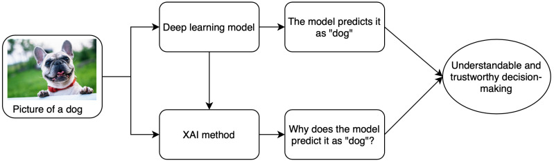
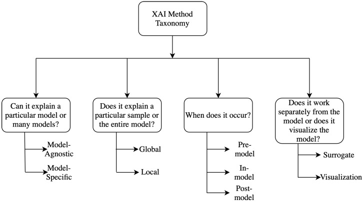
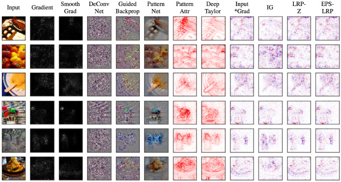

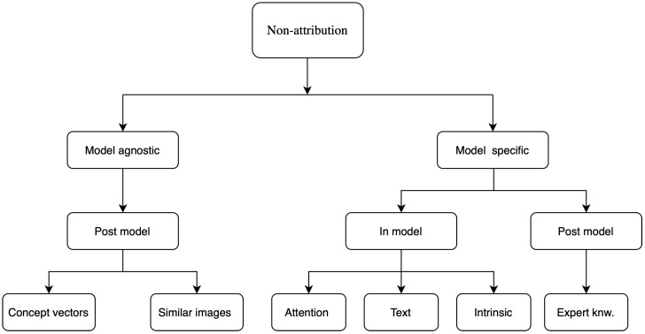
Verification
Claims the paper makes
The claims and statistics of this paper haven’t been checked yet.
Free: it reads the open-access text and checks the first 5 claims that cite a source we can check. Create a free account to check every claim.
Self-check
Percentages, sums and reported differences re-computed from the paper’s own numbers.
The self-check needs the paper’s full text.
Citations
Cites
Vaswani, A., Shazeer, N., Parmar, N., Uszkoreit, J., Jones, L., Gomez, A. N., Kaiser, L., & Polosukhin, I. (2017) · arXiv · doi:10.48550/arxiv.1706.03762 · Cited by 120,404 · cited 1×
“More details about attention mechanisms in neural networks is discussed in [51].”
BackgroundImageNet classification with deep convolutional neural networks
Krizhevsky, A., Sutskever, I., & Hinton, G. E. (2017) · Communications of the ACM · doi:10.1145/3065386 · Cited by 112,155 · cited 1×
“An earlier study for breast mass classification from mammograms [40] using two different CNNs - AlexNet [41] and GoogleNet [42] - employed saliency maps to visualize the image features.”
MethodU-Net: Convolutional Networks for Biomedical Image Segmentation
Ronneberger, O., Fischer, P., & Brox, T. (2015) · Lecture notes in computer science · doi:10.1007/978-3-319-24574-4_28 · Cited by 99,404
ImageNet: A large-scale hierarchical image database
Deng, J., Dong, W., Socher, R., Li, L.-J., Li, K., & Fei-Fei, L. (2009) · 2009 IEEE Conference on Computer Vision and Pattern Recognition · doi:10.1109/cvpr.2009.5206848 · Cited by 65,333 · cited 1×
“Both approaches are beneficial for medical imaging datasets which tend to be relatively smaller than computer vision benchmarks like ImageNet [33].”
BackgroundGoing deeper with convolutions
Szegedy, C., Liu, W., Jia, Y., Sermanet, P., Reed, S., Anguelov, D., Erhan, D., Vanhoucke, V., & Rabinovich, A. (2015) · 2015 IEEE Conference on Computer Vision and Pattern Recognition (CVPR) · doi:10.1109/cvpr.2015.7298594 · Cited by 48,562
(2008) · Journal of Machine Learning Research · Cited by 33,051
Grad-CAM: Visual Explanations from Deep Networks via Gradient-Based Localization
Selvaraju, R. R., Cogswell, M., Das, A., Vedantam, R., Parikh, D., & Batra, D. (2017) · 2017 IEEE International Conference on Computer Vision (ICCV) · doi:10.1109/iccv.2017.74 · Cited by 24,513
Visualizing and understanding convolutional networks
Zeiler, M. D., & Fergus, R. (2014) · Lecture Notes in Computer Science · doi:10.1007/978-3-319-10590-1_53 · Cited by 15,839 · cited 2×
“rey patch and hence occluding them from the system’s view. It can provide both positive and negative evidence by highlighting the responsible features. This technique was applied by Zeiler and Fergus [18] to the CNN for the image classification task. Occlusion is the benchmark for any attribution study as it is a simpl…”
MethodWold, S., Esbensen, K., & Geladi, P. (1987) · Chemometrics and Intelligent Laboratory Systems · doi:10.1016/0169-7439(87)80084-9 · Cited by 11,606 · cited 1×
“Principal component analysis (PCA) [12], t-Distributed Stochastic Neighbor Embedding (t-SNE) [13] are some common examples of these methods.”
MethodArrieta, A. B., Díaz-Rodríguez, N., Del Ser, J., Bennetot, A., Tabik, S., Barbado, A., García, S., Gil-López, S., Molina, D., Benjamins, R., et al. (2019) · arXiv · doi:10.48550/arxiv.1910.10045 · Cited by 11,321 · cited 2×
“Different kinds of taxonomies and classification methods are discussed briefly here and a detailed analysis of the taxonomies can be found in [8,9] and a flow chart for them is shown in 1.”
Background
Research works printed but not linked: 2
- El-Baz, A.S. (2020). Recent Advances in Deep Learning Applications for Retinal Diagnosis using OCT. State of the Art in Neural Networks, Elsevier. in press.
- Goodfellow, I.J., Shlens, J., and Szegedy, C. (2014). Explaining and harnessing adversarial examples. arXiv.
Cited by
Transformers in medical imaging: A survey
Shamshad, F., Khan, S., Zamir, S. W., Khan, M. H., Hayat, M., Khan, F. S., & Fu, H. (2023) · Medical Image Analysis · doi:10.1016/j.media.2023.102802 · Cited by 1,296
Explainable artificial intelligence: a comprehensive review
Minh, D., Wang, H. X., Li, Y. F., & Nguyen, T. N. (2022) · Artificial Intelligence Review · doi:10.1007/s10462-021-10088-y · Cited by 983
Loh, H. W., Ooi, C. P., Seoni, S., Barua, P. D., Molinari, F., & Acharya, U. R. (2022) · Computer Methods and Programs in Biomedicine · doi:10.1016/j.cmpb.2022.107161 · Cited by 734
Interpretable deep learning: interpretation, interpretability, trustworthiness, and beyond
Li, X., Xiong, H., Li, X., Wu, X., Zhang, X., Liu, J., Bian, J., & Dou, D. (2022) · Knowledge and Information Systems · doi:10.1007/s10115-022-01756-8 · Cited by 497
“Explanations for deep learningbased methods are more urged in this field than in other fields, to gain the trust of physicians, regulators as well as the patients [154].”
BackgroundAdvances in medical image analysis with vision Transformers: A comprehensive review
Azad, R., Kazerouni, A., Heidari, M., Aghdam, E. K., Molaei, A., Jia, Y., Jose, A., Roy, R., & Merhof, D. (2024) · Medical Image Analysis · doi:10.1016/j.media.2023.103000 · Cited by 485
Metadata
- Authors
- Singh, A., Sengupta, S., & Lakshminarayanan, V.
- Affiliations
- University of Waterloo (CA)
- Corresponding author
- Singh, Amitojdeep
- Venue
- Journal of Imaging
- Publisher
- MDPI AG
- Published
- 2020-06-20
- Volume
- 6
- Issue
- 6
- Pages
- 52
- ISSN
- 2313-433X (ISSN-L)
- PMC
- PMC8321083
- OpenAlex
- W3031116957
- Semantic Scholar
- af6af40155860ac2fd36680e63686473803b5f52
- Cited by
- 718
- References
- 75
- Citations by year
- 2026: 82 · 2025: 135 · 2024: 132 · 2023: 154 · 2022: 130 · 2021: 77 · 2020: 8
- FWCI
- 3.17
- Type
- journal article
- Language
- en
- Open access
- gold
- dx.doi.org
- Licence
- Locations
- Journal of Imaging (publishedVersion, cc-by)
- PubMed Central (publishedVersion, cc-by)
- PubMed (publishedVersion)
- arXiv (Cornell University) (submittedVersion)
- DOAJ (DOAJ: Directory of Open Access Journals) (submittedVersion, cc-by-sa)
- arXiv (Cornell University) (cc-by)
- Funders
- Nvidia
- Compute Canada
- Natural Sciences and Engineering Research Council of Canada
- Topics
- Physical Sciences › Computer Science › Artificial Intelligence › Explainable Artificial Intelligence (XAI)
- Health Sciences › Medicine › Health Informatics › Artificial Intelligence in Healthcare and Education
- Health Sciences › Medicine › Radiology, Nuclear Medicine and Imaging › Radiomics and Machine Learning in Medical Imaging
- Keywords
- explainable deep learning, medical image analysis, model interpretability, black-box models, clinical deployment, feature importance, diagnosis
About these checks
Registry, retraction, Cites and statistics results are computed, not judged, with one exception: a printed reference that no index lists is read once by a model before it counts as a citation error. Claims are extracted by a model and kept only when their quote appears verbatim in the text; each cited source is then checked, with a model’s help, for whether it says what the claim relies on. Those verdicts can be wrong — read the claim’s quote and the cited source before relying on one.
Statistics and arithmetic are recomputed, not judged.
Confidence bands are shown only for fields calibrated on our annotated test set. None is calibrated yet, so no band is shown.
What counts as a claim ›How we measure confidence ›How we check this ›
Are you an author, or do you see a problem with this page? Request a review
Data sources
Bibliographic data: Semantic Scholar, OpenAlex, Crossref.
Contains information from the Semantic Scholar Academic Graph Datasets, made available under the ODC Attribution License (ODC-BY 1.0).
Semantic Scholar’s data is described in: Kinney, R., et al. (2023). The Semantic Scholar Open Data Platform. arXiv:2301.10140.
