

The DOI matches its Crossref record; 1 of the 1 references it cites were found in a registry or an index.
Ultrasonic Based Tissue Modelling and Engineering
Open accessAbstract
Systems and devices for in vitro tissue modelling and engineering are valuable tools, which combine the strength between the controlled laboratory environment and the complex tissue organization and environment in vivo. Device-based tissue engineering is also a possible avenue for future explant culture in regenerative medicine. The most fundamental requirements on platforms intended for tissue modelling and engineering are their ability to shape and maintain cell aggregates over long-term culture. An emerging technology for tissue shaping and culture is ultrasonic standing wave (USW) particle manipulation, which offers label-free and gentle positioning and aggregation of cells. The pressure nodes defined by the USW, where cells are trapped in most cases, are stable over time and can be both static and dynamic depending on actuation schemes. In this review article, we highlight the potential of USW cell manipulation as a tool for tissue modelling and engineering.
Overview
The claims and statistics of this paper haven’t been checked yet.
Run the check — free, no account needed. It reads the open-access text and checks the sources it cites.
Integrity signals
Registry
Cites
Statistics
Figures
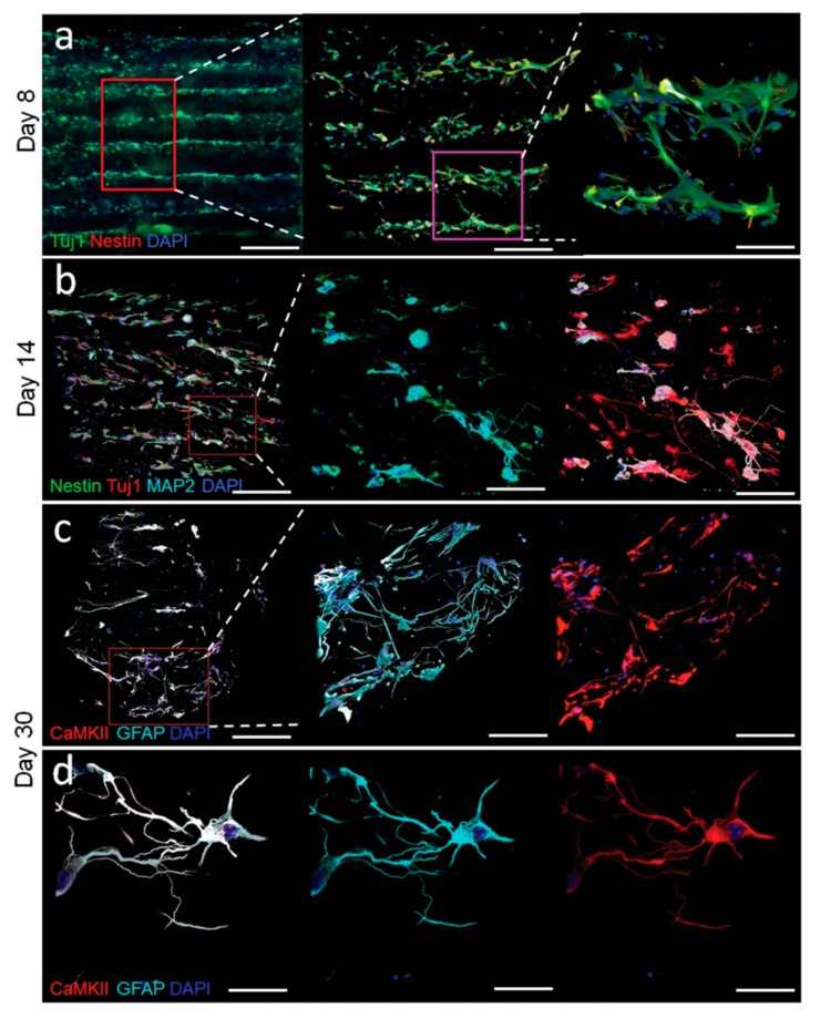
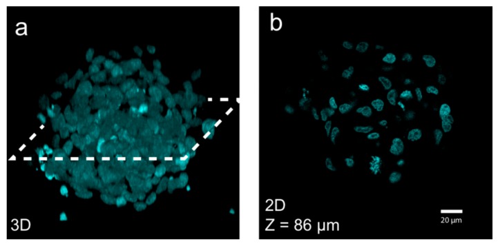
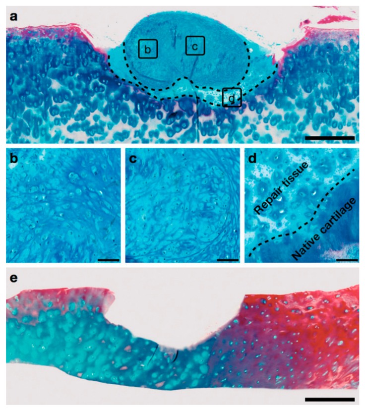
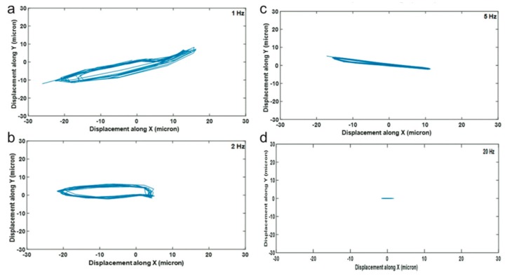
Verification
Claims the paper makes
The claims and statistics of this paper haven’t been checked yet.
Run the check — free, no account needed. It reads the open-access text and checks the sources it cites.
Claims other papers make about this one
Each is checked against this paper’s own text: does it say what the citing paper says it does?
The source backs part of the claim, not all of it — or backs it for a different population, condition or comparison.
In the citing paper
“Moreover, the acoustofluidics also manipulates cell-cell contacts while maintaining cells in their native culture medium or supporting Matrigel gel.39”
From Acoustofluidic assembly of 3D neurospheroids to model Alzheimer's disease (2020) · cites this paper as [39]
Experimental — accuracy not yet measured
Self-check
Percentages, sums and reported differences re-computed from the paper’s own numbers.
The self-check needs the paper’s full text.
Citations
Cites
Three-dimensional culture systems in cancer research: Focus on tumor spheroid model
Nath, S., & Devi, G. R. (2016) · Pharmacology & Therapeutics · doi:10.1016/j.pharmthera.2016.03.013 · Cited by 919
Research works printed but not linked: 74
- Laurell, T., and Lenshof, A. (2014). Microscale Acoustofluidics, Royal Society of Chemistry. · DOI
- Laurell, 2007, Chip integrated strategies for acoustic separation and manipulation of cells and particles, Chem. Soc. Rev., 36, 492, 10.1039/B601326K · DOI
- Shi, 2009, Acoustic tweezers: Patterning cells and microparticles using standing surface acoustic waves (SSAW), Lab Chip, 9, 2890, 10.1039/b910595f · DOI
- Brandt, 2001, Acoustic physics. Suspended by sound, Nature, 413, 474, 10.1038/35097192 · DOI
- Evander, 2012, Acoustofluidics 20: Applications in acoustic trapping, Lab Chip, 12, 4667, 10.1039/c2lc40999b · DOI
- Lenshof, 2012, Acoustofluidics 5: Building microfluidic acoustic resonators, Lab Chip, 12, 684, 10.1039/c1lc20996e · DOI
- Petersson, 2007, Free flow acoustophoresis: Microfluidic-based mode of particle and cell separation, Anal. Chem., 79, 5117, 10.1021/ac070444e · DOI
- Wiklund, 2012, Acoustofluidics 14: Applications of acoustic streaming in microfluidic devices, Lab Chip, 12, 2438, 10.1039/c2lc40203c · DOI
- Karlsen, 2016, Acoustic Force Density Acting on Inhomogeneous Fluids in Acoustic Fields, Phys. Rev. Lett., 117, 114504, 10.1103/PhysRevLett.117.114504 · DOI
- Dyson, 1971, Flow of Red Blood Cells stopped by Ultrasound, Nature, 232, 572, 10.1038/232572a0 · DOI
- Coakley, 2004, Cell-cell contact and membrane spreading in an ultrasound trap, Colloids Surf. B Biointerfaces, 34, 221, 10.1016/j.colsurfb.2004.01.002 · DOI
- Bazou, 2005, Physical enviroment of 2-D animal cell aggregates formed in a short pathlength ultrasound standing wave trap, Ultrasound Med. Biol., 31, 423, 10.1016/j.ultrasmedbio.2004.12.007 · DOI
- Hultstrom, 2007, Proliferation and viability of adherent cells manipulated by standing-wave ultrasound in a microfluidic chip, Ultrasound Med. Biol., 33, 145, 10.1016/j.ultrasmedbio.2006.07.024 · DOI
- Bazou, 2006, Gap junctional intercellular communication and cytoskeletal organization in chondrocytes in suspension in an ultrasound trap, Mol. Membr. Biol., 23, 195, 10.1080/09687860600555906 · DOI
- Bazou, 2008, Long-term viability and proliferation of alginate-encapsulated 3-D HepG2 aggregates formed in an ultrasound trap, Toxicol. In Vitro, 22, 1321, 10.1016/j.tiv.2008.03.014 · DOI
- Christakou, 2013, Live cell imaging in a micro-array of acoustic traps facilitates quantification of natural killer cell heterogeneity, Integr. Biol. Quant. Biosci. Nano Macro, 5, 712
- Bazou, 2005, Molecular adhesion development in a neural cell monolayer forming in an ultrasound trap, Mol. Membr. Biol., 22, 229, 10.1080/09687860500093396 · DOI
- Bazou, 2011, Gene expression analysis of mouse embryonic stem cells following levitation in an ultrasound standing wave trap, Ultrasound Med. Biol., 37, 321, 10.1016/j.ultrasmedbio.2010.10.019 · DOI
- Wiklund, 2012, Acoustofluidics 12: Biocompatibility and cell viability in microfluidic acoustic resonators, Lab Chip, 12, 2018, 10.1039/c2lc40201g · DOI
- Ohlin, 2015, Temperature-controlled MPa-pressure ultrasonic cell manipulation in a microfluidic chip, Lab Chip, 15, 3341, 10.1039/C5LC00490J · DOI
- Kuznetsova, 2009, Multiple three-dimensional mammalian cell aggregates formed away from solid substrata in ultrasound standing waves, Biotechnol. Prog., 25, 834, 10.1002/btpr.164 · DOI
- Bazou, 2004, Characterisation of the morphology of 2-D particle aggregates in different electrolyte concentrations in an ultrasound trap, Colloids Surf. A Physicochem. Eng. Asp., 243, 97, 10.1016/j.colsurfa.2004.04.075 · DOI
- Jonnalagadda, 2018, Acoustically modulated biomechanical stimulation for human cartilage tissue engineering, Lab Chip, 18, 473, 10.1039/C7LC01195D · DOI
- Hawkes, 2004, Ultrasonic deposition of cells on a surface, Biosens. Bioelectron., 19, 1021, 10.1016/j.bios.2003.10.003 · DOI
- Hoyos, 2018, Induced clustering of Escherichia coli by acoustic fields, Sci. Rep., 8, 4668, 10.1038/s41598-018-22960-z · DOI
- Hammarstrom, 2012, Seed particle-enabled acoustic trapping of bacteria and nanoparticles in continuous flow systems, Lab Chip, 12, 4296, 10.1039/c2lc40697g · DOI
- Martin, 2005, Spore and micro-particle capture on an immunosensor surface in an ultrasound standing wave system, Biosens. Bioelectron., 21, 758, 10.1016/j.bios.2005.01.013 · DOI
- Wiklund, 2013, Acoustofluidics 21: Ultrasound-enhanced immunoassays and particle sensors, Lab Chip, 13, 25, 10.1039/C2LC41073G · DOI
- Liu, 2007, Functional three-dimensional HepG2 aggregate cultures generated from an ultrasound trap: Comparison with HepG2 spheroids, J. Cell. Biochem., 102, 1180, 10.1002/jcb.21345 · DOI
- Neild, 2007, Simultaneous positioning of cells into two-dimensional arrays using ultrasound, Biotechnol. Bioeng., 97, 1335, 10.1002/bit.21315 · DOI
- Haake, 2005, Positioning, displacement, and localization of cells using ultrasonic forces, Biotechnol. Bioeng., 92, 8, 10.1002/bit.20540 · DOI
- Chen, 2016, Rapid formation of size-controllable multicellular spheroids via 3D acoustic tweezers, Lab Chip, 16, 2636, 10.1039/C6LC00444J · DOI
- Ohlin, 2013, Influence of acoustic streaming on ultrasonic particle manipulation in a 100-well ring-transducer microplate, J. Micromech. Microeng., 23, 035008, 10.1088/0960-1317/23/3/035008 · DOI
- Vanherberghen, 2010, Ultrasound-controlled cell aggregation in a multi-well chip, Lab Chip, 10, 2727, 10.1039/c004707d · DOI
- Christakou, 2015, Ultrasonic three-dimensional on-chip cell culture for dynamic studies of tumor immune surveillance by natural killer cells, Lab Chip, 15, 3222, 10.1039/C5LC00436E · DOI
- Hsu, 2016, NK cells converge lytic granules to promote cytotoxicity and prevent bystander killing, J. Cell Biol., 215, 875, 10.1083/jcb.201604136 · DOI
- Bernassau, 2012, Direct patterning of mammalian cells in an ultrasonic heptagon stencil, Biomed. Microdevices, 14, 559, 10.1007/s10544-012-9633-z · DOI
- Caleap, 2014, Acoustically trapped colloidal crystals that are reconfigurable in real time, Proc. Natl. Acad. Sci. USA, 111, 6226, 10.1073/pnas.1323048111 · DOI
- Owens, 2016, Highly parallel acoustic assembly of microparticles into well-ordered colloidal crystallites, Soft Matter, 12, 717, 10.1039/C5SM02348C · DOI
- Li, 2014, Standing surface acoustic wave based cell coculture, Anal. Chem., 86, 9853, 10.1021/ac502453z · DOI
- Vuillermet, 2016, Chladni Patterns in a Liquid at Microscale, Phys. Rev. Lett., 116, 184501, 10.1103/PhysRevLett.116.184501 · DOI
- Chen, 2014, Microscale assembly directed by liquid-based template, Adv. Mater., 26, 5936, 10.1002/adma.201402079 · DOI
- Chen, 2015, Biotunable acoustic node assembly of organoids, Adv. Healthc. Mater., 4, 1937, 10.1002/adhm.201500279 · DOI
- Serpooshan, 2017, Bioacoustic-enabled patterning of human iPSC-derived cardiomyocytes into 3D cardiac tissue, Biomaterials, 131, 47, 10.1016/j.biomaterials.2017.03.037 · DOI
- Barnkob, 2012, Acoustic radiation- and streaming-induced microparticle velocities determined by microparticle image velocimetry in an ultrasound symmetry plane, Phys. Rev. E Stat. Nonlinear Soft Matter Phys., 86, 056307, 10.1103/PhysRevE.86.056307 · DOI
- Huang, 2018, A sharp-edge-based acoustofluidic chemical signal generator, Lab Chip, 18, 1411, 10.1039/C8LC00193F · DOI
- Garg, N., Westerhof, T.M., Liu, V., Liu, R., Nelson, E.L., and Lee, A.P. (2018). Whole-blood sorting, enrichment and in situ immunolabeling of cellular subsets using acoustic microstreaming. Microsyst. Nanoeng., 4. · DOI
- Kurashina, 2017, Cell agglomeration in the wells of a 24-well plate using acoustic streaming, Lab Chip, 17, 876, 10.1039/C6LC01310D · DOI
- Melde, 2016, Holograms for acoustics, Nature, 537, 518, 10.1038/nature19755 · DOI
- Melde, K., Choi, E., Wu, Z., Palagi, S., Qiu, T., and Fischer, P. (2018). Acoustic Fabrication via the Assembly and Fusion of Particles. Adv. Mater., 30. · DOI
- Pedde, R.D., Mirani, B., Navaei, A., Styan, T., Wong, S., Mehrali, M., Thakur, A., Mohtaram, N.K., Bayati, A., and Dolatshahi-Pirouz, A. (2017). Emerging Biofabrication Strategies for Engineering Complex Tissue Constructs. Adv. Mater., 29. · DOI
- Caliari, 2016, A practical guide to hydrogels for cell culture, Nat. Methods, 13, 405, 10.1038/nmeth.3839 · DOI · Paper page
- Bouyer, 2016, A Bio-Acoustic Levitational (BAL) Assembly Method for Engineering of Multilayered, 3D Brain-Like Constructs, Using Human Embryonic Stem Cell Derived Neuro-Progenitors, Adv. Mater., 28, 161, 10.1002/adma.201503916 · DOI
- Thoma, 2014, 3D cell culture systems modeling tumor growth determinants in cancer target discovery, Adv. Drug Deliv. Rev., 69–70, 29, 10.1016/j.addr.2014.03.001 · DOI · Paper page
- Moshksayan, 2018, Spheroids-on-a-chip: Recent advances and design considerations in microfluidic platforms for spheroid formation and culture, Sens. Actuators B Chem., 263, 151, 10.1016/j.snb.2018.01.223 · DOI · Paper page
- Olofsson, 2018, Acoustic formation of multicellular tumor spheroids enabling on-chip functional and structural imaging, Lab Chip, 18, 2466, 10.1039/C8LC00537K · DOI
- Olofsson, K., Carannante, V., Önfelt, B., and Wiklund, M. (2018, January 11–15). High-content nucleus based 3D image cytometery of whole multicellular tumour spheroids. Proceedings of the 22nd International Conference on Miniaturized Systems for Chemistry and Life Sciences (µTAS 2018), Kaohsiung, Taiwan.
- Sriphutkiat, Y., Kasetsirikul, S., and Zhou, Y. (2018). Formation of cell spheroids using Standing Surface Acoustic Wave (SSAW). Int. J. Bioprinting, 4. · DOI
- Nagahashi, 2015, Stable surface coating of silicone elastomer with phosphorylcholine and organosilane copolymer with cross-linking for repelling proteins, Colloids Surf. B Biointerfaces, 134, 384, 10.1016/j.colsurfb.2015.07.040 · DOI
- Wiklund, 2012, Acoustofluidics 18: Microscopy for acoustofluidic micro-devices, Lab Chip, 12, 3221, 10.1039/c2lc40757d · DOI
- Frisk, 2011, A silicon-glass microwell platform for high-resolution imaging and high-content screening with single cell resolution, Biomed. Microdevices, 13, 683, 10.1007/s10544-011-9538-2 · DOI
- Li, 2014, Application of an acoustofluidic perfusion bioreactor for cartilage tissue engineering, Lab Chip, 14, 4475, 10.1039/C4LC00956H · DOI
- Hammarstrom, 2014, Frequency tracking in acoustic trapping for improved performance stability and system surveillance, Lab Chip, 14, 1005, 10.1039/C3LC51144H · DOI
- Lata, 2016, Surface Acoustic Waves Grant Superior Spatial Control of Cells Embedded in Hydrogel Fibers, Adv. Mater., 28, 8632, 10.1002/adma.201602947 · DOI
- Garvin, 2010, Controlling the Spatial Organization of Cells and Extracellular Matrix Proteins in Engineered Tissues Using Ultrasound Standing Wave Fields, Ultrasound Med. Biol., 36, 1919, 10.1016/j.ultrasmedbio.2010.08.007 · DOI
- Naseer, 2017, Surface acoustic waves induced micropatterning of cells in gelatin methacryloyl (GelMA) hydrogels, Biofabrication, 9, 015020, 10.1088/1758-5090/aa585e · DOI
- Garvin, 2013, Controlling collagen fiber microstructure in three-dimensional hydrogels using ultrasound, J. Acoust. Soc. Am., 134, 1491, 10.1121/1.4812868 · DOI
- Garvin, 2013, Spatial patterning of endothelial cells and vascular network formation using ultrasound standing wave fields, J. Acoust. Soc. Am., 134, 1483, 10.1121/1.4812867 · DOI
- Dalecki, 2015, Ultrasound technologies for biomaterials fabrication and imaging, Ann. Biomed. Eng., 43, 747, 10.1007/s10439-014-1158-6 · DOI
- Comeau, 2017, Ultrasound patterning technologies for studying vascular morphogenesis in 3D, J. Cell Sci., 130, 232
- Ovsianikov, 2018, The Synergy of Scaffold-Based and Scaffold-Free Tissue Engineering Strategies, Trends Biotechnol., 36, 348, 10.1016/j.tibtech.2018.01.005 · DOI · Paper page
- Rivron, 2009, Tissue assembly and organization: Developmental mechanisms in microfabricated tissues, Biomaterials, 30, 4851, 10.1016/j.biomaterials.2009.06.037 · DOI
- Xu, 2011, The assembly of cell-encapsulating microscale hydrogels using acoustic waves, Biomaterials, 32, 7847, 10.1016/j.biomaterials.2011.07.010 · DOI
- Zhu, Y., Serpooshan, V., Wu, S., Demirci, U., Chen, P., and Guven, S. (2017). Tissue Engineering of 3D Organotypic Microtissues by Acoustic Assembly. Methods Mol. Biol. · DOI
Cited by
Sorted by citations
Ultrasound-responsive systems as components for smart materials
Athanassiadis, A. G., Ma, Z., Moreno-Gomez, N., Melde, K., Choi, E., Goyal, R., & Fischer, P. (2022) · Chemical Reviews · doi:10.1021/acs.chemrev.1c00622 · Cited by 293
Acoustofluidic assembly of 3D neurospheroids to model Alzheimer's disease
Cai, H., Ao, Z., Hu, L., Moon, Y., Wu, Z., Lu, H.-C., Kim, J., & Guo, F. (2020) · The Analyst · doi:10.1039/d0an01373k · Cited by 70
Ultrasound-assisted tissue engineering
Melde, K., Athanassiadis, A. G., Missirlis, D., Shi, M., Seneca, S., & Fischer, P. (2024) · Nature Reviews Bioengineering · doi:10.1038/s44222-024-00166-5 · Cited by 59
Metadata
- Authors
- Olofsson, K., Hammarström, B., & Wiklund, M.
- Affiliations
- KTH Royal Institute of Technology (SE)
- Department of Applied Physics, KTH Royal Institute of Technology, SE-106 91 Stockholm, Sweden
- Corresponding author
- Wiklund, Martin
- Venue
- Micromachines
- Publisher
- MDPI AG
- Published
- 2018-11-14
- Volume
- 9
- Issue
- 11
- Pages
- 594
- ISSN
- 2072-666X (ISSN-L)
- PMC
- PMC6266922
- OpenAlex
- W2900642484
- Cited by
- 39
- References
- 1
- Citations by year
- 2026: 1 · 2025: 4 · 2024: 7 · 2023: 5 · 2022: 8 · 2021: 10 · 2020: 2 · 2019: 1
- FWCI
- 0.26
- Type
- journal article
- Language
- en
- Open access
- gold
- dx.doi.org
- Licence
- Locations
- Micromachines (publishedVersion, cc-by)
- Europe PMC (PubMed Central) (publishedVersion, other-oa)
- PubMed Central (publishedVersion, cc-by)
- PubMed (publishedVersion)
- DOAJ (DOAJ: Directory of Open Access Journals) (submittedVersion)
- Funders
- Stockholms Läns Landsting
- Vetenskapsrådet
- Topics
- Physical Sciences › Engineering › Biomedical Engineering › Microfluidic and Bio-sensing Technologies
- Life Sciences › Biochemistry, Genetics and Molecular Biology › Cell Biology › Cellular Mechanics and Interactions
- Physical Sciences › Engineering › Biomedical Engineering › 3D Printing in Biomedical Research
- Keywords
- ultrasonic standing waves, acoustic cell manipulation, cell aggregation, tissue engineering, pressure nodes, explant culture, regenerative medicine, acoustic levitation
About these checks
Registry, retraction, Cites and statistics results are computed, not judged, with one exception: a printed reference that no index lists is read once by a model before it counts as a citation error. Claims are extracted by a model and kept only when their quote appears verbatim in the text; each cited source is then checked, with a model’s help, for whether it says what the claim relies on. Those verdicts can be wrong — read the claim’s quote and the cited source before relying on one.
Statistics and arithmetic are recomputed, not judged.
Confidence bands are shown only for fields calibrated on our annotated test set. None is calibrated yet, so no band is shown.
What counts as a claim ›How we measure confidence ›How we check this ›
Are you an author, or do you see a problem with this page? Request a review
