

The DOI matches its Crossref record; 2 of the 2 references it cites were found in a registry or an index.
Cancer Targeting and Drug Delivery Using Carbon-Based Quantum Dots and Nanotubes
Abstract
Currently cancer treatment is in large part non-specific with respect to treatment. Medication is often harsh on patients, whereby they suffer several undesired side effects as a result. Carbon-based nanoparticles have attracted attention in recent years due to their ability to act as a platform for the attachment of several drugs and/or ligands. Relatively simple models are often used in cancer research, wherein carbon nanoparticles are conjugated to a ligand that is specific to an overexpressed receptor for imaging and drug delivery in cancer treatment. These carbon nanoparticles confer unique properties to the imaging or delivery vehicle due to their nontoxic nature and their high fluorescence qualities. Chief among the ongoing research within carbon-based nanoparticles emerge carbon dots (C-dots) and carbon nanotubes (CNTs). In this review, the aforementioned carbon nanoparticles will be discussed in their use within doxorubicin and gemcitabine based drug delivery vehicles, as well as the ligand-mediated receptor specific targeted therapy. Further directions of research in current field are also discussed.
Overview
The claims and statistics of this paper haven’t been checked yet.
Run the check — free, no account needed. It reads the open-access text and checks the sources it cites.
Integrity signals
Registry
Cites
Statistics
Figures
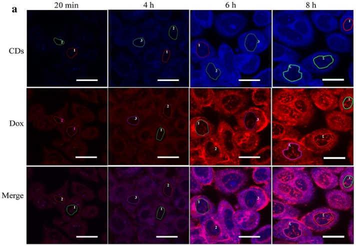
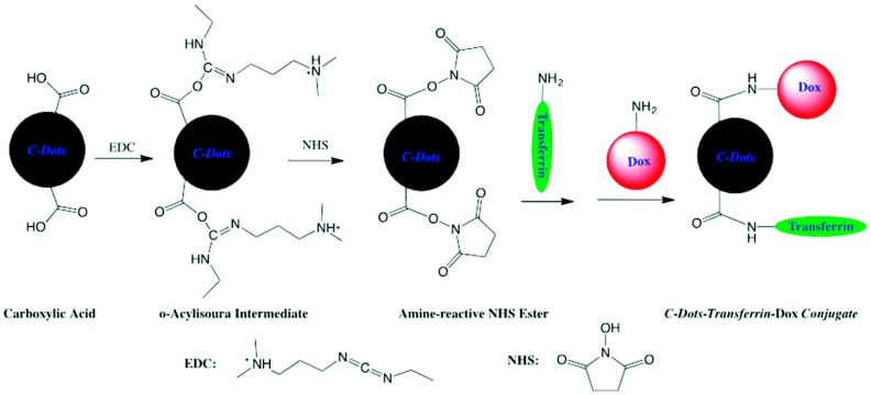
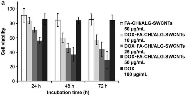
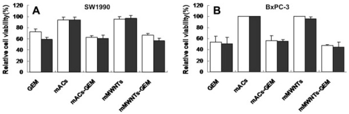
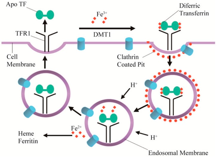
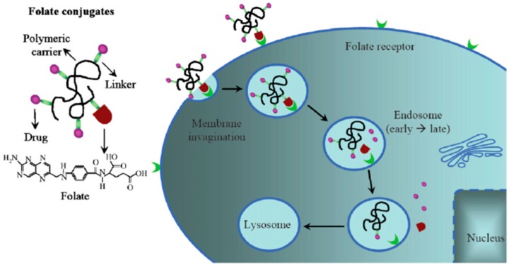
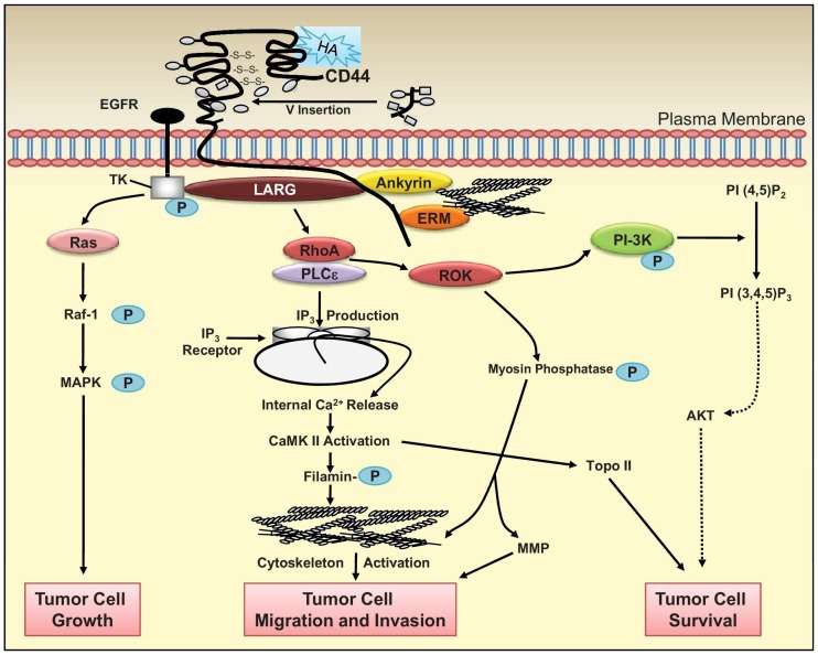
Verification
Claims the paper makes
The claims and statistics of this paper haven’t been checked yet.
Run the check — free, no account needed. It reads the open-access text and checks the sources it cites.
Claims other papers make about this one
Each is checked against this paper’s own text: does it say what the citing paper says it does?
In the citing paper
“Fucose-based Quantum Dots (QDs)can effectively deliver therapeutic agents to BC cells while also demonstrating inherent anticancer properties by generating reactive oxygen species (ROS).”
From Emerging role of exosomes in cancer therapy: progress and challenges (2025) · cites this paper as [217]
Self-check
Percentages, sums and reported differences re-computed from the paper’s own numbers.
The self-check needs the paper’s full text.
Citations
Cites
Crossing the blood-brain barrier with nanoparticles
Zhou, Y., Peng, Z., Seven, E. S., & Leblanc, R. M. (2018) · Journal of Controlled Release · doi:10.1016/j.jconrel.2017.12.015 · Cited by 837
Self-citationde Moor, J. S., Mariotto, A. B., Parry, C., Alfano, C. M., Padgett, L., Kent, E. E., Forsythe, L., Scoppa, S., Hachey, M., & Rowland, J. H. (2013) · Cancer Epidemiology, Biomarkers & Prevention · doi:10.1158/1055-9965.epi-12-1356 · Cited by 670 · cited 1×
“An over 400% increase in cancer survival by 2022 is projected when compared to 1977, the majority of growth occurring in the long-term (15+ years) survival of patients [19].”
Background
Research works printed but not linked: 110
- Xu, 2004, Electrophoretic analysis and purification of fluorescent single-walled carbon nanotube fragments, J. Am. Chem. Soc., 126, 12736, 10.1021/ja040082h · DOI
- Peng, 2017, Carbon dots: Biomacromolecule interaction, bioimaging and nanomedicine, Coord. Chem. Rev., 343, 256, 10.1016/j.ccr.2017.06.001 · DOI
- Margraf, 2015, The electronic structure of amorphous carbon nanodots, J. Phys. Chem. B, 119, 7258, 10.1021/jp510620j · DOI
- Gao, 2016, Carbon quantum dot-based nanoprobes for metal ion detection, J. Mater. Chem. C, 4, 6927, 10.1039/C6TC02055K · DOI
- Zhou, Y.Q., Sharma, S.K., Peng, Z.L., and Leblanc, R.M. (2017). Polymers in carbon dots: A review. Polymers, 9. · DOI
- Li, 2016, “Dark” carbon dots specifically “light-up” calcified zebrafish bones, J. Mater. Chem. B, 4, 7398, 10.1039/C6TB02241C · DOI
- Peng, 2017, Carbon dots: Promising biomaterials for bone-specific imaging and drug delivery, Nanoscale, 9, 17533, 10.1039/C7NR05731H · DOI
- Li, 2015, Method to determine protein concentration in the protein nanoparticle conjugates aqueous solution using circular dichroism spectroscopy, Anal. Chem., 87, 6455, 10.1021/acs.analchem.5b01451 · DOI
- Peng, 2016, Determination of the composition, encapsulation efficiency and loading capacity in protein drug delivery systems using circular dichroism spectroscopy, Anal. Chim. Acta, 937, 113, 10.1016/j.aca.2016.08.014 · DOI
- Jelinek, R. (2017). Carbon Quantum Dots, Springer International Publishing. · DOI
- Han, 2016, Interactions between carbon nanomaterials and biomolecules, J. Oleo Sci., 65, 1, 10.5650/jos.ess15248 · DOI
- Li, 2015, Nontoxic carbon dots potently inhibit human insulin fibrillation, Chem. Mater., 27, 1764, 10.1021/cm504572b · DOI
- Wang, 2016, Highly fluorescent carbon dots for visible sensing of doxorubicin release based on efficient nanosurface energy transfer, Biotechnol. Lett., 38, 191, 10.1007/s10529-015-1965-3 · DOI
- Wang, 2014, Carbon quantum dots: Synthesis, properties and applications, J. Mater. Chem. C, 2, 6921, 10.1039/C4TC00988F · DOI
- Lim, 2015, Carbon quantum dots and their applications, Chem. Soc. Rev., 44, 362, 10.1039/C4CS00269E · DOI
- Liu, 2017, Red emission B, N, S-co-doped carbon dots for colorimetric and fluorescent dual mode detection of Fe3+ ions in complex biological fluids and living cells, ACS Appl. Mater. Interfaces, 9, 12663, 10.1021/acsami.6b15746 · DOI
- Zhang, 2017, A novel mechanism for red emission carbon dots: Hydrogen bond dominated molecular states emission, Nanoscale, 9, 13042, 10.1039/C7NR03570E · DOI
- Yan, 2013, Particle carriers for combating multidrug-resistant cancer, ACS Nano, 7, 9512, 10.1021/nn405632s · DOI
- Safdie, 2009, Fasting and cancer treatment in humans: A case series report, Aging-US, 1, 988, 10.18632/aging.100114 · DOI
- Kano, 2007, Improvement of cancer-targeting therapy, using nanocarriers for intractable solid tumors by inhibition of TGF-beta signaling, Proc. Natl. Acad. Sci. USA, 104, 3460, 10.1073/pnas.0611660104 · DOI
- Li, 2014, Carbon dots prepared from ginger exhibiting efficient inhibition of human hepatocellular carcinoma cells, J. Mater. Chem. B, 2, 4564, 10.1039/c4tb00216d · DOI
- Hsu, 2013, Extremely high inhibition activity of photoluminescent carbon nanodots toward cancer cells, J. Mater. Chem. B, 1, 1774, 10.1039/c3tb00545c · DOI
- Yu, 2010, Receptor-targeted nanocarriers for therapeutic delivery to cancer, Mol. Membr. Biol., 27, 286, 10.3109/09687688.2010.521200 · DOI
- Zhang, 2013, Combating P-glycoprotein-mediated multidrug resistance using therapeutic nanoparticles, Curr. Pharm. Des., 19, 6655, 10.2174/1381612811319370009 · DOI
- Li, 2016, Crossing the blood-brain barrier with transferrin conjugated carbon dots: A zebrafish model study, Colloids Surf. B Biointerfaces, 145, 251, 10.1016/j.colsurfb.2016.05.007 · DOI
- Wicki, 2015, Nanomedicine in cancer therapy: Challenges, opportunities, and clinical applications, J. Control. Release, 200, 138, 10.1016/j.jconrel.2014.12.030 · DOI
- Trang, 2011, Systemic delivery of tumor suppressor microRNA mimics using a neutral lipid emulsion inhibits lung tumors in mice, Mol. Ther., 19, 1116, 10.1038/mt.2011.48 · DOI
- Iijima, 1991, Helical microtubules of graphitic carbon, Nature, 354, 56, 10.1038/354056a0 · DOI
- Yang, 2009, Hydrophilic multi-walled carbon nanotubes decorated with magnetite nanoparticles as lymphatic targeted drug delivery vehicles, Chem. Commun., 2009, 4447, 10.1039/b908012k · DOI
- Niu, 2013, Folate-conjugated peg on single walled carbon nanotubes for targeting delivery of doxorubicin to cancer cells, Macromol. Biosci., 13, 735, 10.1002/mabi.201200475 · DOI
- Pistone, 2016, Tunable doxorubicin release from polymer-gated multiwalled carbon nanotubes, Int. J. Pharm., 515, 30, 10.1016/j.ijpharm.2016.10.010 · DOI
- Zhang, 2009, Targeted delivery and controlled release of doxorubicin to cancer cells using modified single wall carbon nanotubes, Biomaterials, 30, 6041, 10.1016/j.biomaterials.2009.07.025 · DOI
- Lay, 2010, Delivery of paclitaxel by physically loading onto poly(ethyleneglycol) (PEG)-graftcarbon nanotubes for potent cancer therapeutics, Nanotechnology, 21, 065101, 10.1088/0957-4484/21/6/065101 · DOI
- Donnenberg, 2005, Multiple drug resistance in cancer revisited: The cancer stem cell hypothesis, J. Clin. Pharmacol., 45, 872, 10.1177/0091270005276905 · DOI
- Weinberg, R. (2013). The Biology of Cancer, Garland Science. · DOI
- Housman, 2014, Drug resistance in cancer: An overview, Cancers, 6, 1769, 10.3390/cancers6031769 · DOI · Paper page
- Ke, 2014, Co-delivery of thioridazine and doxorubicin using polymeric micelles for targeting both cancer cells and cancer stem cells, Biomaterials, 35, 1096, 10.1016/j.biomaterials.2013.10.049 · DOI
- Jia, 2016, PEGylated oxidized alginate-Dox prodrug conjugate nanoparticles cross-linked with fluorescent carbon dots for tumor theranostics, ACS Biomater. Sci. Eng., 2, 1641, 10.1021/acsbiomaterials.6b00443 · DOI
- Li, 2016, Transferrin conjugated nontoxic carbon dots for doxorubicin delivery to target pediatric brain tumor cells, Nanoscale, 8, 16662, 10.1039/C6NR05055G · DOI
- Singh, 2013, Gemcitabine-loaded smart carbon nanotubes for effective targeting to cancer cells, J. Drug Target., 21, 581, 10.3109/1061186X.2013.778264 · DOI
- Singh, 2016, Effects of transferrin conjugated multi-walled carbon nanotubes in lung cancer delivery, Mater. Sci. Eng. C, 67, 313, 10.1016/j.msec.2016.05.013 · DOI
- Yang, 2016, Doxorubicin conjugated functionalizable carbon dots for nucleus targeted delivery and enhanced therapeutic efficacy, Nanoscale, 8, 6801, 10.1039/C6NR00247A · DOI
- Sun, 2017, Supramolecular hybrids of carbon dots with doxorubicin: Synthesis, stability and cellular trafficking, Mater. Chem. Front., 1, 354, 10.1039/C6QM00042H · DOI
- Mewada, 2014, Swarming carbon dots for folic acid mediated delivery of doxorubicin and biological imaging, J. Mater. Chem. B, 2, 698, 10.1039/C3TB21436B · DOI
- Song, 2012, Fluorescent carbon nanodots conjugated with folic acid for distinguishing folate-receptor-positive cancer cells from normal cells, J. Mater. Chem., 22, 12568, 10.1039/c2jm31582c · DOI
- Zhao, 2017, Folic acid-conjugated carbon dots as green fluorescent probes based on cellular targeting imaging for recognizing cancer cells, RSC Adv., 7, 42159, 10.1039/C7RA07002K · DOI
- Wang, 2017, Hyaluronic acid-based carbon dots for efficient gene delivery and cell imaging, RSC Adv., 7, 15613, 10.1039/C7RA01417A · DOI
- Huang, 2011, A new family of folate-decorated and carbon nanotube-mediated drug delivery system: Synthesis and drug delivery response, Adv. Drug Deliv. Rev., 63, 1332, 10.1016/j.addr.2011.04.001 · DOI
- Datir, 2012, Hyaluronate tethered, “smart” multiwalled carbon nanotubes for tumor-targeted delivery of doxorubicin, Bioconjugate Chem., 23, 2201, 10.1021/bc300248t · DOI
- Yang, 2011, Magnetic functionalised carbon nanotubes as drug vehicles for cancer lymph node metastasis treatment, Eur. J. Cancer, 47, 1873, 10.1016/j.ejca.2011.03.018 · DOI
- Yin, 2012, Water-dispersible multiwalled carbon nanotube/iron oxide hybrids as contrast agents for cellular magnetic resonance imaging, Carbon, 50, 2162, 10.1016/j.carbon.2012.01.026 · DOI
- Lamprecht, 2012, Mapping the intracellular distribution of carbon nanotubes after targeted delivery to carcinoma cells using confocal Raman imaging as a label-free technique, J. Phys. Condens. Matter, 24, 164206, 10.1088/0953-8984/24/16/164206 · DOI
- Kam, 2005, Carbon nanotubes as multifunctional biological transporters and near-infrared agents for selective cancer cell destruction, Proc. Natl. Acad. Sci. USA, 102, 11600, 10.1073/pnas.0502680102 · DOI
- Yao, 2014, The effect of hyaluronic acid functionalized carbon nanotubes loaded with salinomycin on gastric cancer stem cells, Biomaterials, 35, 9208, 10.1016/j.biomaterials.2014.07.033 · DOI
- Pommier, 2010, DNA topoisomerases and their poisoning by anticancer and antibacterial drugs, Chem. Biol., 17, 421, 10.1016/j.chembiol.2010.04.012 · DOI
- Tacar, 2013, Doxorubicin: An update on anticancer molecular action, toxicity and novel drug delivery systems, J. Pharm. Pharmacol., 65, 157, 10.1111/j.2042-7158.2012.01567.x · DOI
- Woods, 1994, Antagonism between Tamoxifen and Doxorubicin in the Mcf-7 Human Breast-Tumor Cell-Line, Biochem. Pharmacol., 47, 1449, 10.1016/0006-2952(94)90346-8 · DOI
- Kim, 2014, Sweet nanodot for biomedical imaging: Carbon dot derived from xylitol, RSC Adv., 4, 23210, 10.1039/c4ra01723d · DOI
- Motlagh, 2016, Fluorescence properties of several chemotherapy drugs: Doxorubicin, paclitaxel and bleomycin, Biomed. Opt. Express, 7, 2400, 10.1364/BOE.7.002400 · DOI
- Ding, 2015, DNA-carbon dots function as fluorescent vehicles for drug delivery, ACS Appl. Mater. Interfaces, 7, 6889, 10.1021/acsami.5b00628 · DOI
- Lee, H.U., Park, S.Y., Park, E.S., Son, B., Lee, S.C., Lee, J.W., Lee, Y.C., Kang, K.S., Kim, M.I., and Park, H.G. (2014). Photoluminescent carbon nanotags from harmful cyanobacteria for drug delivery and imaging in cancer cells. Sci. Rep., 4. · DOI
- Kose, 2006, Gemcitabine plus carboplatin in platinum-sensitive recurrent ovarian carcinoma, Expert Rev. Anticancer Ther., 6, 437, 10.1586/14737140.6.3.437 · DOI
- Cerqueira, 2007, Understanding ribonucleotide reductase inactivation by gemcitabine, Chem. Eur. J., 13, 8507, 10.1002/chem.200700260 · DOI
- Winer, 2009, Clinical cancer advances 2008: Major research advances in cancer treatment, prevention, and screening-a report from the american society of clinical oncology, J. Clin. Oncol., 27, 812, 10.1200/JCO.2008.21.2134 · DOI
- Arsawang, 2011, How do carbon nanotubes serve as carriers for gemcitabine transport in a drug delivery system?, J. Mol. Graph. Model., 29, 591, 10.1016/j.jmgm.2010.11.002 · DOI
- Cavallaro, 2006, Folate-mediated targeting of polymeric conjugates of gemcitabine, Int. J. Pharm., 307, 258, 10.1016/j.ijpharm.2005.10.015 · DOI
- Sharker, 2015, Target delivery of beta-cyclodextrin/paclitaxel complexed fluorescent carbon nanoparticles: Externally NIR light and internally pH sensitive-mediated release of paclitaxel with bio-imaging, J. Mater. Chem. B, 3, 5833, 10.1039/C5TB00779H · DOI
- Liu, 2008, Drug delivery with carbon nanotubes for in vivo cancer treatment, Cancer Res., 68, 6652, 10.1158/0008-5472.CAN-08-1468 · DOI
- Ajima, 2008, Enhancement of in vivo anticancer effects of cisplatin by incorporation inside single-wall carbon nanohorns, ACS Nano, 2, 2057, 10.1021/nn800395t · DOI
- Piccart, M., and Ponde, N. (2016). Cancer drugs, survival and ethics: A critical look from the inside. ESMO Open, 1. · DOI
- Scarano, 2015, Dual-drug delivery of curcumin and platinum drugs in polymeric micelles enhances the synergistic effects: A double act for the treatment of multidrug-resistant cancer, Biomater. Sci., 3, 163, 10.1039/C4BM00272E · DOI
- Cheng, 2011, Poly(ethylene glycol)-conjugated multi-walled carbon nanotubes as an efficient drug carrier for overcoming multidrug resistance, Toxicol. Appl. Pharmacol., 250, 184, 10.1016/j.taap.2010.10.012 · DOI
- Wu, 2014, Thermosensitive hydrogel used in dual drug delivery system with paclitaxel-loaded micelles for in situ treatment of lung cancer, Colloids Surf. B Biointerfaces, 122, 90, 10.1016/j.colsurfb.2014.06.052 · DOI
- Wei, 2009, Dual-drug delivery system based on hydrogel/micelle composites, Biomaterials, 30, 2606, 10.1016/j.biomaterials.2009.01.006 · DOI
- Heister, 2012, Drug loading, dispersion stability, and therapeutic efficacy in targeted drug delivery with carbon nanotubes, Carbon, 50, 622, 10.1016/j.carbon.2011.08.074 · DOI
- Zhang, 2010, Functional graphene oxide as a nanocarrier for controlled loading and targeted delivery of mixed anticancer drugs, Small, 6, 537, 10.1002/smll.200901680 · DOI
- Hou, 2016, Antioxidant enzyme activities as biomarkers of fluvial biofilm to ZnO NPs ecotoxicity and the Integrated Biomarker Responses (IBR) assessment, Ecotoxicol. Environ. Saf., 133, 10, 10.1016/j.ecoenv.2016.06.014 · DOI
- Hardman, 2006, A toxicologic review of quantum dots: Toxicity depends on physicochemical and environmental factors, Environ. Health Perspect., 114, 165, 10.1289/ehp.8284 · DOI
- Young, 2016, Nanoparticle-siRNA: A potential cancer therapy?, Crit. Rev. Oncol. Hemat., 98, 159, 10.1016/j.critrevonc.2015.10.015 · DOI
- Cheng, 2013, Dual and multi-stimuli responsive polymeric nanoparticles for programmed site-specific drug delivery, Biomaterials, 34, 3647, 10.1016/j.biomaterials.2013.01.084 · DOI
- Yang, 2013, EGRF conjugated PEGylated nanographene oxide for targeted chemotherapy and photothermal therapy, Biomaterials, 34, 7204, 10.1016/j.biomaterials.2013.06.007 · DOI
- Georgakilas, 2016, Noncovalent functionalization of graphene and graphene oxide for energy materials, biosensing, catalytic, and biomedical applications, Chem. Rev., 116, 5464, 10.1021/acs.chemrev.5b00620 · DOI
- Abbaspour, 2014, Review on iron and its importance for human health, J. Res. Med. Sci., 19, 164
- Elliott, 2012, Cancer: Tumor iron metabolism, mitochondrial dysfunction and tumor immunosuppression; “A tight partnership—was Warburg correct?”, J. Cancer Ther., 3, 278, 10.4236/jct.2012.34039 · DOI
- Warner, 1978, Electron paramagnetic resonance investigation of high-spin iron (III) in cancer, Med. Phys., 5, 100, 10.1118/1.594471 · DOI
- Qian, 2002, Targeted drug delivery via the transferrin receptor-mediated endocytosis pathway, Pharmacol. Rev., 54, 561, 10.1124/pr.54.4.561 · DOI
- Tortorella, 2014, Transferrin receptor-mediated endocytosis: A useful target for cancer therapy, J. Membr. Biol., 247, 291, 10.1007/s00232-014-9637-0 · DOI
- Fritzer, 1992, Cytotoxicity of a transferrin-adriamycin conjugate to anthracycline-resistant cells, Int. J. Cancer, 52, 619, 10.1002/ijc.2910520421 · DOI
- Fritzer, 1996, Cytotoxic effects of a doxorubicin-transferrin conjugate in multidrug-resistant KB cells, Biochem. Pharmacol., 51, 489, 10.1016/0006-2952(95)02225-2 · DOI
- Lubgan, 2009, Doxorubicin-transferrin conjugate selectively overcomes multidrug resistance in leukaemia cells, Cell. Mol. Biol. Lett., 14, 113, 10.2478/s11658-008-0037-2 · DOI
- Thevis, 2003, Mass spectrometric characterization of transferrins and their fragments derived by reduction of disulfide bonds, J. Am. Soc. Mass. Spectrom., 14, 635, 10.1016/S1044-0305(03)00199-5 · DOI
- Wood, 2007, Magnetic resonance imaging measurement of iron overload, Curr. Opin. Hematol., 14, 183, 10.1097/MOH.0b013e3280d2b76b · DOI
- Wang, 2016, Role of hemoglobin and transferrin in multi-wall carbon nanotube-induced mesothelial injury and carcinogenesis, Cancer Sci., 107, 250, 10.1111/cas.12865 · DOI
- Park, 2008, Polymeric nanomedicine for cancer therapy, Prog. Polym. Sci., 33, 113, 10.1016/j.progpolymsci.2007.09.003 · DOI · Paper page
- Leamon, 1991, Delivery of macromolecules into living cells—A method that exploits folate receptor endocytosis, Proc. Natl. Acad. Sci. USA, 88, 5572, 10.1073/pnas.88.13.5572 · DOI
- Low, 2008, Discovery and development of folic-acid-based receptor targeting for Imaging and therapy of cancer and inflammatory diseases, Acc. Chem. Res., 41, 120, 10.1021/ar7000815 · DOI
- Garinchesa, 1993, Trophoblast and ovarian-cancer antigen-Lk26—Sensitivity and specificity in immunopathology and molecular-identification as a folate-binding protein, Am. J. Pathol., 142, 557
- Parker, 2005, Folate receptor expression in carcinomas and normal tissues determined by a quantitative radioligand binding assay, Anal. Biochem., 338, 284, 10.1016/j.ab.2004.12.026 · DOI
- Zwicke, G.L., Mansoori, G.A., and Jeffery, C.J. (2012). Utilizing the folate receptor for active targeting of cancer nanotherapeutics. Nano Rev., 3. · DOI · Paper page
- Quere, 2011, High levels of the adhesion molecule CD44 on leukemic cells generate acute myeloid leukemia relapse after withdrawal of the initial transforming event, Leukemia, 25, 515, 10.1038/leu.2010.281 · DOI
- Fraser, 1997, Hyaluronan: Its nature, distribution, functions and turnover, J. Intern. Med., 242, 27, 10.1046/j.1365-2796.1997.00170.x · DOI · Paper page
- Wang, 2011, Role of hyaluronan-mediated CD44 signaling in head and neck squamous cell carcinoma progression and chemoresistance, Am. J. Pathol., 178, 956, 10.1016/j.ajpath.2010.11.077 · DOI
- Wang, 2013, Role of hyaluronan synthase 2 to promote CD44-dependent oral cavity squamous cell carcinoma progression, Head Neck, 35, 511, 10.1002/hed.22991 · DOI
- Josefsson, 2011, Prostate cancer increases hyaluronan in surrounding nonmalignant stroma, and this response is associated with tumor growth and an unfavorable outcome, Am. J. Pathol., 179, 1961, 10.1016/j.ajpath.2011.06.005 · DOI
- Gritsenko, 2012, Interstitial guidance of cancer invasion, J. Pathol., 226, 185, 10.1002/path.3031 · DOI
- Basakran, 2015, CD44 as a potential diagnostic tumor marker, Saudi Med. J., 36, 273, 10.15537/smj.2015.3.9622 · DOI
- Zwick, 2001, Receptor tyrosine kinase signalling as a target for cancer intervention strategies, Endocr.-Relat. Cancer, 8, 161, 10.1677/erc.0.0080161 · DOI
- Shi, 2013, PEGylated fullerene/iron oxide nanocomposites for photodynamic therapy, targeted drug delivery and MR imaging, Biomaterials, 34, 9666, 10.1016/j.biomaterials.2013.08.049 · DOI
- Lee, 2013, Target delivery and cell imaging using hyaluronic acid-functionalized graphene quantum dots, Mol. Pharm., 10, 3736, 10.1021/mp400219u · DOI
- Fischer, J., and Ganellin, C.R. (2010). Analogue-Based Drug Discovery II, John Wiley & Sons. · DOI
Cited by
Sorted by citations
Emerging role of exosomes in cancer therapy: progress and challenges
Li, J., Wang, J., & Chen, Z. (2025) · Molecular Cancer · doi:10.1186/s12943-024-02215-4 · Cited by 297
Toxicity of Carbon Nanotubes as Anti-Tumor Drug Carriers
Yan, H., Xue, Z., Xie, J., Dong, Y., Ma, Z., Sun, X., Kebebe Borga, D., Liu, Z., & Li, J. (2019) · International Journal of Nanomedicine · doi:10.2147/ijn.s220087 · Cited by 113
Mishra, S., das, K., Chatterjee, S., Sahoo, P., Kundu, S., Pal, M., Bhaumik, A., & Ghosh, C. K. (2023) · ACS Omega · doi:10.1021/acsomega.2c04964 · Cited by 65
Metadata
- Authors
- Pardo, J., Peng, Z., & Leblanc, R.
- Venue
- Molecules
- Publisher
- MDPI AG
- Published
- 2018-02-10
- Volume
- 23
- Issue
- 2
- Pages
- 378
- ISSN
- 1420-3049
- PMC
- PMC6017112
- Cited by
- 226
- References
- 2
- Language
- en
- Funders
- National Institutes of Health (R21 AR07226)
About these checks
Registry, retraction, Cites and statistics results are computed, not judged, with one exception: a printed reference that no index lists is read once by a model before it counts as a citation error. Claims are extracted by a model and kept only when their quote appears verbatim in the text; each cited source is then checked, with a model’s help, for whether it says what the claim relies on. Those verdicts can be wrong — read the claim’s quote and the cited source before relying on one.
Statistics and arithmetic are recomputed, not judged.
Confidence bands are shown only for fields calibrated on our annotated test set. None is calibrated yet, so no band is shown.
What counts as a claim ›How we measure confidence ›How we check this ›
Are you an author, or do you see a problem with this page? Request a review
