

The DOI matches its Crossref record; 1 of the 1 references it cites were found in a registry or an index.
Enhancement of Magnetic Hyperthermia by Mixing Synthetic Inorganic and Biomimetic Magnetic Nanoparticles
Abstract
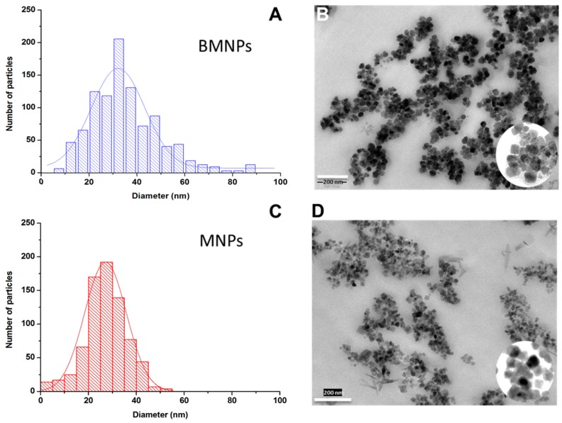
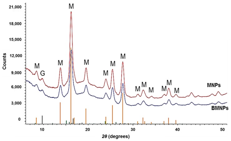
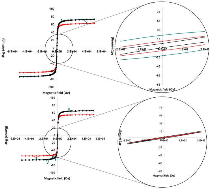
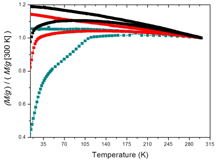
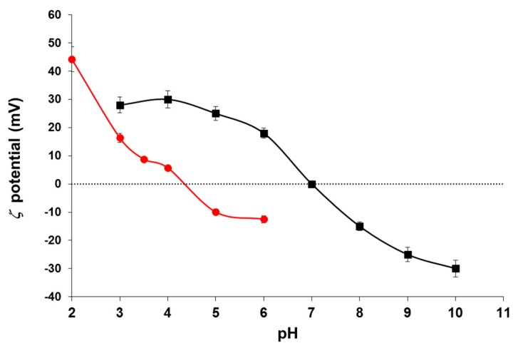
In this work we report on the synthesis and characterization of magnetic nanoparticles of two distinct origins, one inorganic (MNPs) and the other biomimetic (BMNPs), the latter based on a process of bacterial synthesis. Each of these two kinds of particles has its own advantages when used separately with biomedical purposes. Thus, BMNPs present an isoelectric point below neutrality (around pH 4.4), while MNPs show a zero-zeta potential at pH 7, and appear to be excellent agents for magnetic hyperthermia. This means that the biomimetic particles are better suited to be loaded with drug molecules positively charged at neutral pH (notably, doxorubicin, for instance) and releasing it at the acidic tumor environment. In turn, MNPs may provide their transport capabilities under a magnetic field. In this study it is proposed to use a mixture of both kinds of particles at two different concentrations, trying to get the best from each of them. We study which mixture performs better from different points of view, like stability and magnetic hyperthermia response, while keeping suitable drug transport capabilities. This composite system is proposed as a close to ideal drug vehicle with added enhanced hyperthermia response.
Overview
The claims and statistics of this paper haven’t been checked yet.
Run the check — free, no account needed. It reads the open-access text and checks the sources it cites.
Integrity signals
Registry
Cites
Statistics
Figures
Verification
Claims the paper makes
The claims and statistics of this paper haven’t been checked yet.
Run the check — free, no account needed. It reads the open-access text and checks the sources it cites.
Claims other papers make about this one
Each is checked against this paper’s own text: does it say what the citing paper says it does?
In the citing paper
“and as hyperthermia agents [16,17]”
From Biomimetic Magnetoliposomes as Oxaliplatin Nanocarriers: In Vitro Study for Potential Application in Colon Cancer (2020) · cites this paper as [16]
Self-check
Percentages, sums and reported differences re-computed from the paper’s own numbers.
The self-check needs the paper’s full text.
Citations
Cites
Magnetic fluid hyperthermia: focus on superparamagnetic iron oxide nanoparticles
Laurent, S., Dutz, S., Häfeli, U. O., & Mahmoudi, M. (2011) · Advances in Colloid and Interface Science · doi:10.1016/j.cis.2011.04.003 · Cited by 1,326
Research works printed but not linked: 56
- Rezayan, 2016, Monodisperse magnetite (Fe3O4) nanoparticles modified with water soluble polymers for the diagnosis of breast cancer by MRI method, J. Magn. Magn. Mater., 420, 210, 10.1016/j.jmmm.2016.07.003 · DOI
- Argibay, 2016, Multicore magnetic Fe3O4@C beads with enhanced magnetic response for MRI in brain biomedical applications, IEEE Trans. Magn., 52, 1
- Portillo, 2017, Magnetic nanoparticles as a redispersing additive in magnetorheological fluid, J. Nanomater., 2017, 8, 10.1155/2017/9026219 · DOI
- Cardoso, 2018, Advances in magnetic nanoparticles for biomedical applications, Adv. Healthc. Mater., 7, 1700845, 10.1002/adhm.201700845 · DOI
- Ghazi, 2018, Rietveld refinement, morphology analysis, optical and magnetic properties of magnesium-zinc ferrite nanofibers, J. Magn. Magn. Mater., 468, 132, 10.1016/j.jmmm.2018.07.084 · DOI
- Lisjak, 2018, Anisotropic magnetic nanoparticles: A review of their properties, syntheses and potential applications, Prog. Mater. Sci., 95, 286, 10.1016/j.pmatsci.2018.03.003 · DOI
- Muhlberger, 2019, Functionalization of T lymphocytes for magnetically controlled immune therapy: Selection of suitable superparamagnetic iron oxide nanoparticles, J. Magn. Magn. Mater., 473, 61, 10.1016/j.jmmm.2018.10.022 · DOI
- Danilushkina, 2018, Influence of magnetic nanoparticles stabilized with polyelectrolytes on 2D and 3D cell cultures formation, Hum. Gene Ther., 29, A71
- Chen, 2016, Influence of magnetoplasmonic gamma-Fe2O3/Au core/shell nanoparticles on low-field nuclear magnetic resonance, Sci. Rep., 6, 35477, 10.1038/srep35477 · DOI
- Wang, 2016, A magnetic nanoparticles relaxation sensor for protein-protein interaction detection at ultra-low magnetic field, Biosens. Bioelectron., 80, 661, 10.1016/j.bios.2016.02.037 · DOI
- Angelakeris, 2018, Magnetic nanoparticles: A multifunctional vehicle for modern theranostics, Biochim. Biophys. Acta Gen. Subj., 1861, 1642, 10.1016/j.bbagen.2017.02.022 · DOI
- Winter, A., Engels, S., Reinhardt, L., Wasylow, C., Gerullis, H., and Wawroschek, F. (2017). Magnetic marking and intraoperative detection of primary draining lymph nodes in high-risk prostate cancer using superparamagnetic iron oxide nanoparticles: Additional diagnostic value. Molecules, 22. · DOI
- Duran, 2008, Magnetic colloids as drug vehicles, J. Pharm. Sci., 97, 2948, 10.1002/jps.21249 · DOI
- Patitsa, 2017, Magnetic nanoparticles coated with polyarabic acid demonstrate enhanced drug delivery and imaging properties for cancer theranostic applications, Sci. Rep., 7, 775, 10.1038/s41598-017-00836-y · DOI
- Gao, 2018, A theranostic nanocomposite system based on radial mesoporous silica hybridized with Fe3O4 nanoparticles for targeted magnetic field responsive chemotherapy of breast cancer, Rsc Adv., 8, 4321, 10.1039/C7RA12446E · DOI
- Moskvin, 2018, Biological evaluation of surface-modified magnetic nanoparticles as a platform for colon cancer cell theranostics, Colloids Surf. B Biointerfaces, 161, 35, 10.1016/j.colsurfb.2017.10.034 · DOI
- Semkina, 2018, Multimodal doxorubicin loaded magnetic nanoparticles for VEGF targeted theranostics of breast cancer, Nanomed. Nanoechnol., 14, 1733, 10.1016/j.nano.2018.04.019 · DOI
- Vakilinezhad, 2018, Fabrication and in vitro evaluation of magnetic PLGA nanoparticles as a potential Methotrexate delivery system for breast cancer, J. Drug Deliv. Sci. Technol., 44, 467, 10.1016/j.jddst.2018.01.002 · DOI
- Obaidat, 2015, Magnetic properties of magnetic nanoparticles for efficient hyperthermia, Nanomaterials (Basel), 5, 63, 10.3390/nano5010063 · DOI
- Balidemaj, 2016, Hyperthermia treatment planning for cervical cancer patients based on electrical conductivity tissue properties acquired in vivo with EPT at 3 T MRI, Int. J. Hyperth., 32, 558, 10.3109/02656736.2015.1129440 · DOI
- Walter, 2016, Design of iron oxide-based nanoparticles for MRI and magnetic hyperthermia, Nanomedicine (Lond), 11, 1889, 10.2217/nnm-2016-5001 · DOI · Paper page
- Iglesias, 2016, Magnetic hyperthermia with magnetite nanoparticles: Electrostatic and polymeric stabilization, Colloid Polym. Sci., 294, 1541, 10.1007/s00396-016-3918-3 · DOI
- Sato, 2016, Simultaneous hyperthermia-chemotherapy with controlled drug delivery using single-drug nanoparticles, Sci. Rep., 6, 24629, 10.1038/srep24629 · DOI
- Hedayatnasab, 2017, Review on magnetic nanoparticles for magnetic nanofluid hyperthermia application, Mater. Des., 123, 174, 10.1016/j.matdes.2017.03.036 · DOI
- Nemati, 2017, Superparamagnetic iron oxide nanodiscs for hyperthermia therapy: Does size matter?, J. Alloy. Compd., 714, 709, 10.1016/j.jallcom.2017.04.211 · DOI
- Vamvakidis, 2018, Magnetic hyperthermia efficiency and MRI contrast sensitivity of colloidal soft/hard ferrite nanoclusters, J. Colloid Interface Sci., 511, 101, 10.1016/j.jcis.2017.10.001 · DOI
- Pankhurst, 2003, Applications of magnetic nanoparticles in biomedicine, J. Phys. D, 36, R167, 10.1088/0022-3727/36/13/201 · DOI
- Soares, 2012, Application of hyperthermia for cancer treatment: Recent patents review, Recent Pat. Anticancer Drug Discov., 7, 64, 10.2174/157489212798358038 · DOI
- Alphandery, 2011, Chains of magnetosomes extracted from AMB-1 magnetotactic bacteria for application in alternative magnetic field cancer therapy, Acs Nano, 5, 6279, 10.1021/nn201290k · DOI
- 2018, Magnetic iron oxide nanoparticles as drug carriers: Preparation, conjugation and delivery, Nanomedicine (Lond), 13, 929, 10.2217/nnm-2017-0320 · DOI
- Prozorov, 2015, Size control of in vitro synthesized magnetite crystals by the MamC protein of Magnetococcus marinus strain MC-1, Appl. Microbiol. Biotechnol., 99, 5109, 10.1007/s00253-014-6326-y · DOI · Paper page
- Peigneux, 2018, pH-dependent adsorption release of doxorubicin on MamC-biomimetic magnetite nanoparticles, Langmuir, 34, 13713, 10.1021/acs.langmuir.8b03109 · DOI · Paper page
- Lacroix, 2009, Magnetic hyperthermia in single-domain monodisperse FeCo nanoparticles: Evidences for Stoner-Wohlfarth behavior and large losses, J. Appl. Phys., 105, 023911, 10.1063/1.3068195 · DOI
- Carrey, 2011, Simple models for dynamic hysteresis loop calculations of magnetic single-domain nanoparticles: Application to magnetic hyperthermia optimization, J. Appl. Phys., 110, 083921, 10.1063/1.3551582 · DOI
- Mehdaoui, 2011, Optimal size of nanoparticles for magnetic hyperthermia: A combined theoretical and experimental study, Adv. Funct. Mater., 21, 4573, 10.1002/adfm.201101243 · DOI
- Stoner, 1991, A mechanism of magnetic hysteresis in heterogeneous alloys, IEEE Trans. Magn., 27, 3475, 10.1109/TMAG.1991.1183750 · DOI
- Pankhurst, 2009, Progress in applications of magnetic nanoparticles in biomedicine, J. Phys. D Appl. Phys., 42, 224001, 10.1088/0022-3727/42/22/224001 · DOI
- 2013, Magnetic hyperthermia, Nanoscience: Nanostructures through Chemistry, Volume 1, 60
- Surowiec, 2017, Investigation of magnetite Fe3O4 nanoparticles for magnetic hyperthermia, Nukleonika, 62, 183, 10.1515/nuka-2017-0028 · DOI
- Coffey, 1995, Constant-magnetic-field effect in Néel relaxation of single-domain ferromagnetic particles, Phys. Rev. B Condens. Matter., 51, 15947, 10.1103/PhysRevB.51.15947 · DOI
- Hergt, 2010, Validity limits of the Neel relaxation model of magnetic nanoparticles for hyperthermia, Nanotechnology, 21, 015706, 10.1088/0957-4484/21/1/015706 · DOI
- Bakoglidis, 2012, Size-Dependent mechanisms in AC magnetic hyperthermia response of iron-oxide nanoparticles, IEEE Trans. Magn., 48, 1320, 10.1109/TMAG.2011.2173474 · DOI
- Dutz, 2013, Magnetic nanoparticle heating and heat transfer on a microscale: Basic principles, realities and physical limitations of hyperthermia for tumour therapy, Int. J. Hyperth., 29, 790, 10.3109/02656736.2013.822993 · DOI
- Dutz, 2014, Magnetic particle hyperthermia-a promising tumour therapy?, Nanotechnology, 25, 452001, 10.1088/0957-4484/25/45/452001 · DOI
- Guibert, 2015, Hyperthermia of magnetic nanoparticles: Experimental study of the role of aggregation, J. Phys. Chem. C, 119, 28148, 10.1021/acs.jpcc.5b07796 · DOI
- Fortin, 2007, Size-sorted anionic iron oxide nanomagnets as colloidal mediators for magnetic hyperthermia, J. Am. Chem. Soc., 129, 2628, 10.1021/ja067457e · DOI
- 2011, Inorganic magnetite precipitation at 25 °C: A low-cost inorganic coprecipitation method, J. Supercond. Nov. Magn., 24, 549, 10.1007/s10948-010-0999-y · DOI
- Gas, P., and Kurgan, E. (2017, January 25–30). Cooling effects inside water-cooled inductors for magnetic fluid hyperthermia. Proceedings of the 2017 Progress in Applied Electrical Engineering (PAEE), Koscielisko, Poland. · DOI
- Gas, P., and Miaskowski, A. (2015, January 17–19). Specifying the ferrofluid parameters important from the viewpoint of Magnetic Fluid Hyperthermia. Proceedings of the 2015 Selected Problems of Electrical Engineering and Electronics (WZEE), Kielce, Poland. · DOI
- Wildeboer, 2014, On the reliable measurement of specific absorption rates and intrinsic loss parameters in magnetic hyperthermia materials, J. Phys. D Appl. Phys., 47, 495003, 10.1088/0022-3727/47/49/495003 · DOI
- Muniz, 2016, The Scherrer equation and the dynamical theory of X-ray diffraction, Acta Crystallogr. A Found. Adv., A72, 385, 10.1107/S205327331600365X · DOI
- Zarivach, 2017, Magnetite nanoparticles biomineralization in the presence of the magnetosome membrane protein MamC: Effect of protein aggregation and protein atructure on magnetite formation, Cryst. Growth Des., 17, 1620, 10.1021/acs.cgd.6b01643 · DOI · Paper page
- Fonseca, 2002, Superparamagnetism and magnetic properties of Ni nanoparticles embedded in SiO2, Phys. Rev. B, 66, 104406, 10.1103/PhysRevB.66.104406 · DOI
- Obrien, 1978, Electrophoretic mobility of a spherical colloidal particle, J. Chem. Soc. Faraday Trans. 2, 74, 1607, 10.1039/f29787401607 · DOI
- Nudelman, 2016, Structure-function studies of the magnetite-biomineralizing magnetosome-associated protein MamC, J. Struct. Biol., 194, 244, 10.1016/j.jsb.2016.03.001 · DOI
- Jabalera, 2019, Protein conservation method affects MamC-mediated biomineralization of magnetic nanoparticles, Cryst. Growth Des., 19, 1064, 10.1021/acs.cgd.8b01590 · DOI
Cited by
Sorted by citations
Garcia-Pinel, B., Jabalera, Y., Ortiz, R., Cabeza, L., Jimenez-Lopez, C., Melguizo, C., & Prados, J. (2020) · Pharmaceutics · doi:10.3390/pharmaceutics12060589 · Cited by 40
Self-citation
Metadata
- Authors
- Iglesias, G. R., Jabalera, Y., Peigneux, A., Checa Fernández, B. L., Delgado, Á. V., & Jimenez-Lopez, C.
- Venue
- Pharmaceutics
- Publisher
- MDPI AG
- Published
- 2019-06-11
- Volume
- 11
- Issue
- 6
- Pages
- 273
- ISSN
- 1999-4923
- PMC
- PMC6631210
- Cited by
- 36
- References
- 1
- Language
- en
About these checks
Registry, retraction, Cites and statistics results are computed, not judged, with one exception: a printed reference that no index lists is read once by a model before it counts as a citation error. Claims are extracted by a model and kept only when their quote appears verbatim in the text; each cited source is then checked, with a model’s help, for whether it says what the claim relies on. Those verdicts can be wrong — read the claim’s quote and the cited source before relying on one.
Statistics and arithmetic are recomputed, not judged.
Confidence bands are shown only for fields calibrated on our annotated test set. None is calibrated yet, so no band is shown.
What counts as a claim ›How we measure confidence ›How we check this ›
Are you an author, or do you see a problem with this page? Request a review
How to cite
Cite CoChat for the checks on this page, and the paper for its own findings.
- This page
- CoChat. (2026). Citation check: Enhancement of Magnetic Hyperthermia by Mixing Synthetic Inorganic and Biomimetic Magnetic Nanoparticles. https://app.cochat.ai/papers/10.3390/pharmaceutics11060273
- The paper
- Iglesias, G. R., Jabalera, Y., Peigneux, A., Checa Fernández, B. L., Delgado, Á. V., & Jimenez-Lopez, C. (2019). Enhancement of Magnetic Hyperthermia by Mixing Synthetic Inorganic and Biomimetic Magnetic Nanoparticles. Pharmaceutics. https://doi.org/10.3390/pharmaceutics11060273
