

The DOI matches its Crossref record; 63 of the 63 references it cites were found in a registry or an index; 1 of the 68 sources its claims cite were checked for support.
Biomimetic Magnetoliposomes as Oxaliplatin Nanocarriers: In Vitro Study for Potential Application in Colon Cancer
Open accessAbstract
Current chemotherapy for colorectal cancer (CRC) includes the use of oxaliplatin (Oxa), a first-line cytotoxic drug which, in combination with irinotecan/5-fluorouracil or biologic agents, increases the survival rate of patients. However, the administration of this drug induces side effects that limit its application in patients, making it necessary to develop new tools for targeted chemotherapy. MamC-mediated biomimetic magnetic nanoparticles coupled with Oxa (Oxa-BMNPs) have been previously demonstrated to efficiently reduce the IC50 compared to that of soluble Oxa. However, their strong interaction with the macrophages revealed toxicity and possibility of aggregation. In this scenario, a further improvement of this nanoassembly was necessary. In the present study, Oxa-BMNPs nanoassemblies were enveloped in phosphatidylcholine unilamellar liposomes (both pegylated and non-pegylated). Our results demonstrate that the addition of both a lipid cover and further pegylation improves the biocompatibility and cellular uptake of the Oxa-BMNPs nanoassemblies without significantly reducing their cytotoxic activity in colon cancer cells. In particular, with the pegylated magnetoliposome nanoformulation (a) hemolysis was reduced from 5% to 2%, being now hematocompatibles, (b) red blood cell agglutination was reduced, (c) toxicity in white blood cells was eliminated. This study represents a truly stepforward in this area as describes the production of one of the very few existing nanoformulations that could be used for a local chemotherapy to treat CRC.
Overview
AI snapshot
Written from the paper’s full text.
TL;DR
Coating oxaliplatin-loaded MamC-biomimetic magnetic nanoparticles in phosphatidylcholine liposomes, especially pegylated ones, improved blood compatibility and cellular uptake while preserving or improving cytotoxicity in colon cancer cells in vitro. The pegylated magnetoliposome reduced hemolysis to <2%, eliminated white blood cell toxicity, and reduced macrophage toxicity, supporting further development for local colorectal cancer chemotherapy.
Key findings
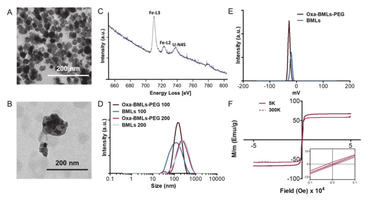
- Characterization: BMNPs were ~30–40 nm; Oxa-BMLs-PEG were 100–200 nm by TEM. DLS sizes were 98 ± 2 nm and 134.1 ± 0.9 nm for 100 nm extruder samples, and 227 ± 4 nm and 235 ± 5 nm for 200 nm extruder samples, with PDIs of 0.253 ± 0.009, 0.162 ± 0.004, 0.38 ± 0.01, and 0.36 ± 0.02. BMLs and Oxa-BMLs-PEG had ζ potentials of −20.4 ± 0.9 mV and −27.3 ± 0.4 mV; Oxa-MBLs-PEG showed superparamagnetic behavior at 300 K with Ms = 60 emu/g.
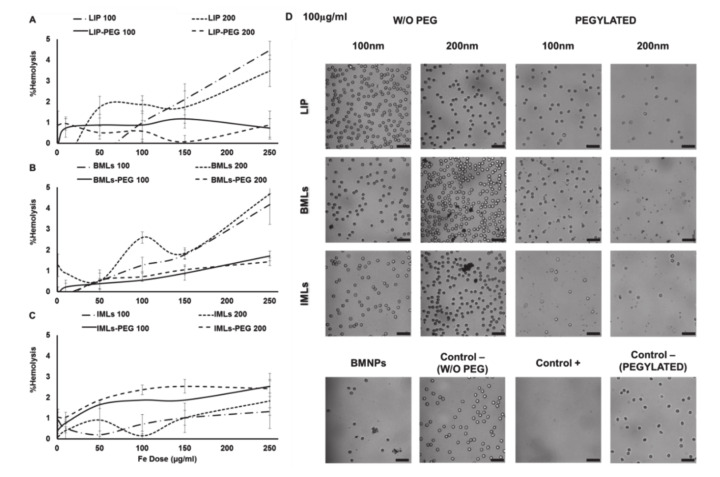
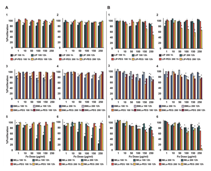
- Blood compatibility: LIP and BMLs caused ~5% hemolysis; pegylation reduced this to ~1% and <2%, respectively, while IML pegylation increased hemolysis from ~1% to ~2%. Pegylated formulations eliminated red blood cell agglutination. Pegylated nanoformulations showed no WBC toxicity at any dose at 1 h or 12 h; non-pegylated IMLs caused dose-dependent WBC toxicity at 1 h, with recovery at 12 h except for 100 nm BMLs.
- Macrophage and cytotoxicity: Pegylated nanoformulations were more toxic than non-pegylated ones in RAW 264.7, especially IMLs, but BMLs-PEG showed a 40% toxicity decrease versus previously reported uncoated BMNPs. Oxa-BMLs (100 and 200 nm), pegylated or not, had similar or higher effects than soluble Oxa; IC50 reductions versus free Oxa occurred in most tumor lines with pegylated formulations, especially HT29, though less pronounced than with previous Oxa-BMNPs.
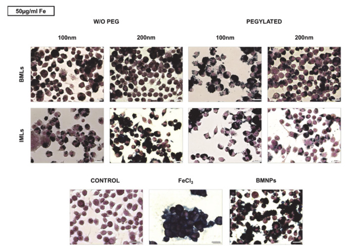
- Uptake and migration: BMLs and IMLs promoted magnetic migration, enhanced by pegylation. Prussian blue staining showed pegylation promoted internalization in T84, more for BMLs than IMLs and at 200 nm; CCD18 internalized within 15 min, while SW480 BMLs-PEG accumulations appeared after 2 h.
Methods
- BMNPs were synthesized using recombinant MamC expressed in E. coli, purified by FPLC/IMAC, with magnetite precipitated under anoxic conditions. Oxa coupling used 5 mg BMNPs mixed with 1 mL Oxa (2 mg/mL) for 72 h, with encapsulation efficiency of 80 ± 10%.
- Magnetoliposomes were made from egg phosphatidylcholine with or without DSPE-PEG (8% molar ratio), at a lipid/NP ratio of ~10,000, and extruded through 200 and 100 nm filters. Characterization used TEM/EELS, DLS, ζ potential, and SQUID.
- In vitro assays used colon cancer lines T84, HT29, SW480, HCT15, and MC38, plus normal RAW 264.7 and CCD18 cells. Assays included human RBC hemolysis, WBC proliferation (CCK-8), RAW 264.7 MTT, SRB proliferation after 72 h, Prussian blue iron staining, TEM, and magnetic migration; statistics used Student’s t-test with p < 0.05.
Limitations
- The paper does not include an explicit limitations section; the study is in vitro only, with no in vivo efficacy, biodistribution, or safety data, and the authors describe it as a proof of concept for future potential application.
- The authors state they cannot explain why pegylated nanoplatforms were slightly more toxic in macrophages than non-pegylated ones.
- The IC50 reduction with Oxa-BMLs/Oxa-BMLs-PEG was less pronounced than with previous Oxa-BMNPs, attributed to lower internalization; blood compatibility used human blood from a healthy donor (and WBCs from healthy donors), so donor variability is not addressed.
Model-written summary. Check it against the paper before relying on it.
Integrity signals
Registry
Cites
Statistics
Figures
Verification
Claims the paper makes
64 Own result · 31 Not checked yet · 14 Own interpretation · 12 No full text of the sources · 10 No basis given · 1 Partly supported · 1 Own method · accuracy not yet measured
The cited sources support only part of this claim — read each source’s verdict below.
In the paper
“On the other hand, pegylation also significantly reduce the interaction between the nanoformulations and the reticuloendothelial system cells such as lymphocytes and macrophages [49,51].”
§ Biomimetic Magnetoliposomes as Oxaliplatin Nanocarriers: In Vitro Study for Potential Application in Colon Cancer > 4. Discussion
What the sources say
- Partly supportedcites [49] Zhang, 2019, MWCNT interactions with protein: Surface-induced changes in protein adsorption and the impact of protein corona on cellular uptake and cytotoxicity, Int. J. Nanomed. sourceExperimental — accuracy not yet measured
The Discussion reports less uptake of PEGylated multiwalled carbon nanotubes by RAW264.7 macrophages, supporting the macrophage portion of the claim. It does not establish a significant reduction in interactions for nanoformulations generally or address lymphocytes or reticuloendothelial-system cells as a group. Page numbers are unavailable in our copy. (LLM, conf=0.86)
- Not checked — no full text of the sourcecites [51] Orlando, 2015, Iron oxide nanoparticles surface coating and cell uptake affect biocompatibility and inflammatory responses of endothelial cells and macrophages, J. Nanopart. Res. source
plaindirect- Asserted: passes — The authors put it forward as true.
- Checkable: passes — It can be checked against evidence.
- Load-bearing: passes — The paper’s conclusions depend on it.
- Based: passes — The paper gives a reason why it holds.
- Partly supported
A result the paper reports from its own data — it cites no other source for it, so there is none to check. The quote shows it is in the paper.
In the paper
“Magnetic nanoparticles carrying oxaliplatin (Oxa-BMNPs) were entrapped inside the liposome, as demonstrated by TEM and EELS analyses (Figure 1A–C).”
§ Biomimetic Magnetoliposomes as Oxaliplatin Nanocarriers: In Vitro Study for Potential Application in Colon Cancer > 3. Results > 3.1. Characterization of the Nanoformulations
boosted- Asserted: passes — The authors put it forward as true.
- Checkable: passes — It can be checked against evidence.
- Load-bearing: passes — The paper’s conclusions depend on it.
- Based: passes — The paper gives a reason why it holds.
A result the paper reports from its own data — it cites no other source for it, so there is none to check. The quote shows it is in the paper.
In the paper
“Our results demonstrate that the addition of both a lipid cover and further pegylation improves the biocompatibility and cellular uptake of the Oxa-BMNPs nanoassemblies without significantly reducing their cytotoxic activity in colon cancer cells.”
§ Biomimetic Magnetoliposomes as Oxaliplatin Nanocarriers: In Vitro Study for Potential Application in Colon Cancer > Abstract
boosteddirect- Asserted: passes — The authors put it forward as true.
- Checkable: passes — It can be checked against evidence.
- Load-bearing: passes — The paper’s conclusions depend on it.
- Based: passes — The paper gives a reason why it holds.
A result the paper reports from its own data — it cites no other source for it, so there is none to check. The quote shows it is in the paper.
In the paper
“Our results demonstrate that the addition of both a lipid cover and further pegylation improves the biocompatibility and cellular uptake of the Oxa-BMNPs nanoassemblies without significantly reducing their cytotoxic activity in colon cancer cells.”
§ Biomimetic Magnetoliposomes as Oxaliplatin Nanocarriers: In Vitro Study for Potential Application in Colon Cancer > Abstract
boosted- Asserted: passes — The authors put it forward as true.
- Checkable: passes — It can be checked against evidence.
- Load-bearing: passes — The paper’s conclusions depend on it.
- Based: passes — The paper gives a reason why it holds.
A result the paper reports from its own data — it cites no other source for it, so there is none to check. The quote shows it is in the paper.
In the paper
“(b) red blood cell agglutination was reduced”
§ Biomimetic Magnetoliposomes as Oxaliplatin Nanocarriers: In Vitro Study for Potential Application in Colon Cancer > Abstract
plaindirect- Asserted: passes — The authors put it forward as true.
- Checkable: passes — It can be checked against evidence.
- Load-bearing: passes — The paper’s conclusions depend on it.
- Based: passes — The paper gives a reason why it holds.
A result the paper reports from its own data — it cites no other source for it, so there is none to check. The quote shows it is in the paper.
In the paper
“(c) toxicity in white blood cells was eliminated.”
§ Biomimetic Magnetoliposomes as Oxaliplatin Nanocarriers: In Vitro Study for Potential Application in Colon Cancer > Abstract
plaindirect- Asserted: passes — The authors put it forward as true.
- Checkable: passes — It can be checked against evidence.
- Load-bearing: passes — The paper’s conclusions depend on it.
- Based: passes — The paper gives a reason why it holds.
A result the paper reports from its own data — it cites no other source for it, so there is none to check. The quote shows it is in the paper.
In the paper
“Our results demonstrate that the addition of both a lipid cover and further pegylation improves the biocompatibility and cellular uptake of the Oxa-BMNPs nanoassemblies without significantly reducing their cytotoxic activity in colon cancer cells.”
§ Biomimetic Magnetoliposomes as Oxaliplatin Nanocarriers: In Vitro Study for Potential Application in Colon Cancer > Abstract
boosteddirect- Asserted: passes — The authors put it forward as true.
- Checkable: passes — It can be checked against evidence.
- Load-bearing: passes — The paper’s conclusions depend on it.
- Based: passes — The paper gives a reason why it holds.
A result the paper reports from its own data — it cites no other source for it, so there is none to check. The quote shows it is in the paper.
In the paper
“In fact, EELS show the presence of iron, as expected for magnetite, inside the defined layer observed by TEM.”
§ Biomimetic Magnetoliposomes as Oxaliplatin Nanocarriers: In Vitro Study for Potential Application in Colon Cancer > 3. Results > 3.1. Characterization of the Nanoformulations
plain- Asserted: passes — The authors put it forward as true.
- Checkable: passes — It can be checked against evidence.
- Load-bearing: unclear — It is unclear whether the paper’s conclusions depend on it.
- Based: passes — The paper gives a reason why it holds.
A result the paper reports from its own data — it cites no other source for it, so there is none to check. The quote shows it is in the paper.
In the paper
“The encapsulation efficiency was 80 ± 10%.”
§ Biomimetic Magnetoliposomes as Oxaliplatin Nanocarriers: In Vitro Study for Potential Application in Colon Cancer > 2. Materials and Methods > 2.1. Preparation of Oxa-BMNP Nanoassemblies
plain- Asserted: passes — The authors put it forward as true.
- Checkable: passes — It can be checked against evidence.
- Load-bearing: unclear — It is unclear whether the paper’s conclusions depend on it.
- Based: passes — The paper gives a reason why it holds.
A result the paper reports from its own data — it cites no other source for it, so there is none to check. The quote shows it is in the paper.
In the paper
“and of 227 ± 4 nm and 235 ± 5 nm, respectively for BMLs and Oxa-BMLs-PEG passed through a 200 nm extruder (Figure 1D).”
§ Biomimetic Magnetoliposomes as Oxaliplatin Nanocarriers: In Vitro Study for Potential Application in Colon Cancer > 3. Results > 3.1. Characterization of the Nanoformulations
plain- Asserted: passes — The authors put it forward as true.
- Checkable: passes — It can be checked against evidence.
- Load-bearing: unclear — It is unclear whether the paper’s conclusions depend on it.
- Based: passes — The paper gives a reason why it holds.
- It may state two separate claims as one. 40
- Probably not a substantial claim. 10
A result the paper reports from its own data — it cites no other source for it, so there is none to check. The quote shows it is in the paper.
In the paper
“(a) hemolysis was reduced from 5% to 2%, being now hematocompatibles”
§ Biomimetic Magnetoliposomes as Oxaliplatin Nanocarriers: In Vitro Study for Potential Application in Colon Cancer > Abstract
plaindirect- Asserted: passes — The authors put it forward as true.
- Checkable: passes — It can be checked against evidence.
- Load-bearing: passes — The paper’s conclusions depend on it.
- Based: passes — The paper gives a reason why it holds.
It may state two separate claims as one.
A result the paper reports from its own data — it cites no other source for it, so there is none to check. The quote shows it is in the paper.
In the paper
“Identically, MNPs showed an average size value of 20 nm, while that for IMLs and IMLs-PEG was of 100 and 200 nm (Figure S1).”
§ Biomimetic Magnetoliposomes as Oxaliplatin Nanocarriers: In Vitro Study for Potential Application in Colon Cancer > 3. Results > 3.1. Characterization of the Nanoformulations
plain- Asserted: passes — The authors put it forward as true.
- Checkable: passes — It can be checked against evidence.
- Load-bearing: unclear — It is unclear whether the paper’s conclusions depend on it.
- Based: passes — The paper gives a reason why it holds.
It may state two separate claims as one.
A result the paper reports from its own data — it cites no other source for it, so there is none to check. The quote shows it is in the paper.
In the paper
“while TEM analyses on Oxa-BMLs-PEG show particles with a diameter of 100–200 nm, enclosing several magnetic nanoparticles per liposome (Figure 1B).”
§ Biomimetic Magnetoliposomes as Oxaliplatin Nanocarriers: In Vitro Study for Potential Application in Colon Cancer > 3. Results > 3.1. Characterization of the Nanoformulations
plain- Asserted: passes — The authors put it forward as true.
- Checkable: passes — It can be checked against evidence.
- Load-bearing: passes — The paper’s conclusions depend on it.
- Based: passes — The paper gives a reason why it holds.
It may state two separate claims as one.
A result the paper reports from its own data — it cites no other source for it, so there is none to check. The quote shows it is in the paper.
In the paper
“The polydispersity index (PDI) of these samples were 0.253 ± 0.009, 0.162 ± 0.004, 0.38 ± 0.01 and 0.36 ± 0.02, respectively, which indicates the good dispersity of the samples.”
§ Biomimetic Magnetoliposomes as Oxaliplatin Nanocarriers: In Vitro Study for Potential Application in Colon Cancer > 3. Results > 3.1. Characterization of the Nanoformulations
hedged- Asserted: passes — The authors put it forward as true.
- Checkable: passes — It can be checked against evidence.
- Load-bearing: unclear — It is unclear whether the paper’s conclusions depend on it.
- Based: passes — The paper gives a reason why it holds.
It may state two separate claims as one.
A result the paper reports from its own data — it cites no other source for it, so there is none to check. The quote shows it is in the paper.
In the paper
“Dynamic light scattering (DLS) analysis show that the hydrodynamic diameters (Table S1) of the BMLs and Oxa-BMLs-PEG passed through a 100 nm extruder were of 98 ± 2 nm and 134.1 ± 0.9 nm, respectively”
§ Biomimetic Magnetoliposomes as Oxaliplatin Nanocarriers: In Vitro Study for Potential Application in Colon Cancer > 3. Results > 3.1. Characterization of the Nanoformulations
plain- Asserted: passes — The authors put it forward as true.
- Checkable: passes — It can be checked against evidence.
- Load-bearing: unclear — It is unclear whether the paper’s conclusions depend on it.
- Based: passes — The paper gives a reason why it holds.
It may state two separate claims as one.
A result the paper reports from its own data — it cites no other source for it, so there is none to check. The quote shows it is in the paper.
In the paper
“BMNPs (Figure 1A) were uniform well-defined nanoparticles with a diameter of approximately 30–40 nm”
§ Biomimetic Magnetoliposomes as Oxaliplatin Nanocarriers: In Vitro Study for Potential Application in Colon Cancer > 3. Results > 3.1. Characterization of the Nanoformulations
plain- Asserted: passes — The authors put it forward as true.
- Checkable: passes — It can be checked against evidence.
- Load-bearing: unclear — It is unclear whether the paper’s conclusions depend on it.
- Based: passes — The paper gives a reason why it holds.
It may state two separate claims as one.
A result the paper reports from its own data — it cites no other source for it, so there is none to check. The quote shows it is in the paper.
In the paper
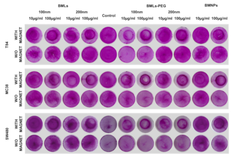
“when looking closely to the dark halo resulting from the magnet treatment in non-pegylated nanoformulations, it can be concluded that it is created by non-internalized ones, which does not occur in pegylated nanoformulations.”
§ Biomimetic Magnetoliposomes as Oxaliplatin Nanocarriers: In Vitro Study for Potential Application in Colon Cancer > 3. Results > 3.4. Cell Migration under a Magnetic Field In Vitro
plaindirect- Asserted: passes — The authors put it forward as true.
- Checkable: passes — It can be checked against evidence.
- Load-bearing: unclear — It is unclear whether the paper’s conclusions depend on it.
- Based: passes — The paper gives a reason why it holds.
It may state two separate claims as one.
A result the paper reports from its own data — it cites no other source for it, so there is none to check. The quote shows it is in the paper.
In the paper
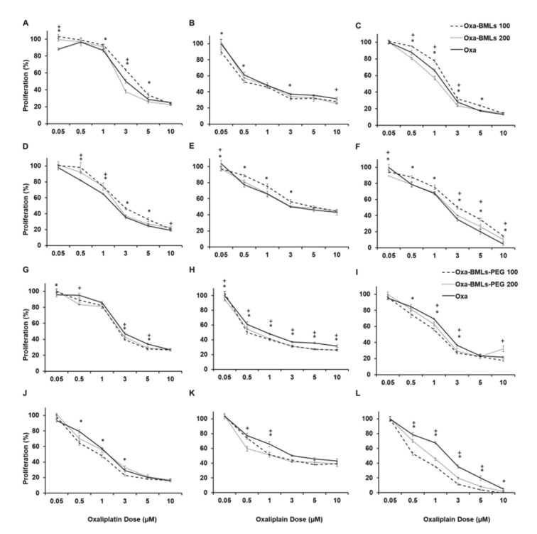
“In all cases there are significant differences (p < 0.05) between the IC50 of the treatments in the different lines except for CCD18 (all the treatments), Oxa-BMLs 200 nm in HT29 and the Oxa-BMLs-PEG 200 nm in SW480.”
§ Biomimetic Magnetoliposomes as Oxaliplatin Nanocarriers: In Vitro Study for Potential Application in Colon Cancer > 3. Results > 3.3. In Vitro Proliferation Assays
plain- Asserted: passes — The authors put it forward as true.
- Checkable: passes — It can be checked against evidence.
- Load-bearing: unclear — It is unclear whether the paper’s conclusions depend on it.
- Based: passes — The paper gives a reason why it holds.
It may state two separate claims as one.
A result the paper reports from its own data — it cites no other source for it, so there is none to check. The quote shows it is in the paper.
In the paper
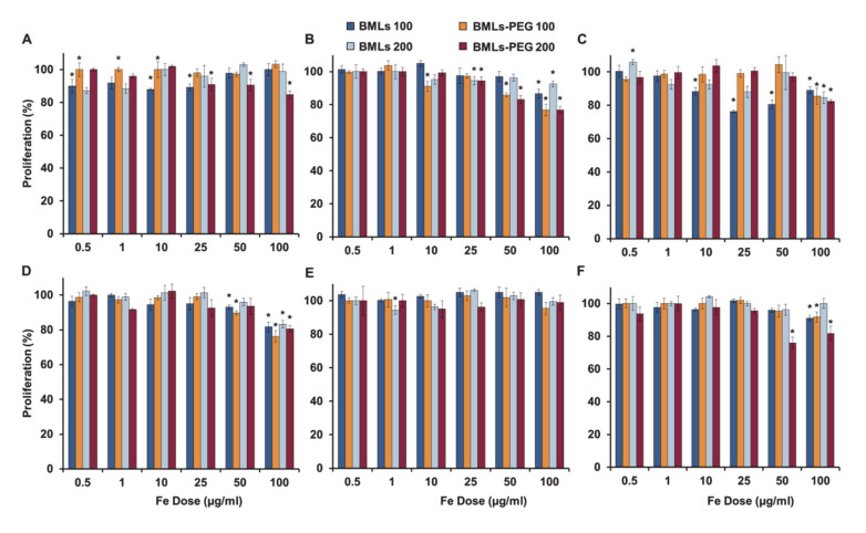
“In contrast, LIP (Figure 3(A1),(A2)) and BMLs (Figure 3(A3)) induce a ~ 20% toxicity regardless of the dose (with the exception of 200 nm BMLs in which a dose dependent toxicity is observed; Figure 3(A4)).”
§ Biomimetic Magnetoliposomes as Oxaliplatin Nanocarriers: In Vitro Study for Potential Application in Colon Cancer > 3. Results > 3.2. BMLs and IMLs Biocompatibility in Blood Cells
plaindirect- Asserted: passes — The authors put it forward as true.
- Checkable: passes — It can be checked against evidence.
- Load-bearing: passes — The paper’s conclusions depend on it.
- Based: passes — The paper gives a reason why it holds.
It may state two separate claims as one.
A result the paper reports from its own data — it cites no other source for it, so there is none to check. The quote shows it is in the paper.
In the paper
“the administration of BMNPs reduces such Fe internalization, because the nanoformulations adhere to the surface and only a small proportion is internalized.”
§ Biomimetic Magnetoliposomes as Oxaliplatin Nanocarriers: In Vitro Study for Potential Application in Colon Cancer > 3. Results > 3.2. BMLs and IMLs Biocompatibility in Blood Cells
plaindirect- Asserted: passes — The authors put it forward as true.
- Checkable: passes — It can be checked against evidence.
- Load-bearing: unclear — It is unclear whether the paper’s conclusions depend on it.
- Based: passes — The paper gives a reason why it holds.
It may state two separate claims as one.
A result the paper reports from its own data — it cites no other source for it, so there is none to check. The quote shows it is in the paper.
In the paper
“The hysteresis loop of Oxa-MBLs-PEG showed a typical ferromagnetic behavior at 5 K, while at 300 K, these formulations showed zero coercivity, which indicates their superparamagnetic character”
§ Biomimetic Magnetoliposomes as Oxaliplatin Nanocarriers: In Vitro Study for Potential Application in Colon Cancer > 3. Results > 3.1. Characterization of the Nanoformulations
hedgedcorrelational- Asserted: passes — The authors put it forward as true.
- Checkable: passes — It can be checked against evidence.
- Load-bearing: passes — The paper’s conclusions depend on it.
- Based: passes — The paper gives a reason why it holds.
It may state two separate claims as one.
None of the sources cited for this claim have been checked yet.
In the paper
“our results demonstrated that BMLs-PEG were non-toxic to WBC both at 1 and 12 h of exposure by contrast to uncovered NPs in addition to observing a recovery of viability in most treatments after 12 h of exposure such as has been previously described [14].”
§ Biomimetic Magnetoliposomes as Oxaliplatin Nanocarriers: In Vitro Study for Potential Application in Colon Cancer > 4. Discussion
What the sources say
- Not checked yetcites [14] Jabalera, Y., Garcia-Pinel, B., Ortiz, R., Iglesias, G., Cabeza, L., Prados, J., Jimenez-Lopez, C., and Melguizo, C. (2019). Oxaliplatin–Biomimetic Magnetic Nanoparticle Assemblies for Colon Cancer-Targeted Chemotherapy: An In Vitro Study. Pharmaceutics, 11. source
Checked before — its earlier verdict is held back until a new check runs.
boosted- Asserted: passes — The authors put it forward as true.
- Checkable: passes — It can be checked against evidence.
- Load-bearing: passes — The paper’s conclusions depend on it.
- Based: passes — The paper gives a reason why it holds.
It may state two separate claims as one.
- Not checked yet
The paper states this without a source or data behind it here, so there is nothing to check it against. The quote shows it is in the paper.
In the paper
“A reduction in the IC50 of free OXA is observed in most tumor cell lines when treated with pegylated nanoformulations, being especially remarkable such reduction in HT29.”
§ Biomimetic Magnetoliposomes as Oxaliplatin Nanocarriers: In Vitro Study for Potential Application in Colon Cancer > 4. Discussion
plaindirect- Asserted: passes — The authors put it forward as true.
- Checkable: passes — It can be checked against evidence.
- Load-bearing: passes — The paper’s conclusions depend on it.
- Based: passes — The paper gives a reason why it holds.
It may state two separate claims as one.
A result the paper reports from its own data — it cites no other source for it, so there is none to check. The quote shows it is in the paper.
In the paper
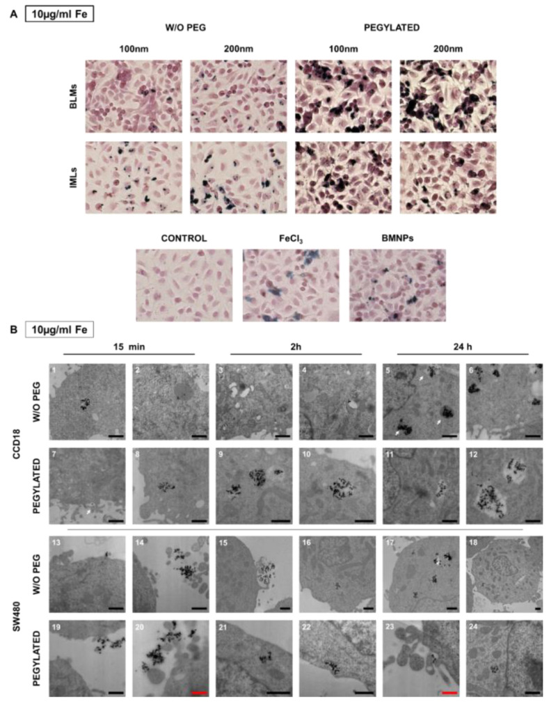
“This event can be observed more noticeably for BMLs than for IMLs, and for the 200 nm size.”
§ Biomimetic Magnetoliposomes as Oxaliplatin Nanocarriers: In Vitro Study for Potential Application in Colon Cancer > 3. Results > 3.5. BMLs Internalization in Colon Cell Lines
plaindirect- Asserted: passes — The authors put it forward as true.
- Checkable: passes — It can be checked against evidence.
- Load-bearing: unclear — It is unclear whether the paper’s conclusions depend on it.
- Based: passes — The paper gives a reason why it holds.
It may state two separate claims as one.
A result the paper reports from its own data — it cites no other source for it, so there is none to check. The quote shows it is in the paper.
In the paper
“On the other hand, in MC38, the administration of IMLs-PEG at high doses is toxic, however this toxicity is lower than that observed with the administration of free iron.”
§ Biomimetic Magnetoliposomes as Oxaliplatin Nanocarriers: In Vitro Study for Potential Application in Colon Cancer > 3. Results > 3.3. In Vitro Proliferation Assays
plaindirect- Asserted: passes — The authors put it forward as true.
- Checkable: passes — It can be checked against evidence.
- Load-bearing: unclear — It is unclear whether the paper’s conclusions depend on it.
- Based: passes — The paper gives a reason why it holds.
It may state two separate claims as one.
A result the paper reports from its own data — it cites no other source for it, so there is none to check. The quote shows it is in the paper.
In the paper
“Both BMLs and Oxa-BMLs-PEG are negatively charged at pH 7.4, (ζ potential of −20.4 ± 0.9 mV and –27.3 ± 0.4, respectively, Figure 1E and Table S1).”
§ Biomimetic Magnetoliposomes as Oxaliplatin Nanocarriers: In Vitro Study for Potential Application in Colon Cancer > 3. Results > 3.1. Characterization of the Nanoformulations
plain- Asserted: passes — The authors put it forward as true.
- Checkable: passes — It can be checked against evidence.
- Load-bearing: unclear — It is unclear whether the paper’s conclusions depend on it.
- Based: passes — The paper gives a reason why it holds.
It may state two separate claims as one.
In the paper
“however, with pegylation, their percentage of hemolysis decreases to ~1% and <2% (respectively), being then considered biocompatible (Figure 2A,B) [39].”
§ Biomimetic Magnetoliposomes as Oxaliplatin Nanocarriers: In Vitro Study for Potential Application in Colon Cancer > 3. Results > 3.2. BMLs and IMLs Biocompatibility in Blood Cells
What the sources say
- Not checked — no full text of the sourcecites [39] Schlenk, 2017, Comprehensive analysis of the in vitro and ex ovo hemocompatibility of surface engineered iron oxide nanoparticles for biomedical applications, Arch. Toxicol. source
plaindirect- Asserted: passes — The authors put it forward as true.
- Checkable: passes — It can be checked against evidence.
- Load-bearing: passes — The paper’s conclusions depend on it.
- Based: passes — The paper gives a reason why it holds.
It may state two separate claims as one.
A result the paper reports from its own data — it cites no other source for it, so there is none to check. The quote shows it is in the paper.
In the paper
“In fact, BMLs-PEG accumulations start being observed inside the cell after 2 h of exposure and continue at 24 h.”
§ Biomimetic Magnetoliposomes as Oxaliplatin Nanocarriers: In Vitro Study for Potential Application in Colon Cancer > 3. Results > 3.5. BMLs Internalization in Colon Cell Lines
plaincorrelational- Asserted: passes — The authors put it forward as true.
- Checkable: passes — It can be checked against evidence.
- Load-bearing: unclear — It is unclear whether the paper’s conclusions depend on it.
- Based: passes — The paper gives a reason why it holds.
It may state two separate claims as one.
A result the paper reports from its own data — it cites no other source for it, so there is none to check. The quote shows it is in the paper.
In the paper
“In the context of toxicity for RAW 264.7 cell line (Figure 3B), our results demonstrate that the pegylated nanoformulations are more toxic than non-pegylated ones, being that especially true for IMLs.”
§ Biomimetic Magnetoliposomes as Oxaliplatin Nanocarriers: In Vitro Study for Potential Application in Colon Cancer > 3. Results > 3.2. BMLs and IMLs Biocompatibility in Blood Cells
boostedcorrelational- Asserted: passes — The authors put it forward as true.
- Checkable: passes — It can be checked against evidence.
- Load-bearing: unclear — It is unclear whether the paper’s conclusions depend on it.
- Based: passes — The paper gives a reason why it holds.
It may state two separate claims as one.
In the paper
“Only IML pegylation induced a slight increase on their hemolysis capacity (1% to ~2%), although within the normal range of hemolysis that is considered biocompatible (0–2%) [47].”
§ Biomimetic Magnetoliposomes as Oxaliplatin Nanocarriers: In Vitro Study for Potential Application in Colon Cancer > 4. Discussion
What the sources say
- Not checked — no full text of the sourcecites [47] Raguraman, 2020, Comparative ecotoxicity assessment of magnetosomes and magnetite nanoparticles, Int. J. Environ. Health Res. source
plaindirect- Asserted: passes — The authors put it forward as true.
- Checkable: passes — It can be checked against evidence.
- Load-bearing: unclear — It is unclear whether the paper’s conclusions depend on it.
- Based: passes — The paper gives a reason why it holds.
It may state two separate claims as one.
None of the sources cited for this claim have been checked yet.
In the paper
“In comparison to our previous studies [14], the addition of a lipid cover to the nanoparticles improve their dispersion, eliminating the agglutination of erythrocytes previously observed.”
§ Biomimetic Magnetoliposomes as Oxaliplatin Nanocarriers: In Vitro Study for Potential Application in Colon Cancer > 4. Discussion
What the sources say
- Not checked yetcites [14] Jabalera, Y., Garcia-Pinel, B., Ortiz, R., Iglesias, G., Cabeza, L., Prados, J., Jimenez-Lopez, C., and Melguizo, C. (2019). Oxaliplatin–Biomimetic Magnetic Nanoparticle Assemblies for Colon Cancer-Targeted Chemotherapy: An In Vitro Study. Pharmaceutics, 11. source
Checked before — its earlier verdict is held back until a new check runs.
plaindirect- Asserted: passes — The authors put it forward as true.
- Checkable: passes — It can be checked against evidence.
- Load-bearing: passes — The paper’s conclusions depend on it.
- Based: passes — The paper gives a reason why it holds.
It may state two separate claims as one.
- Not checked yet
The paper states this without a source or data behind it here, so there is nothing to check it against. The quote shows it is in the paper.
In the paper
“While LIPs and BMLs show a slight toxicity at very high doses (250 µg/mL) at 12 h of exposure, IMLs show a dramatic reduction of viability.”
§ Biomimetic Magnetoliposomes as Oxaliplatin Nanocarriers: In Vitro Study for Potential Application in Colon Cancer > 4. Discussion
plaindirect- Asserted: passes — The authors put it forward as true.
- Checkable: passes — It can be checked against evidence.
- Load-bearing: passes — The paper’s conclusions depend on it.
- Based: passes — The paper gives a reason why it holds.
It may state two separate claims as one.
A result the paper reports from its own data — it cites no other source for it, so there is none to check. The quote shows it is in the paper.
In the paper
“In the context of the Oxa-BMNPs nanoassemblies, the addition of both a lipid shell and further pegylation not only improves the biocompatibility of the nanoassembly, but also the antiproliferative effect compared to that of soluble OXA.”
§ Biomimetic Magnetoliposomes as Oxaliplatin Nanocarriers: In Vitro Study for Potential Application in Colon Cancer > 4. Discussion
plaindirect- Asserted: passes — The authors put it forward as true.
- Checkable: passes — It can be checked against evidence.
- Load-bearing: passes — The paper’s conclusions depend on it.
- Based: passes — The paper gives a reason why it holds.
It may state two separate claims as one.
A result the paper reports from its own data — it cites no other source for it, so there is none to check. The quote shows it is in the paper.
In the paper
“it can be observed that non-pegylated LIP present toxicity in the SW480, HCT15 and MC38 lines in a dose-dependent manner, being higher the toxicity in the MC38 cell line.”
§ Biomimetic Magnetoliposomes as Oxaliplatin Nanocarriers: In Vitro Study for Potential Application in Colon Cancer > 3. Results > 3.3. In Vitro Proliferation Assays
plaincorrelational- Asserted: passes — The authors put it forward as true.
- Checkable: passes — It can be checked against evidence.
- Load-bearing: unclear — It is unclear whether the paper’s conclusions depend on it.
- Based: passes — The paper gives a reason why it holds.
It may state two separate claims as one.
A conclusion the paper draws from its own results — it cites no other source for it, so there is none to check. The quote shows it is in the paper.
In the paper
“while (ii) increase the intensity of the BMNPs response to an external magnetic field, thus increasing the efficiency of their magnetic guidance.”
§ Biomimetic Magnetoliposomes as Oxaliplatin Nanocarriers: In Vitro Study for Potential Application in Colon Cancer > 1. Introduction
plaindirect- Asserted: passes — The authors put it forward as true.
- Checkable: passes — It can be checked against evidence.
- Load-bearing: passes — The paper’s conclusions depend on it.
- Based: passes — The paper gives a reason why it holds.
It may state two separate claims as one.
In the paper
“MamC confers new surface properties to these BMNPs, in particular, an isoelectric point (pI) at pH 4.4 which allows functionalization based on electrostatic interactions and drug release being pH dependent.”
§ Biomimetic Magnetoliposomes as Oxaliplatin Nanocarriers: In Vitro Study for Potential Application in Colon Cancer > 1. Introduction
What the sources say
- Not checked — no full text of the sourcecites [5] Peigneux, 2018, PH-Dependent Adsorption Release of Doxorubicin on MamC-Biomimetic Magnetite Nanoparticles, Langmuir source
plaindirect- Asserted: passes — The authors put it forward as true.
- Checkable: passes — It can be checked against evidence.
- Load-bearing: passes — The paper’s conclusions depend on it.
- Based: passes — The paper gives a reason why it holds.
It may state two separate claims as one.
None of the sources cited for this claim have been checked yet.
In the paper
“In addition, a camptothecin nanoformulation achieved a pathologic complete response with only a grade 4 toxicity event in patients with advanced rectal cancer (phase Ib/II study) [26].”
§ Biomimetic Magnetoliposomes as Oxaliplatin Nanocarriers: In Vitro Study for Potential Application in Colon Cancer > 1. Introduction
What the sources say
- Not checked yetcites [26] Sanoff, 2019, Phase I/II trial of nano-camptothecin CRLX101 with capecitabine and radiotherapy as neoadjuvant treatment for locally advanced rectal cancer, Nanomed. Nanotechnol. Biol. Med. source
Checked before — its earlier verdict is held back until a new check runs.
plaindirect- Asserted: passes — The authors put it forward as true.
- Checkable: passes — It can be checked against evidence.
- Load-bearing: fails — The paper’s conclusions may not depend on it.
- Based: passes — The paper gives a reason why it holds.
Probably not a substantial claim. The paper’s conclusions may not depend on it.
- Not checked yet
None of the sources cited for this claim have been checked yet.
In the paper
“In fact, a previous proof of concept showed that BMNPs were biocompatible and could be coupled with Oxaliplatin (Oxa) [14].”
§ Biomimetic Magnetoliposomes as Oxaliplatin Nanocarriers: In Vitro Study for Potential Application in Colon Cancer > 1. Introduction
What the sources say
- Not checked yetcites [14] Jabalera, Y., Garcia-Pinel, B., Ortiz, R., Iglesias, G., Cabeza, L., Prados, J., Jimenez-Lopez, C., and Melguizo, C. (2019). Oxaliplatin–Biomimetic Magnetic Nanoparticle Assemblies for Colon Cancer-Targeted Chemotherapy: An In Vitro Study. Pharmaceutics, 11. source
Checked before — its earlier verdict is held back until a new check runs.
plaindirect- Asserted: passes — The authors put it forward as true.
- Checkable: passes — It can be checked against evidence.
- Load-bearing: passes — The paper’s conclusions depend on it.
- Based: passes — The paper gives a reason why it holds.
It may state two separate claims as one.
- Not checked yet
A conclusion the paper draws from its own results — it cites no other source for it, so there is none to check. The quote shows it is in the paper.
In the paper
“Since BMNPs showed much better performance as Oxa carriers (detailed below), MNPs-bearing nanoformulations were not further used for the present study.”
§ Biomimetic Magnetoliposomes as Oxaliplatin Nanocarriers: In Vitro Study for Potential Application in Colon Cancer > 3. Results > 3.1. Characterization of the Nanoformulations
plaindirect- Asserted: passes — The authors put it forward as true.
- Checkable: passes — It can be checked against evidence.
- Load-bearing: passes — The paper’s conclusions depend on it.
- Based: passes — The paper gives a reason why it holds.
It may state two separate claims as one.
None of the sources cited for this claim have been checked yet.
In the paper
“However, as a drawback, there was a strong interaction of the nanoassemblies with the macrophages thus reducing the viability of these cells.”
§ Biomimetic Magnetoliposomes as Oxaliplatin Nanocarriers: In Vitro Study for Potential Application in Colon Cancer > 1. Introduction
What the sources say
- Not checked yetcites [14] Jabalera, Y., Garcia-Pinel, B., Ortiz, R., Iglesias, G., Cabeza, L., Prados, J., Jimenez-Lopez, C., and Melguizo, C. (2019). Oxaliplatin–Biomimetic Magnetic Nanoparticle Assemblies for Colon Cancer-Targeted Chemotherapy: An In Vitro Study. Pharmaceutics, 11. source
Checked before — its earlier verdict is held back until a new check runs.
boosteddirect- Asserted: passes — The authors put it forward as true.
- Checkable: passes — It can be checked against evidence.
- Load-bearing: passes — The paper’s conclusions depend on it.
- Based: passes — The paper gives a reason why it holds.
It may state two separate claims as one.
- Not checked yet
None of the sources cited for this claim have been checked yet.
In the paper
“However, their strong interaction with the macrophages revealed toxicity and possibility of aggregation.”
§ Biomimetic Magnetoliposomes as Oxaliplatin Nanocarriers: In Vitro Study for Potential Application in Colon Cancer > Abstract
What the sources say
- Not checked yetcites [14] Jabalera, Y., Garcia-Pinel, B., Ortiz, R., Iglesias, G., Cabeza, L., Prados, J., Jimenez-Lopez, C., and Melguizo, C. (2019). Oxaliplatin–Biomimetic Magnetic Nanoparticle Assemblies for Colon Cancer-Targeted Chemotherapy: An In Vitro Study. Pharmaceutics, 11. source
Checked before — its earlier verdict is held back until a new check runs.
boosteddirect- Asserted: passes — The authors put it forward as true.
- Checkable: passes — It can be checked against evidence.
- Load-bearing: passes — The paper’s conclusions depend on it.
- Based: passes — The paper gives a reason why it holds.
It may state two separate claims as one.
- Not checked yet
In the paper
“(1) a higher blocking temperature while being superparamagnetic at room temperature”
§ Biomimetic Magnetoliposomes as Oxaliplatin Nanocarriers: In Vitro Study for Potential Application in Colon Cancer > 1. Introduction
What the sources say
- Not checked — no full text of the sourcecites [5] Peigneux, 2018, PH-Dependent Adsorption Release of Doxorubicin on MamC-Biomimetic Magnetite Nanoparticles, Langmuir source
plain- Asserted: passes — The authors put it forward as true.
- Checkable: passes — It can be checked against evidence.
- Load-bearing: passes — The paper’s conclusions depend on it.
- Based: passes — The paper gives a reason why it holds.
It may state two separate claims as one.
The paper states this without a source or data behind it here, so there is nothing to check it against. The quote shows it is in the paper.
In the paper
“and it is also one of the first tryout for the local delivery of this drug”
§ Biomimetic Magnetoliposomes as Oxaliplatin Nanocarriers: In Vitro Study for Potential Application in Colon Cancer > 1. Introduction
plain- Asserted: passes — The authors put it forward as true.
- Checkable: passes — It can be checked against evidence.
- Load-bearing: fails — The paper’s conclusions may not depend on it.
- Based: fails — The paper may give no reason why it holds.
Probably not a substantial claim. The paper’s conclusions may not depend on it. The paper may give no reason why it holds.
None of the sources cited for this claim have been checked yet.
In the paper
“In particular, Oxa induces peripheral sensory neurotoxicity [19] which may be the cause to discontinuation of drug treatment.”
§ Biomimetic Magnetoliposomes as Oxaliplatin Nanocarriers: In Vitro Study for Potential Application in Colon Cancer > 1. Introduction
What the sources say
- Not checked yetcites [19] Bender, 2019, Adjuvant therapy for stages II and III colon cancer: Risk stratification, treatment duration, and future directions, Curr. Oncol. source
Checked before — its earlier verdict is held back until a new check runs.
hedgedconditional- Asserted: passes — The authors put it forward as true.
- Checkable: passes — It can be checked against evidence.
- Load-bearing: passes — The paper’s conclusions depend on it.
- Based: passes — The paper gives a reason why it holds.
It may state two separate claims as one.
- Not checked yet
None of the sources cited for this claim have been checked yet.
In the paper
“Harada et al. recently demonstrated a significant reduction of adverse effects (AEs) using a paclitaxel (PTX) nanoformulation (nab-PTX) in a phase I/II study of advanced non-small cell lung cancer (NSCLC) [24].”
§ Biomimetic Magnetoliposomes as Oxaliplatin Nanocarriers: In Vitro Study for Potential Application in Colon Cancer > 1. Introduction
What the sources say
- Not checked yetcites [24] Harada, 2019, A phase I/II trial of weekly nab-paclitaxel for pretreated non-small-cell lung cancer patients without epidermal growth factor receptor mutations and anaplastic lymphoma kinase rearrangement, Asia Pac. J. Clin. Oncol. source
Checked before — its earlier verdict is held back until a new check runs.
boosteddirect- Asserted: passes — The authors put it forward as true.
- Checkable: passes — It can be checked against evidence.
- Load-bearing: fails — The paper’s conclusions may not depend on it.
- Based: passes — The paper gives a reason why it holds.
Probably not a substantial claim. The paper’s conclusions may not depend on it.
- Not checked yet
In the paper
“remaining attached to the nanoparticles and forming a nanocomposite (95 wt% magnetite + 5 wt% MamC)”
§ Biomimetic Magnetoliposomes as Oxaliplatin Nanocarriers: In Vitro Study for Potential Application in Colon Cancer > 1. Introduction
What the sources say
- Not checked — no full text of the sourcecites [5] Peigneux, 2018, PH-Dependent Adsorption Release of Doxorubicin on MamC-Biomimetic Magnetite Nanoparticles, Langmuir source
plaindirect- Asserted: passes — The authors put it forward as true.
- Checkable: passes — It can be checked against evidence.
- Load-bearing: unclear — It is unclear whether the paper’s conclusions depend on it.
- Based: passes — The paper gives a reason why it holds.
It may state two separate claims as one.
In the paper
“Fujiwara et al. showed a decrease (81.3%) of the peripheral sensory neuropathy with the use of polymeric micelles loaded with PTX (NK105) in patients with breast cancer (phase III) [25].”
§ Biomimetic Magnetoliposomes as Oxaliplatin Nanocarriers: In Vitro Study for Potential Application in Colon Cancer > 1. Introduction
What the sources say
- Not checked yetcites [25] Fujiwara, 2019, A multi-national, randomised, open-label, parallel, phase III non-inferiority study comparing NK105 and paclitaxel in metastatic or recurrent breast cancer patients, Br. J. Cancer source
plaindirect- Asserted: passes — The authors put it forward as true.
- Checkable: passes — It can be checked against evidence.
- Load-bearing: fails — The paper’s conclusions may not depend on it.
- Based: passes — The paper gives a reason why it holds.
Probably not a substantial claim. The paper’s conclusions may not depend on it.
None of the sources cited for this claim have been checked yet.
In the paper
“Finally, NAPOLI-1 trial (phase III) assayed the administration of nal-IRI+5-FU/LV which increases the time without AEs ≥ 3 grade compared to 5-FU/LV in pancreatic cancer [27].”
§ Biomimetic Magnetoliposomes as Oxaliplatin Nanocarriers: In Vitro Study for Potential Application in Colon Cancer > 1. Introduction
What the sources say
- Not checked yetcites [27] Pelzer, 2017, Quality-adjusted survival with combination nal-IRI+5-FU/LV vs 5-FU/LV alone in metastatic pancreatic cancer patients previously treated with gemcitabine-based therapy: A Q-TWiST analysis, Br. J. Cancer source
Checked before — its earlier verdict is held back until a new check runs.
plaindirect- Asserted: passes — The authors put it forward as true.
- Checkable: passes — It can be checked against evidence.
- Load-bearing: fails — The paper’s conclusions may not depend on it.
- Based: passes — The paper gives a reason why it holds.
Probably not a substantial claim. The paper’s conclusions may not depend on it.
- Not checked yet
None of the sources cited for this claim have been checked yet.
In the paper
“In fact, only magnetoliposomes containing Mms6-mediated BMNPs have been described so far, and their production is intended for magnetic resonance imaging [28,29,30].”
§ Biomimetic Magnetoliposomes as Oxaliplatin Nanocarriers: In Vitro Study for Potential Application in Colon Cancer > 1. Introduction
What the sources say
- Not checked yetcites [28] Bakhshi, 2016, Manufacturing Man-Made Magnetosomes: High-Throughput In Situ Synthesis of Biomimetic Magnetite Loaded Nanovesicles, Macromol. Biosci. source
Checked before — its earlier verdict is held back until a new check runs.
- Not checked yetcites [29] Bain, 2015, In situ formation of magnetopolymersomes via electroporation for MRI, Sci. Rep. source
Checked before — its earlier verdict is held back until a new check runs.
- Not checked yetcites [30] Bain, 2019, A biomimetic magnetosome: Formation of iron oxide within carboxylic acid terminated polymersomes, Nanoscale source
Checked before — its earlier verdict is held back until a new check runs.
plain- Asserted: passes — The authors put it forward as true.
- Checkable: passes — It can be checked against evidence.
- Load-bearing: passes — The paper’s conclusions depend on it.
- Based: passes — The paper gives a reason why it holds.
It may state two separate claims as one.
- Not checked yet
None of the sources cited for this claim have been checked yet.
In the paper
“The Oxa-BMNPs nanoassemblies used in the present study have been previously characterized in Jabalera et al. [14].”
§ Biomimetic Magnetoliposomes as Oxaliplatin Nanocarriers: In Vitro Study for Potential Application in Colon Cancer > 2. Materials and Methods > 2.1. Preparation of Oxa-BMNP Nanoassemblies
What the sources say
- Not checked yetcites [14] Jabalera, Y., Garcia-Pinel, B., Ortiz, R., Iglesias, G., Cabeza, L., Prados, J., Jimenez-Lopez, C., and Melguizo, C. (2019). Oxaliplatin–Biomimetic Magnetic Nanoparticle Assemblies for Colon Cancer-Targeted Chemotherapy: An In Vitro Study. Pharmaceutics, 11. source
Checked before — its earlier verdict is held back until a new check runs.
plain- Asserted: passes — The authors put it forward as true.
- Checkable: passes — It can be checked against evidence.
- Load-bearing: fails — The paper’s conclusions may not depend on it.
- Based: passes — The paper gives a reason why it holds.
Probably not a substantial claim. The paper’s conclusions may not depend on it.
- Not checked yet
A conclusion the paper draws from its own results — it cites no other source for it, so there is none to check. The quote shows it is in the paper.
In the paper
“This study represents a truly stepforward in this area as describes the production of one of the very few existing nanoformulations that could be used for a local chemotherapy to treat CRC.”
§ Biomimetic Magnetoliposomes as Oxaliplatin Nanocarriers: In Vitro Study for Potential Application in Colon Cancer > Abstract
hedged- Asserted: passes — The authors put it forward as true.
- Checkable: passes — It can be checked against evidence.
- Load-bearing: passes — The paper’s conclusions depend on it.
- Based: fails — The paper may give no reason why it holds.
Probably not a substantial claim. The paper may give no reason why it holds.
- Rests on an assumption: In vitro results in cell lines and blood cells predict in vivo safety and efficacy for local chemotherapy in colorectal cancer.
What the paper did — it cites no source for this, so there is none to check. The quote shows it is in the paper.
In the paper
“All measurements, done in triplicate for each sample, were carried out at 25 °C using disposable plastic cuvettes.”
§ Biomimetic Magnetoliposomes as Oxaliplatin Nanocarriers: In Vitro Study for Potential Application in Colon Cancer > 2. Materials and Methods > 2.3. Characterization of Magnetoliposomes
plain- Asserted: passes — The authors put it forward as true.
- Checkable: passes — It can be checked against evidence.
- Load-bearing: fails — The paper’s conclusions may not depend on it.
- Based: passes — The paper gives a reason why it holds.
Probably not a substantial claim. The paper’s conclusions may not depend on it.
In the paper
“MamC controls magnetite nucleation and growth by both template and ionotropic effects [18]”
§ Biomimetic Magnetoliposomes as Oxaliplatin Nanocarriers: In Vitro Study for Potential Application in Colon Cancer > 1. Introduction
What the sources say
- Not checked — no full text of the sourcecites [18] Zarivach, 2017, Magnetite Nanoparticles Biomineralization in the Presence of the Magnetosome Membrane Protein MamC: Effect of Protein Aggregation and Protein Structure on Magnetite Formation, Cryst. Growth Des. source
plaindirect- Asserted: passes — The authors put it forward as true.
- Checkable: passes — It can be checked against evidence.
- Load-bearing: fails — The paper’s conclusions may not depend on it.
- Based: passes — The paper gives a reason why it holds.
Probably not a substantial claim. The paper’s conclusions may not depend on it.
None of the sources cited for this claim have been checked yet.
In the paper
“it is well documented that SPIONs can accumulate in large amounts inside the cell, both in cancer cells and in normal cells [61,62].”
§ Biomimetic Magnetoliposomes as Oxaliplatin Nanocarriers: In Vitro Study for Potential Application in Colon Cancer > 4. Discussion
What the sources say
- Not checked — no full text of the sourcecites [61] Han, 2019, The synthesis and characterization of glutathione-modified superparamagnetic iron oxide nanoparticles and their distribution in rat brains after injection in substantia nigra, J. Mater. Sci. Mater. Med. source
- Not checked yetcites [62] Iacovita, C., Florea, A., Dudric, R., Pall, E., Moldovan, A., Tetean, R., Stiufiuc, R., and Lucaciu, C. (2016). Small versus Large Iron Oxide Magnetic Nanoparticles: Hyperthermia and Cell Uptake Properties. Molecules, 21. source
Checked before — its earlier verdict is held back until a new check runs.
boosted- Asserted: passes — The authors put it forward as true.
- Checkable: passes — It can be checked against evidence.
- Load-bearing: fails — The paper’s conclusions may not depend on it.
- Based: passes — The paper gives a reason why it holds.
Probably not a substantial claim. The paper’s conclusions may not depend on it.
In the paper
“However, BMLs have a biomimetic core with the MamC protein strongly adhered to its surface [5] while IMLs have a core with a free surface.”
§ Biomimetic Magnetoliposomes as Oxaliplatin Nanocarriers: In Vitro Study for Potential Application in Colon Cancer > 4. Discussion
What the sources say
- Not checked — no full text of the sourcecites [5] Peigneux, 2018, PH-Dependent Adsorption Release of Doxorubicin on MamC-Biomimetic Magnetite Nanoparticles, Langmuir source
plain- Asserted: passes — The authors put it forward as true.
- Checkable: passes — It can be checked against evidence.
- Load-bearing: passes — The paper’s conclusions depend on it.
- Based: passes — The paper gives a reason why it holds.
It may state two separate claims as one.
None of the sources cited for this claim have been checked yet.
In the paper
“It is widely reported in the literature that the accessibility of chelating agents that degrade iron oxide crystal depends on its functionalization and that this degradation begins first on surfaces where functionalization is less dense [53,54].”
§ Biomimetic Magnetoliposomes as Oxaliplatin Nanocarriers: In Vitro Study for Potential Application in Colon Cancer > 4. Discussion
What the sources say
- Not checked yetcites [53] Romero, 2015, Degradation of magnetic nanoparticles mimicking lysosomal conditions followed by {AC} susceptibility, Biomed. Tech.
- Not checked — no full text of the sourcecites [54] Lartigue, 2013, Biodegradation of iron oxide nanocubes: High-resolution in situ monitoring, ACS Nano source
boosteddirect- Asserted: passes — The authors put it forward as true.
- Checkable: passes — It can be checked against evidence.
- Load-bearing: passes — The paper’s conclusions depend on it.
- Based: passes — The paper gives a reason why it holds.
It may state two separate claims as one.
A conclusion the paper draws from its own results — it cites no other source for it, so there is none to check. The quote shows it is in the paper.
In the paper
“the presence of the MamC protein on the surface of the core of BMLs not only slows down the degradation of the core, making the release of iron ions more gradual and therefore less toxic, but also increases their useful life allowing that they can be used for hyperthermia at longer exposure times without repeating their inoculation.”
§ Biomimetic Magnetoliposomes as Oxaliplatin Nanocarriers: In Vitro Study for Potential Application in Colon Cancer > 4. Discussion
plaindirect- Asserted: passes — The authors put it forward as true.
- Checkable: passes — It can be checked against evidence.
- Load-bearing: passes — The paper’s conclusions depend on it.
- Based: passes — The paper gives a reason why it holds.
It may state two separate claims as one.
A conclusion the paper draws from its own results — it cites no other source for it, so there is none to check. The quote shows it is in the paper.
In the paper
“The study of the biocompatibility in blood cells, cell internalization capability and cytotoxicity assays performed in the present work show that the enveloping in a lipid shell and the further pegylation improves the Oxa-BMNPs biocompatibility and cellular uptake without significantly reducing the Oxa cytotoxic activity in colon cancer cell lines.”
§ Biomimetic Magnetoliposomes as Oxaliplatin Nanocarriers: In Vitro Study for Potential Application in Colon Cancer > 5. Conclusions
plaindirect- Asserted: passes — The authors put it forward as true.
- Checkable: passes — It can be checked against evidence.
- Load-bearing: passes — The paper’s conclusions depend on it.
- Based: passes — The paper gives a reason why it holds.
It may state two separate claims as one.
A conclusion the paper draws from its own results — it cites no other source for it, so there is none to check. The quote shows it is in the paper.
In the paper
“it is the first study in which nanoformulations are designed to locally release Oxa in which both a reduction of the IC50 compared to that for free Oxa, and an improvement of their biocompatibility and cellular uptake have been demonstrated.”
§ Biomimetic Magnetoliposomes as Oxaliplatin Nanocarriers: In Vitro Study for Potential Application in Colon Cancer > 4. Discussion
boosteddirect- Asserted: passes — The authors put it forward as true.
- Checkable: passes — It can be checked against evidence.
- Load-bearing: passes — The paper’s conclusions depend on it.
- Based: passes — The paper gives a reason why it holds.
It may state two separate claims as one.
None of the sources cited for this claim have been checked yet.
In the paper
“Our results are in agreement with the findings of other authors, who documented that pegylation reduced blood proteins binding [48] such as BSA and IgG [49] improving the dispersibility of the nanoparticles and reducing their interactions [50].”
§ Biomimetic Magnetoliposomes as Oxaliplatin Nanocarriers: In Vitro Study for Potential Application in Colon Cancer > 4. Discussion
What the sources say
- Not checked yetcites [48] Stepien, 2018, Effect of Surface Chemistry and Associated Protein Corona on the Long-Term Biodegradation of Iron Oxide Nanoparticles In Vivo, ACS Appl. Mater. Interfaces source
Checked before — its earlier verdict is held back until a new check runs.
- Not checked yetcites [49] Zhang, 2019, MWCNT interactions with protein: Surface-induced changes in protein adsorption and the impact of protein corona on cellular uptake and cytotoxicity, Int. J. Nanomed. source
Checked before — its earlier verdict is held back until a new check runs.
- Not checked yetcites [50] Chiu, 2019, Effects of PEGylation on capture of dextran-coated magnetic nanoparticles in microcirculation, Int. J. Nanomed. source
Checked before — its earlier verdict is held back until a new check runs.
plaindirect- Asserted: passes — The authors put it forward as true.
- Checkable: passes — It can be checked against evidence.
- Load-bearing: unclear — It is unclear whether the paper’s conclusions depend on it.
- Based: passes — The paper gives a reason why it holds.
It may state two separate claims as one.
- Not checked yet
The paper relies on this without support.
In vitro results in cell lines and blood cells predict in vivo safety and efficacy for local chemotherapy in colorectal cancer.
“This study represents a truly stepforward in this area as describes the production of one of the very few existing nanoformulations that could be used for a local chemotherapy to treat CRC.”
The authors state this as an assumption.
Magnetoliposomes containing BMNPs perform better in terms of drug delivery and magnetic guidance than magnetoliposomes containing synthetic magnetic nanoparticles.
“Therefore, magnetoliposomes containing BMNPs are expected to perform better in terms of drug delivery and magnetic guidance.”
The paper relies on this without support.
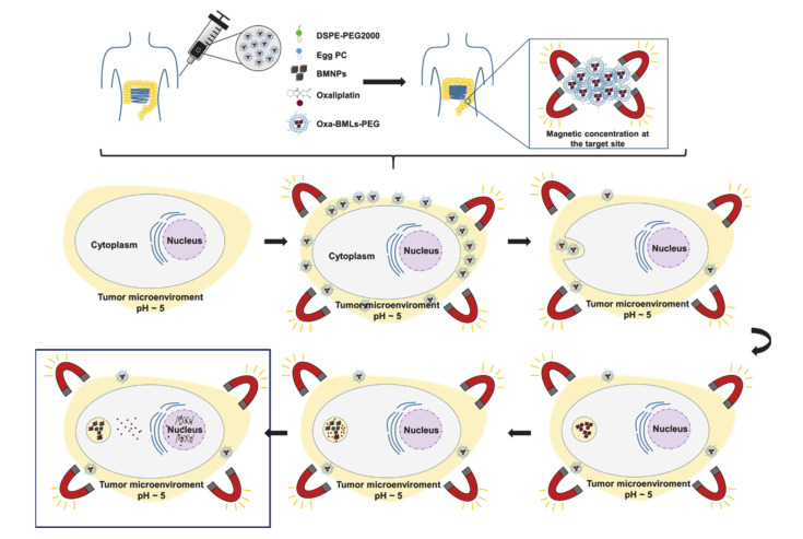
Magnetic concentration at the target site after intravenous or direct injection is achievable.
“After intravenous injection (or direct injection at the tumor site) and magnetic concentration at the target site, the magnetoliposomes could release their content by different mechanisms”
The authors state this as an assumption.
Magnetoliposomes release their content by one or more of three mechanisms: interaction with the plasma membrane, alteration of the bilayer, or endocytosis.
“the magnetoliposomes could release their content by different mechanisms: (a) magnetoliposomes could interact with the plasma membrane and discharge the nanoassemblies into the cytoplasm, (b) the interaction with the cell could alter the structure of the bilayer and release the nanoassemblies [40], and (c) magnetoliposomes could enter the cell by endocytosis (probably clathrin-mediated endocytosis”
The authors state this as an assumption.
Once liberated, Oxa-BMNPs nanoassemblies are enclosed in endosomal/lysosomal compartments at pH 4–5.
“Once liberated, Oxa-BMNPs nanoassemblies would be enclosed in endosomal/lysosomal compartments at a pH value of 4–5 [43,44,45,46].”
The authors state this as an assumption.
At acidic pH, the lipid membrane is degraded and the BMNPs becomes nearly uncharged (pI = 4.4), releasing the drug previously attached by electrostatic interaction.
“At this acidic pH value, the lipid membrane is degraded [40] and the BMNPs becomes nearly uncharged (pI = 4.4, [5]), releasing the drug that was previously attached by electrostatic interaction [14].”
The authors state this as an assumption.
Released Oxa diffuses toward the cytoplasm and a percentage enters the cell nuclei.
“The released Oxa diffuses toward the cytoplasm and a percentage enters the cell nuclei, as previously shown to occur for doxorubicin when administered as DOXO-BMNPs nanoassemblies [6].”
Self-check
Percentages, sums and reported differences re-computed from the paper’s own numbers.
Each contradiction between two passages listed here was confirmed by an independent check.
Rated major: it would touch a result — a value a result rests on, reported and recomputed p-values on opposite sides of the significance level, two passages that conflict with no sign which is right, or a cited work retracted before this paper was published.
In the paper
§ Biomimetic Magnetoliposomes as Oxaliplatin Nanocarriers: In Vitro Study for Potential Application in Colon Cancer > 4. Discussion
“The reduction of the IC50 with the new nanoformulations (Oxa-BMLs and Oxa-BMLs-PEG) is not as pronounced as it was with the Oxa-BMNPs nanoassemblies of previous works described by Jabalera et al. [14], which is probably due to the lower internalization of the former”
§ Biomimetic Magnetoliposomes as Oxaliplatin Nanocarriers: In Vitro Study for Potential Application in Colon Cancer > 4. Discussion
“Therefore, our results clearly show that the addition of both, lipid envelope and pegylation, to our nanoassemblies (Oxa-BMLs) or to the biomimetic nanoparticles (BMLs) increased their cell uptake compared to that of raw Oxa-BMNPs and/or BMNPs.”
Rated minor: it would not touch a result — a value no result rests on, a p-value that stays on the same side of the significance level, or a conflict that is clerical, resolved elsewhere in the paper, or outside its own findings.
In the paper
§ Biomimetic Magnetoliposomes as Oxaliplatin Nanocarriers: In Vitro Study for Potential Application in Colon Cancer > 3. Results > 3.3. In Vitro Proliferation Assays
“In HCT15, CCD18 and MC38 cell lines only slight toxicity occurs at the higher doses.”
§ Biomimetic Magnetoliposomes as Oxaliplatin Nanocarriers: In Vitro Study for Potential Application in Colon Cancer > 3. Results > 3.3. In Vitro Proliferation Assays
“In addition, an absence of toxicity was observed in the administration of BMLs and BMLs-PEG in this cell line.”
Citations
Cites
Bray, F., Ferlay, J., Soerjomataram, I., Siegel, R. L., Torre, L. A., & Jemal, A. (2018) · CA: A Cancer Journal for Clinicians · doi:10.3322/caac.21492 · Cited by 84,109
Rejman, J., Oberle, V., Zuhorn, I. S., & Hoekstra, D. (2004) · Biochemical Journal · doi:10.1042/bj20031253 · Cited by 2,872
Liposomes as nanomedical devices
Bozzuto, G., & Molinari, A. (2015) · International Journal of Nanomedicine · doi:10.2147/ijn.s68861 · Cited by 2,382
Magnetosome formation in prokaryotes
Bazylinski, D., & Frankel, R. (2004) · Nature Reviews Microbiology · doi:10.1038/nrmicro842 · Cited by 1,115
NCCN Guidelines Insights: Colon Cancer, Version 2.2018
Benson, A. B., Venook, A. P., Al-Hawary, M. M., Cederquist, L., Chen, Y.-J., Ciombor, K. K., Cohen, S., Cooper, H. S., Deming, D., Engstrom, P. F., et al. (2018) · Journal of the National Comprehensive Cancer Network · doi:10.6004/jnccn.2018.0021 · Cited by 806
Biodegradation of Iron Oxide Nanocubes: High-Resolution In Situ Monitoring
Lartigue, L., Alloyeau, D., Kolosnjaj-Tabi, J., Javed, Y., Guardia, P., Riedinger, A., Péchoux, C., Pellegrino, T., Wilhelm, C., & Gazeau, F. (2013) · ACS Nano · doi:10.1021/nn305719y · Cited by 274
Amemiya, Y., Arakaki, A., Staniland, S. S., Tanaka, T., & Matsunaga, T. (2007) · Biomaterials · doi:10.1016/j.biomaterials.2007.07.051 · Cited by 257
Controlled Release from Bilayer-Decorated Magnetoliposomes via Electromagnetic Heating
Chen, Y., Bose, A., & Bothun, G. D. (2010) · ACS Nano · doi:10.1021/nn100274v · Cited by 226
Geisow, M. J., & Evans, W. H. (1984) · Experimental Cell Research · doi:10.1016/0014-4827(84)90699-2 · Cited by 183
Protein‐Mediated Synthesis of Uniform Superparamagnetic Magnetite Nanocrystals
Prozorov, T., Mallapragada, S. K., Narasimhan, B., Wang, L., Palo, P. E., Nilsen‐Hamilton, M., Williams, T. J., Bazylinski, D. A., Prozorov, R., & Canfield, P. C. (2007) · Advanced Functional Materials · doi:10.1002/adfm.200600448 · Cited by 167
Cited by
Sorted by citations
Nanomaterials in cancer: Reviewing the combination of hyperthermia and triggered chemotherapy
Ribeiro, T. P., Moreira, J. A., Monteiro, F. J., & Laranjeira, M. S. (2022) · Journal of Controlled Release · doi:10.1016/j.jconrel.2022.04.045 · Cited by 90
Photonic and magnetic materials for on-demand local drug delivery
Armenia, I., Cuestas-Ayllón, C., Torres‐Herrero, B., Bussolari, F., Alfranca, G., Grazú, V., & de la Fuente, J. M. (2022) · Advanced Drug Delivery Reviews · doi:10.1016/j.addr.2022.114584 · Cited by 82
Malehmir, S., Esmaili, M. A., Mahabady, M. K., Nasab, A. S., Atapour, A., Ganjali, M. R., Ghasemi, A., & Hasan‐Abad, A. M. (2023) · Frontiers in Chemistry · doi:10.3389/fchem.2023.1249134 · Cited by 82
Darroudi, M., Gholami, M., Rezayi, M., & Khazaei, M. (2021) · Journal of Nanobiotechnology · doi:10.1186/s12951-021-01150-6 · Cited by 79
Oltolina, F., Peigneux, A., Colangelo, D., Clemente, N., D’Urso, A., Valente, G., Iglesias, G. R., Jiménez-López, C., & Prat, M. (2020) · Cancers · doi:10.3390/cancers12092564 · Cited by 57
Metadata
- Authors
- Garcia-Pinel, B., Jabalera, Y., Ortiz, R., Cabeza, L., Jimenez-Lopez, C., Melguizo, C., & Prados, J.
- Affiliations
- Institute of Biopathology and Regenerative Medicine (IBIMER), Center of Biomedical Research (CIBM), University of Granada, 18100 Granada, Spain
- Universidad de Granada (ES)
- Department of Microbiology, Science School, University of Granada, 18002 Granada, Spain
- Corresponding author
- Melguizo, Consolación
- ORCID
- Venue
- Pharmaceutics
- Publisher
- MDPI AG
- Published
- 2020-06-24
- Volume
- 12
- Issue
- 6
- Pages
- 589
- ISSN
- 1999-4923 (ISSN-L)
- PMC
- PMC7356838
- OpenAlex
- W3037496532
- Cited by
- 40
- References
- 63
- Citations by year
- 2026: 7 · 2025: 2 · 2024: 5 · 2023: 11 · 2022: 7 · 2021: 7 · 2020: 1
- FWCI
- 1.57
- Type
- journal article
- Language
- en
- Open access
- gold
- dx.doi.org
- Licence
- Locations
- Pharmaceutics (publishedVersion, cc-by)
- PubMed (publishedVersion)
- PubMed Central (publishedVersion, cc-by)
- Institutional Repository of the University of Granada (University of Granada) (submittedVersion, cc-by)
- LA Referencia (Red Federada de Repositorios Institucionales de Publicaciones Científicas) (submittedVersion, cc-by)
- DOAJ (DOAJ: Directory of Open Access Journals) (submittedVersion, cc-by-sa)
- RISalud Institutional Health Repository of Andalucía (submittedVersion, cc-by)
- Funders
- Ministerio de Economía y Competitividad (CGL2016-76723)
- Junta de Andalucia (A-BIO-376-UGR18)
- Conserjería de Salud de la Junta de Andalucía (PI19/01478)
- Instituto de Salud Carlos III (PI19/01478)
- Topics
- Physical Sciences › Materials Science › Biomaterials › Nanoparticle-Based Drug Delivery
- Life Sciences › Pharmacology, Toxicology and Pharmaceutics › Drug Discovery › Pharmacology and Nanomedicine Research
- Life Sciences › Biochemistry, Genetics and Molecular Biology › Molecular Biology › RNA Interference and Gene Delivery
- Keywords
- oxaliplatin, magnetoliposomes, PEGylated liposomes, colorectal cancer, targeted chemotherapy, drug nanocarriers, hemocompatibility, cellular uptake, macrophage toxicity, local chemotherapy
About these checks
Registry, retraction, Cites and statistics results are computed, not judged, with one exception: a printed reference that no index lists is read once by a model before it counts as a citation error. Claims are extracted by a model and kept only when their quote appears verbatim in the text; each cited source is then checked, with a model’s help, for whether it says what the claim relies on. Those verdicts can be wrong — read the claim’s quote and the cited source before relying on one.
A language model drew the claims from the paper on 2026-11-10, under version 3 of the claim definition (model deepseek-v4.1-flash, prompt extract-v3).
A second model labelled each claim on 2026-11-10 (model jev-1.13-20260917, definition revision gold-guideline-v0.2).
Cited sources are checked with a language model’s help (gpt-6-sol).
Statistics and arithmetic are recomputed, not judged.
Confidence bands are shown only for fields calibrated on our annotated test set. None is calibrated yet, so no band is shown.
What counts as a claim ›How we measure confidence ›How we check this ›
Are you an author, or do you see a problem with this page? Request a review
