

The DOI matches its Crossref record; 1 of the 1 references it cites were found in a registry or an index.
Cyclic RGD-Functionalized pH/ROS Dual-Responsive Nanoparticle for Targeted Breast Cancer Therapy
Abstract
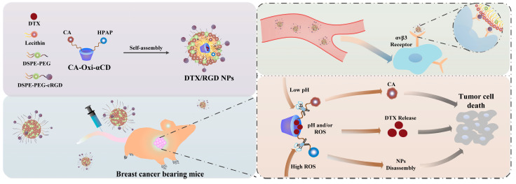
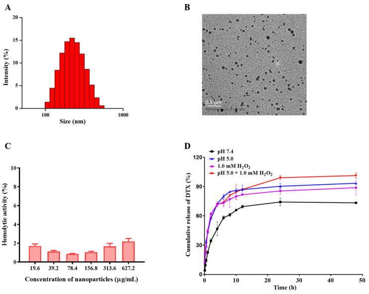
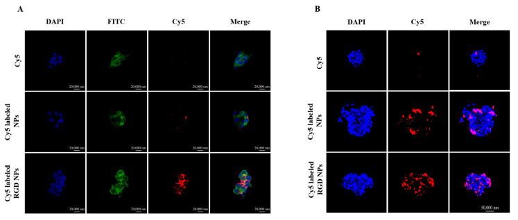
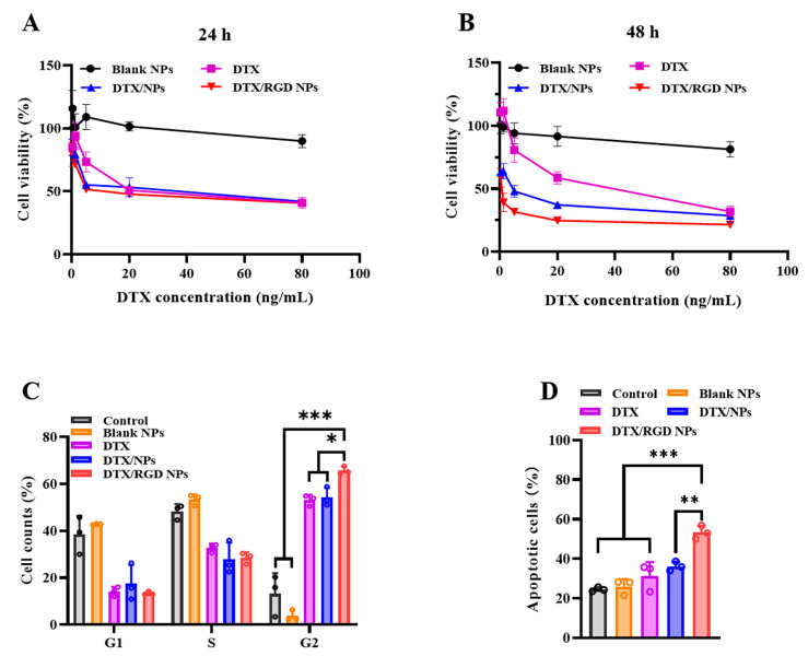
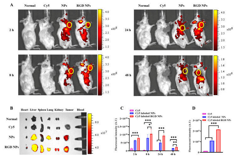
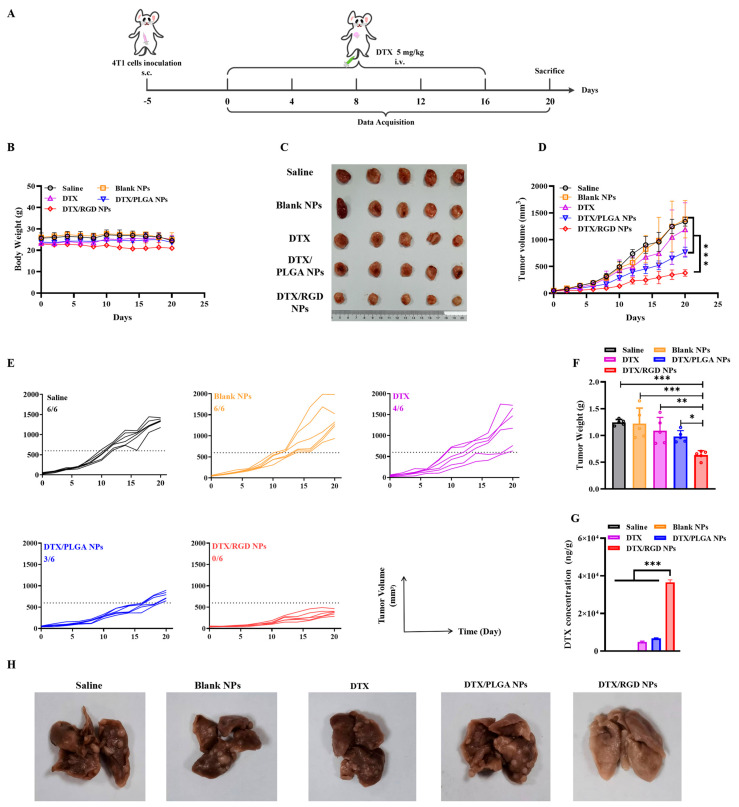
Breast cancer is the most common malignant tumor in women and is a big challenge to clinical treatment due to the high morbidity and mortality. The pH/ROS dual-responsive nanoplatforms may be an effective way to significantly improve the therapeutic efficacy of breast cancer. Herein, we report a docetaxel (DTX)-loaded pH/ROS-responsive NP that could achieve active targeting of cancer cells and selective and complete drug release for effective drug delivery. The pH/ROS-responsive NPs were fabricated using nanocarriers that consist of an ROS-responsive moiety (4-hydroxymethylphenylboronic acid pinacol ester, HPAP), cinnamaldehyde (CA, an aldehyde organic compound with anticancer activities) and cyclodextrin (α-CD). The NPs were loaded with DTX, modified with a tumor-penetration peptide (circular RGD, cRGD) and named DTX/RGD NPs. The cRGD could promote DTX/RGD NPs penetration into deep tumor tissue and specifically target cancer cells. After internalization by cancer cells through receptor-mediated endocytosis, the pH-responsive acetal was cleaved to release CA in the lysosomal acidic environment. Meanwhile, the high ROS in tumor cells induced the disassembly of NPs with complete release of DTX. In vitro cellular assays verified that DTX/RGD NPs could be effectively internalized by 4T1 cells, obviously inducing apoptosis, blocking the cell cycle of 4T1 cells and consequently, killing tumor cells. In vivo animal experiments demonstrated that the NPs could target to the tumor sites and significantly inhibit the tumor growth in 4T1 breast cancer mice. Both in vitro and in vivo investigations demonstrated that DTX/RGD NPs could significantly improve the antitumor effect compared to free DTX. Thus, the DTX/RGD NPs provide a promising strategy for enhancing drug delivery and cancer therapy.
Overview
The claims and statistics of this paper haven’t been checked yet.
Run the check — free, no account needed. It reads the open-access text and checks the sources it cites.
Integrity signals
Registry
Cites
Statistics
Figures
Verification
Claims the paper makes
The claims and statistics of this paper haven’t been checked yet.
Run the check — free, no account needed. It reads the open-access text and checks the sources it cites.
Claims other papers make about this one
Each is checked against this paper’s own text: does it say what the citing paper says it does?
In the citing paper
“Notably, in 4T1 tumor-bearing mice, DTX/RGD NPs yielded 0/6 tumors exceeding 600 mm3 at day 20 (vs 6/6 saline, 6/6 blank NPs, 4/6 free DTX, 3/6 DTX/PLGA NPs), and no metastatic clots were observed in lung after DTX/RGD NP treatment (vs abundant nodules in saline/blank groups)”
From Overcoming tumor microenvironment barriers: transformable and bioinspired nanomedicine strategies for deep tumor penetration (2026) · cites this paper as [83]
In the citing paper
“Cyclic RGD-modified, pH/ROS dual-responsive docetaxel nanoparticles align targeting with stimuli-triggered disassembly to enhance intratumoral distribution and tumor control in 4T1 models [83].”
From Overcoming tumor microenvironment barriers: transformable and bioinspired nanomedicine strategies for deep tumor penetration (2026) · cites this paper as [83]
Self-check
Percentages, sums and reported differences re-computed from the paper’s own numbers.
The self-check needs the paper’s full text.
Citations
Cites
Targeting Free Radicals in Oxidative Stress-Related Human Diseases
Poprac, P., Jomova, K., Simunkova, M., Kollar, V., Rhodes, C. J., & Valko, M. (2017) · Trends in Pharmacological Sciences · doi:10.1016/j.tips.2017.04.005 · Cited by 1,107
Research works printed but not linked: 43
- Davodabadi, 2022, Breast cancer vaccines: New insights into immunomodulatory and nano-therapeutic approaches, J. Control. Release Off. J. Control. Release Soc., 349, 844, 10.1016/j.jconrel.2022.07.036 · DOI
- Bahreyni, 2020, Emerging nanomedicines for effective breast cancer immunotherapy, J. Nanobiotechnol., 18, 180, 10.1186/s12951-020-00741-z · DOI
- Britt, 2020, Key steps for effective breast cancer prevention, Nat. Rev. Cancer, 20, 417, 10.1038/s41568-020-0266-x · DOI · Paper page
- Lau, K.H., Tan, A.M., and Shi, Y. (2022). New and Emerging Targeted Therapies for Advanced Breast Cancer. Int. J. Mol. Sci., 23. · DOI
- Yao, 2016, Ligand-targeted theranostic nanomedicines against cancer, J. Control. Release Off. J. Control. Release Soc., 240, 267, 10.1016/j.jconrel.2016.01.002 · DOI
- Aleksandrowicz, 2017, Drug delivery systems improving chemical and physical properties of anticancer drugs currently investigated for treatment of solid tumors, J. Physiol. Pharmacol. Off. J. Pol. Physiol. Soc., 68, 165
- de la Torre, P., Perez-Lorenzo, M.J., Alcazar-Garrido, A., and Flores, A.I. (2020). Cell-Based Nanoparticles Delivery Systems for Targeted Cancer Therapy: Lessons from Anti-Angiogenesis Treatments. Molecules, 25. · DOI
- Seyyednia, 2021, Nanoparticles modified with vasculature-homing peptides for targeted cancer therapy and angiogenesis imaging, J. Control. Release Off. J. Control. Release Soc., 338, 367, 10.1016/j.jconrel.2021.08.044 · DOI
- Yu, 2022, Polymeric PD-L1 blockade nanoparticles for cancer photothermal-immunotherapy, Biomaterials, 280, 121312, 10.1016/j.biomaterials.2021.121312 · DOI
- Amreddy, 2018, Recent Advances in Nanoparticle-Based Cancer Drug and Gene Delivery, Adv. Cancer Res., 137, 115, 10.1016/bs.acr.2017.11.003 · DOI
- Wang, 2010, Targeting nanoparticles to cancer, Pharmacol. Res., 62, 90, 10.1016/j.phrs.2010.03.005 · DOI
- Jiang, 2019, Erythrocyte-cancer hybrid membrane-camouflaged melanin nanoparticles for enhancing photothermal therapy efficacy in tumors, Biomaterials, 192, 292, 10.1016/j.biomaterials.2018.11.021 · DOI
- Zhang, 2022, Recent advances in dual- and multi-responsive nanomedicines for precision cancer therapy, Biomaterials, 291, 121906, 10.1016/j.biomaterials.2022.121906 · DOI
- Zhou, 2018, Stimuli-Responsive Nanomedicines for Overcoming Cancer Multidrug Resistance, Theranostics, 8, 1059, 10.7150/thno.22679 · DOI
- Antoniou, 2021, Stimulus-responsive liposomes for biomedical applications, Drug Discov. Today, 26, 1794, 10.1016/j.drudis.2021.05.010 · DOI
- Huu, 2015, Light-responsive nanoparticle depot to control release of a small molecule angiogenesis inhibitor in the posterior segment of the eye, J. Control. Release Off. J. Control. Release Soc., 200, 71, 10.1016/j.jconrel.2015.01.001 · DOI
- Li, 2021, Magnetic nanoparticles for cancer theranostics: Advances and prospects, J. Control. Release Off. J. Control. Release Soc., 335, 437, 10.1016/j.jconrel.2021.05.042 · DOI
- Sun, 2022, Self-Illuminating Triggered Release of Therapeutics from Photocleavable Nanoprodrug for the Targeted Treatment of Breast Cancer, ACS Appl. Mater. Interfaces, 14, 8766, 10.1021/acsami.1c21665 · DOI
- Cheng, 2022, A pH-Responsive Cluster Metal-Organic Framework Nanoparticle for Enhanced Tumor Accumulation and Antitumor Effect, Adv. Mater., 34, e2203915, 10.1002/adma.202203915 · DOI
- Shahriari, 2019, Enzyme responsive drug delivery systems in cancer treatment, J. Control. Release Off. J. Control. Release Soc., 308, 172, 10.1016/j.jconrel.2019.07.004 · DOI
- Xu, 2021, Development of ROS-responsive amino acid-based poly(ester amide) nanoparticle for anticancer drug delivery, J. Biomed. Mater. Res. Part A, 109, 524, 10.1002/jbm.a.37035 · DOI
- Ye, 2017, A Tumor-Specific Cascade Amplification Drug Release Nanoparticle for Overcoming Multidrug Resistance in Cancers, Adv. Mater., 29, 1702342, 10.1002/adma.201702342 · DOI
- Wang, 2019, Self-Strengthened Oxidation-Responsive Bioactivating Prodrug Nanosystem with Sequential and Synergistically Facilitated Drug Release for Treatment of Breast Cancer, ACS Appl. Mater. Interfaces, 11, 18914, 10.1021/acsami.9b03056 · DOI
- Zhang, H., Ma, W., Wang, Z., Wu, X., Zhang, H., Fang, W., Yan, R., and Jin, Y. (2022). Self-Supply Oxygen ROS Reactor via Fenton-like Reaction and Modulating Glutathione for Amplified Cancer Therapy Effect. Nanomaterials, 12. · DOI
- Tu, 2022, Cinnamaldehyde-based poly(thioacetal): A ROS-awakened self-amplifying degradable polymer for enhanced cancer immunotherapy, Biomaterials, 289, 121795, 10.1016/j.biomaterials.2022.121795 · DOI
- Zhang, 2018, An Adenosine Triphosphate-Responsive Autocatalytic Fenton Nanoparticle for Tumor Ablation with Self-Supplied H2O2 and Acceleration of Fe(III)/Fe(II) Conversion, Nano Lett., 18, 7609, 10.1021/acs.nanolett.8b03178 · DOI
- Miyanaga, 2016, Concentration-dependent radiosensitizing effect of docetaxel in esophageal squamous cell carcinoma cells, Int. J. Oncol., 48, 517, 10.3892/ijo.2015.3291 · DOI
- Xu, 2015, Shrapnel nanoparticles loading docetaxel inhibit metastasis and growth of breast cancer, Biomaterials, 64, 10, 10.1016/j.biomaterials.2015.06.017 · DOI
- Torchilin, 2011, Tumor delivery of macromolecular drugs based on the EPR effect, Adv. Drug Deliv. Rev., 63, 131, 10.1016/j.addr.2010.03.011 · DOI
- Chen, 2016, Clinical Application of Radiolabeled RGD Peptides for PET Imaging of Integrin αvβ3, Theranostics, 6, 78, 10.7150/thno.13242 · DOI
- Liu, 2016, Integrin (αvβ3) Targeted RGD Peptide Based Probe for Cancer Optical Imaging, Curr. Protein Pept. Sci., 17, 570, 10.2174/1389203717666160101124015 · DOI
- Perrins, R.D., McCarthy, L.A., Robinson, A., Spry, K.L., Cognet, V., Ferreira, A., Porter, J., Garciotaa, C.E., Rodriguez, M.A., and Lopez, D. (2022). Targeting Ultrasmall Gold Nanoparticles with cRGD Peptide Increases the Uptake and Efficacy of Cytotoxic Payload. Nanomaterials, 12. · DOI
- Yang, 2011, cRGD-functionalized, DOX-conjugated, and 64Cu-labeled superparamagnetic iron oxide nanoparticles for targeted anticancer drug delivery and PET/MR imaging, Biomaterials, 32, 4151, 10.1016/j.biomaterials.2011.02.006 · DOI
- Wang, 2019, cRGD-functionalized nanoparticles for combination therapy of anti-endothelium dependent vessels and anti-vasculogenic mimicry to inhibit the proliferation of ovarian cancer, Acta Biomater., 94, 495, 10.1016/j.actbio.2019.06.039 · DOI
- Capello, 2006, Anticancer activity of targeted proapoptotic peptides, J. Nucl. Med. Off. Publ. Soc. Nucl. Med., 47, 122
- Gu, 2022, αvβ3 integrin-specific exosomes engineered with cyclopeptide for targeted delivery of triptolide against malignant melanoma, J. Nanobiotechnol., 20, 384, 10.1186/s12951-022-01597-1 · DOI · Paper page
- Zhao, 2015, A preloaded amorphous calcium carbonate/doxorubicin@silica nanoreactor for pH-responsive delivery of an anticancer drug, Angew. Chem., 54, 919, 10.1002/anie.201408510 · DOI
- Yang, 2016, Dual Stimuli-Responsive Hybrid Polymeric Nanoparticles Self-Assembled from POSS-Based Starlike Copolymer-Drug Conjugates for Efficient Intracellular Delivery of Hydrophobic Drugs, ACS Appl. Mater. Interfaces, 8, 13251, 10.1021/acsami.6b02403 · DOI
- Deng, 2019, Rational Design of Nanoparticles to Overcome Poor Tumor Penetration and Hypoxia-Induced Chemotherapy Resistance: Combination of Optimizing Size and Self-Inducing High Level of Reactive Oxygen Species, ACS Appl. Mater. Interfaces, 11, 31743, 10.1021/acsami.9b12129 · DOI
- Wang, 2021, Folic acid-modified ROS-responsive nanoparticles encapsulating luteolin for targeted breast cancer treatment, Drug Deliv., 28, 1695, 10.1080/10717544.2021.1963351 · DOI
- Morarasu, S., Morarasu, B.C., Ghiarasim, R., Coroaba, A., Tiron, C., Iliescu, R., and Dimofte, G.M. (2022). Targeted Cancer Therapy via pH-Functionalized Nanoparticles: A Scoping Review of Methods and Outcomes. Gels, 8. · DOI
- Farjadian, 2022, Physically stimulus-responsive nanoparticles for therapy and diagnosis, Front. Chem., 10, 952675, 10.3389/fchem.2022.952675 · DOI
- Zhang, 2019, Mitochondria-specific drug release and reactive oxygen species burst induced by polyprodrug nanoreactors can enhance chemotherapy, Nat. Commun., 10, 1704, 10.1038/s41467-019-09566-3 · DOI
Cited by
Sorted by citations
Sheng, J., Yuan, W., Zhang, M., Chen, Z., Qi, Y., Zhao, Y., & Zhang, S. (2026) · Journal of Nanobiotechnology · doi:10.1186/s12951-026-04028-7 · Cited by 35
Metadata
- Authors
- Yao, P., Wang, X., Wang, Q., Dai, Q., Peng, Y., Yuan, Q., Mou, N., Lv, S., Weng, B., Wang, Y., & Sun, F.
- Venue
- Pharmaceutics
- Publisher
- MDPI AG
- Published
- 2023-06-26
- Volume
- 15
- Issue
- 7
- Pages
- 1827
- ISSN
- 1999-4923
- PMC
- PMC10386338
- Cited by
- 22
- References
- 1
- Language
- en
- Funders
- National Natural Science Foundation of China (82102448, cstc2015zdcy-ztzx120005)
- Special Project of Common Key Technology Innovation for Key Industries in Chongqing (82102448, cstc2015zdcy-ztzx120005)
About these checks
Registry, retraction, Cites and statistics results are computed, not judged, with one exception: a printed reference that no index lists is read once by a model before it counts as a citation error. Claims are extracted by a model and kept only when their quote appears verbatim in the text; each cited source is then checked, with a model’s help, for whether it says what the claim relies on. Those verdicts can be wrong — read the claim’s quote and the cited source before relying on one.
Statistics and arithmetic are recomputed, not judged.
Confidence bands are shown only for fields calibrated on our annotated test set. None is calibrated yet, so no band is shown.
What counts as a claim ›How we measure confidence ›How we check this ›
Are you an author, or do you see a problem with this page? Request a review
