

The DOI matches its Crossref record; 1 of the 1 references it cites were found in a registry or an index.
Bioresorbable Stent in Anterior Cruciate Ligament Reconstruction
Open accessAbstract
The exact causes of failure of anterior cruciate ligament (ACL) reconstruction are still unknown. A key to successful ACL reconstruction is the prevention of bone tunnel enlargement (BTE). In this study, a new strategy to improve the outcome of ACL reconstruction was analyzed using a bioresorbable polylactide (PLA) stent as a catalyst for the healing process. The study included 24 sheep with 12 months of age. The animals were randomized to the PLA group (n = 16) and control group (n = 8), subjected to the ACL reconstruction with and without the implantation of the PLA tube, respectively. The sheep were sacrificed 6 or 12 weeks post-procedure, and their knee joints were evaluated by X-ray microcomputed tomography with a 50 μm resolution. While the analysis of tibial and femoral tunnel diameters and volumes demonstrated the presence of BTE in both groups, the enlargement was less evident in the PLA group. Also, the microstructural parameters of the bone adjacent to the tunnels tended to be better in the PLA group. This suggested that the implantation of a bioresorbable PLA tube might facilitate osteointegration of the tendon graft after the ACL reconstruction. The beneficial effects of the stent were likely associated with osteogenic and osteoconductive properties of polylactide.
Overview
The claims and statistics of this paper haven’t been checked yet.
Run the check — free, no account needed. It reads the open-access text and checks the sources it cites.
Integrity signals
Registry
Cites
Statistics
Figures
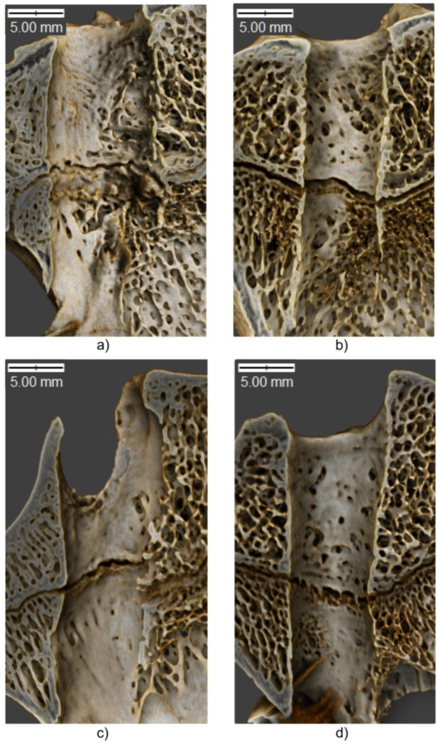
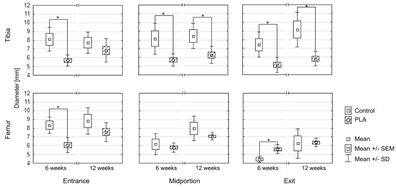
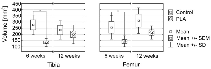
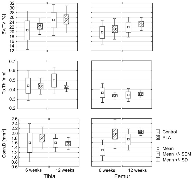
Verification
Claims the paper makes
The claims and statistics of this paper haven’t been checked yet.
Run the check — free, no account needed. It reads the open-access text and checks the sources it cites.
Claims other papers make about this one
Each is checked against this paper’s own text: does it say what the citing paper says it does?
The check wasn’t confident either way, so it gives no verdict — never disputed.
In the citing paper
“in the early postoperative period, limited ingrowth of the newly regenerated connective tissue in the knee cavity restricted ligament regeneration, and bone tissue in the bone tunnel disrupted graft–bone healing [13, 14].”
From Bone Mesenchymal Stem Cells Contribute to Ligament Regeneration and Graft–Bone Healing after Anterior Cruciate Ligament Reconstruction with Silk–Collagen Scaffold (2021) · cites this paper as [14]
Experimental — accuracy not yet measuredThe check wasn’t confident either way, so it gives no verdict — never disputed.
In the citing paper
“in the early postoperative period, limited ingrowth of the newly regenerated connective tissue in the knee cavity restricted ligament regeneration, and bone tissue in the bone tunnel disrupted graft–bone healing [13, 14].”
From Bone Mesenchymal Stem Cells Contribute to Ligament Regeneration and Graft–Bone Healing after Anterior Cruciate Ligament Reconstruction with Silk–Collagen Scaffold (2021) · cites this paper as [14]
Experimental — accuracy not yet measured
Self-check
Percentages, sums and reported differences re-computed from the paper’s own numbers.
The self-check needs the paper’s full text.
Citations
Cites
Li, S. M., Garreau, H., & Vert, M. (1990) · Journal of Materials Science: Materials in Medicine · doi:10.1007/bf00700871 · Cited by 733
Research works printed but not linked: 56
- Kuskucu, 2008, Comparison of short-term results of bone tunnel enlargement between EndoButton CL and cross-pin fixation systems after chronic anterior cruciate ligament reconstruction with autologous quadrupled hamstring tendons, J. Int. Med. Res., 36, 23, 10.1177/147323000803600104 · DOI
- Moisala, 2008, Comparison of the bioabsorbable and metal screw fixation after ACL reconstruction with a hamstring autograft in MRI and clinical outcome: A prospective randomized study, Knee Surg. Sports Traumatol. Arthrosc., 16, 1080, 10.1007/s00167-008-0593-z · DOI
- Siebold, 2008, Effect of compaction drilling during ACL reconstruction with hamstrings on postoperative tunnel widening, Arch. Orthop. Trauma Surg., 128, 461, 10.1007/s00402-007-0443-3 · DOI
- Jaureguito, 1996, Why grafts fail, Clin. Orthop. Relat. Res., 325, 25, 10.1097/00003086-199604000-00005 · DOI
- Meyers, 2010, Imaging of anterior cruciate ligament repair and its complications, AJR Am. J. Roentgenol., 194, 476, 10.2214/AJR.09.3200 · DOI
- Beynnon, 2005, Treatment of anterior cruciate ligament injuries, part 2, Am. J. Sports Med., 33, 1751, 10.1177/0363546505279922 · DOI
- Beynnon, 2005, Treatment of anterior cruciate ligament injuries, part I, Am. J. Sports Med., 33, 1579, 10.1177/0363546505279913 · DOI
- Sundar, 2009, Tendon bone healing can be enhanced by demineralized bone matrix: A functional and histological study, J. Biomed. Mater. Res. B Appl. Biomater., 88, 115, 10.1002/jbm.b.31157 · DOI
- Chen, 2009, Strategies to enhance tendon graft-bone healing in anterior cruciate ligament reconstruction, Chang Gung Med. J., 32, 483
- Chen, 2010, Arthroscopic single-bundle anterior cruciate ligament reconstruction with periosteum-enveloping hamstring tendon graft: Clinical outcome at 2 to 7 years, Arthroscopy, 26, 907, 10.1016/j.arthro.2009.11.011 · DOI
- Chen, 2004, Arthroscopic anterior cruciate ligament reconstruction with periosteum-enveloping hamstring tendon graft, Knee Surg. Sports Traumatol. Arthrosc., 12, 398, 10.1007/s00167-004-0498-4 · DOI
- Karaoglu, 2009, The effects of bone marrow or periosteum on tendon-to-bone tunnel healing in a rabbit model, Knee Surg. Sports Traumatol. Arthrosc., 17, 170, 10.1007/s00167-008-0646-3 · DOI
- Rodeo, 1999, Use of recombinant human bone morphogenetic protein-2 to enhance tendon healing in a bone tunnel, Am. J. Sports Med., 27, 476, 10.1177/03635465990270041201 · DOI
- Baxter, 2010, Augmentation of bone tunnel healing in anterior cruciate ligament grafts: Application of calcium phosphates and other materials, J. Tissue Eng., 2010, 712370, 10.4061/2010/712370 · DOI
- Soon, 2007, An analysis of soft tissue allograft anterior cruciate ligament reconstruction in a rabbit model: A short-term study of the use of mesenchymal stem cells to enhance tendon osteointegration, Am. J. Sports Med., 35, 962, 10.1177/0363546507300057 · DOI · Paper page
- Yeh, 2007, Effects of hyperbaric oxygen treatment on tendon graft and tendon-bone integration in bone tunnel: Biochemical and histological analysis in rabbits, J. Orthop. Res., 25, 636, 10.1002/jor.20360 · DOI
- Yamazaki, 2005, The effect of transforming growth factor-beta1 on intraosseous healing of flexor tendon autograft replacement of anterior cruciate ligament in dogs, Arthroscopy, 21, 1034, 10.1016/j.arthro.2005.05.011 · DOI
- Sasaki, 2008, Enhancement of tendon-bone osteointegration of anterior cruciate ligament graft using granulocyte colony-stimulating factor, Am. J. Sports Med., 36, 1519, 10.1177/0363546508316282 · DOI
- Darabos, 2011, Intraarticular application of autologous conditioned serum (ACS) reduces bone tunnel widening after ACL reconstructive surgery in a randomized controlled trial, Knee Surg. Sports Traumatol. Arthrosc., 19, S36, 10.1007/s00167-011-1458-4 · DOI
- Gokce, 2009, Does bone impaction technique reduce tunnel enlargement in ACL reconstruction?, Int. Orthop., 33, 407, 10.1007/s00264-007-0496-5 · DOI
- Livesay, 1999, Hamstring graft motion in the femoral bone tunnel when using titanium button/polyester tape fixation, Knee Surg. Sports Traumatol. Arthrosc., 7, 215, 10.1007/s001670050151 · DOI
- Scheffler, 2000, Mechanical behavior of two hamstring graft constructs for reconstruction of the anterior cruciate ligament, J. Orthop. Res., 18, 456, 10.1002/jor.1100180319 · DOI
- Iorio, 2007, Bone tunnel enlargement after ACL reconstruction using autologous hamstring tendons: A CT study, Int. Orthop., 31, 49, 10.1007/s00264-006-0118-7 · DOI
- Jagodzinski, 2005, Analysis of forces of ACL reconstructions at the tunnel entrance: Is tunnel enlargement a biomechanical problem?, J. Biomech., 38, 23, 10.1016/j.jbiomech.2004.03.021 · DOI · Paper page
- Klatt, 1997, Tunnel expansion following anterior cruciate ligament reconstruction: A comparison of hamstring and patellar tendon autografts, Knee Surg. Sports Traumatol. Arthrosc., 5, 234, 10.1007/s001670050056 · DOI
- Agarwal, 2004, Osteolysis—Basic science, incidence and diagnosis, Curr. Orthop., 18, 220, 10.1016/j.cuor.2004.03.002 · DOI
- Irie, 2003, Intraarticular inflammatory cytokines in acute anterior cruciate ligament injured knee, Knee, 10, 93, 10.1016/S0968-0160(02)00083-2 · DOI
- Zysk, 2004, Tunnel enlargement and changes in synovial fluid cytokine profile following anterior cruciate ligament reconstruction with patellar tendon and hamstring tendon autografts, Knee Surg. Sports Traumatol. Arthrosc., 12, 98, 10.1007/s00167-003-0426-z · DOI
- Darabos, 2009, Correlation between synovial fluid and serum IL-1beta levels after ACL surgery-preliminary report, Int. Orthop., 33, 413, 10.1007/s00264-008-0649-1 · DOI
- Clatworthy, 1999, Tunnel widening in anterior cruciate ligament reconstruction: A prospective evaluation of hamstring and patella tendon grafts, Knee Surg. Sports Traumatol. Arthrosc., 7, 138, 10.1007/s001670050138 · DOI
- Iorio, 2008, Tunnel enlargement after anterior cruciate ligament reconstruction in patients with post-operative septic arthritis, Knee Surg. Sports Traumatol. Arthrosc., 16, 921, 10.1007/s00167-008-0575-1 · DOI
- Auras, R., Lim, L.T., Selke, S.E.M., and Tsuji, H. (2010). Industrial Production of High Molecular Weight Poly (Lactic Acid). Poly (Lactic Acid). Synthesis, Structures, Properties, Processing, and Application, John Wiley & Sons. · DOI
- Gorrasi, 2013, Effect of PLA grades and morphologies on hydrolytic degradation at composting temperature: Assessment of structural modification and kinetic parameters, Polym. Degrad. Stab., 98, 1006, 10.1016/j.polymdegradstab.2013.02.005 · DOI
- Hakim, 2017, PLA/SiO2 composites: Influence of the filler modifications on the morphology, crystallization behavior, and mechanical properties, J. Appl. Polym. Sci., 134, 45367, 10.1002/app.45367 · DOI
- Gleadall, 2014, Degradation mechanisms of bioresorbable polyesters. Part 1.Effects of random scission, end scission and autocatalysis, Acta Biomater., 10, 2223, 10.1016/j.actbio.2013.12.039 · DOI · Paper page
- Szaraniec, 2015, Effect of clay mineral on the accelerated hydrolytic degradation of polylactide in the polymer/clay nanocomposites, Mater. Lett., 146, 73, 10.1016/j.matlet.2015.01.135 · DOI
- Ficek, 2012, Bioresorbable Polylactide Implant used in orthopaedic surgery—Case report, PrzypadkiMedyczne.pl, 22, 84
- Gugala, 2005, The in vitro growth and activity of sheep osteoblasts on three-dimensional scaffolds from poly(L/DL-lactide) 80/20%, J. Biomed. Mater. Res. A, 75, 702, 10.1002/jbm.a.30479 · DOI
- Gugala, 2004, Differentiation, growth and activity of rat bone marrow stromal cells on resorbable poly(L/DL-lactide) membranes, Biomaterials, 25, 2299, 10.1016/j.biomaterials.2003.09.009 · DOI
- Gugala, 2007, New approaches in the treatment of critical-size segmental defects in long bones, Macromol. Symp., 253, 147, 10.1002/masy.200750722 · DOI
- Leiggener, 2006, Influence of copolymer composition of polylactide implants on cranial bone regeneration, Biomaterials, 27, 202, 10.1016/j.biomaterials.2005.05.068 · DOI
- Ficek, 2012, A revised surgical concept of anterior cruciate ligament replacement in a rabbit model, Eng. Biomater., 113, 33
- Nuss, K.M., Auer, J.A., Boos, A., and von Rechenberg, B. (2006). An animal model in sheep for biocompatibility testing of biomaterials in cancellous bones. BMC Musculoskelet. Disord., 7. · DOI
- Turner, 2002, The sheep as a model for osteoporosis in humans, Vet. J., 163, 232, 10.1053/tvjl.2001.0642 · DOI
- Szumera, 2013, Osteoconductive nanocomposite materials for bone regeneration, Mater. Sci. Forum, 730–732, 38
- Menaszek, 2014, Resorbable polymer membranes for medical applications, J. Biomim. Biomater. Tissue Eng., 19, 99, 10.4028/www.scientific.net/JBBTE.19.99 · DOI
- Schneider, 2012, NIH Image to ImageJ: 25 years of image analysis, Nat. Methods, 9, 671, 10.1038/nmeth.2089 · DOI · Paper page
- Doube, 2010, BoneJ: Free and extensible bone image analysis in ImageJ, Bone, 47, 1076, 10.1016/j.bone.2010.08.023 · DOI
- Stock, S.R. (2012). Drishti: A volume exploration and presentation tool. Developments in X-ray Tomography VIII, Proceedings of the SPIE Optical Engineering + Applications, San Diego, CA, USA, 12–16 August 2012, Society of Photo-Optical Instrumentation Engineers.
- Ruimerman, 2005, A theoretical framework for strain-related trabecular bone maintenance and adaptation, J. Biomech., 38, 931, 10.1016/j.jbiomech.2004.03.037 · DOI
- Iorio, 2013, ACL reconstruction with hamstrings: How different technique and fixation devices influence bone tunnel enlargement, Eur. Rev. Med. Pharmacol. Sci., 17, 2956
- Silva, 2010, Femoral tunnel enlargement after anatomic ACL reconstruction: A biological problem?, Knee Surg. Sports Traumatol. Arthrosc., 18, 1189, 10.1007/s00167-010-1046-z · DOI
- Weiler, 2002, Tendon healing in a bone tunnel. Part II: Histologic analysis after biodegradable interference fit fixation in a model of anterior cruciate ligament reconstruction in sheep, Arthroscopy, 18, 124, 10.1053/jars.2002.30657 · DOI
- Weiler, 2002, Tendon healing in a bone tunnel. Part I: Biomechanical results after biodegradable interference fit fixation in a model of anterior cruciate ligament reconstruction in sheep, Arthroscopy, 18, 113, 10.1053/jars.2002.30656 · DOI
- Buelow, 2002, A prospective evaluation of tunnel enlargement in anterior cruciate ligament reconstruction with hamstrings: Extracortical versus anatomical fixation, Knee Surg. Sports Traumatol. Arthrosc., 10, 80, 10.1007/s00167-001-0267-6 · DOI
- Iorio, 2007, The effect of accelerated, brace free, rehabilitation on bone tunnel enlargement after ACL reconstruction using hamstring tendons: A CT study, Knee Surg. Sports Traumatol. Arthrosc., 15, 365, 10.1007/s00167-006-0219-2 · DOI
Cited by
Sorted by citations
Bi, F., Chen, Y., Liu, J., Hu, W., & Tian, K. (2021) · Stem Cells International · doi:10.1155/2021/6697969 · Cited by 18
“However, in the early postoperative period, limited ingrowth of the newly regenerated connective tissue in the knee cavity restricted ligament regeneration, and bone tissue in the bone tunnel disrupted graft–bone healing [13, 14].”
Background
Metadata
- Authors
- Ficek, K., Rajca, J., Stolarz, M., Stodolak-Zych, E., Wieczorek, J., Muzalewska, M., Wyleżoł, M., Wróbel, Z., Binkowski, M., & Błażewicz, S.
- Affiliations
- Department of Science, Innovation and Development, Galen-Orthopaedics, 43-150 Bierun, Poland
- Department of Biomaterials and Composites, Faculty of Materials Science and Ceramics, AGH University of Science and Technology, 30-059 Krakow, Poland
- University of Agriculture in Krakow (PL)
- Silesian University of Technology (PL)
- University of Silesia in Katowice (PL)
- AGH University of Krakow (PL)
- Corresponding author
- Rajca, Jolanta
- ORCID
- Venue
- Polymers
- Publisher
- MDPI AG
- Published
- 2019-11-29
- Volume
- 11
- Issue
- 12
- Pages
- 1961
- ISSN
- 2073-4360 (ISSN-L)
- PMC
- PMC6960761
- OpenAlex
- W2990697124
- Cited by
- 20
- References
- 1
- Citations by year
- 2026: 1 · 2025: 2 · 2024: 4 · 2023: 2 · 2022: 3 · 2021: 7 · 2020: 1
- FWCI
- 2.63
- Type
- journal article
- Language
- en
- Open access
- gold
- doi.org
- Licence
- Locations
- Polymers (publishedVersion, cc-by)
- Europe PMC (PubMed Central) (publishedVersion, cc-by)
- PubMed Central (publishedVersion, cc-by)
- PubMed (publishedVersion)
- Topics
- Health Sciences › Medicine › Surgery › Knee injuries and reconstruction techniques
- Health Sciences › Medicine › Orthopedics and Sports Medicine › Tendon Structure and Treatment
- Health Sciences › Medicine › Surgery › Surgical Sutures and Adhesives
- Keywords
- anterior cruciate ligament reconstruction, biodegradable stents, polylactic acid, bone tunnel enlargement, osseointegration, sheep model, micro-computed tomography, bone microarchitecture, osteoconduction
About these checks
Registry, retraction, Cites and statistics results are computed, not judged, with one exception: a printed reference that no index lists is read once by a model before it counts as a citation error. Claims are extracted by a model and kept only when their quote appears verbatim in the text; each cited source is then checked, with a model’s help, for whether it says what the claim relies on. Those verdicts can be wrong — read the claim’s quote and the cited source before relying on one.
Statistics and arithmetic are recomputed, not judged.
Confidence bands are shown only for fields calibrated on our annotated test set. None is calibrated yet, so no band is shown.
What counts as a claim ›How we measure confidence ›How we check this ›
Are you an author, or do you see a problem with this page? Request a review
