

The DOI matches its Crossref record.
Integration of Human Papillomavirus Genomes in Head and Neck Cancer: Is It Time to Consider a Paradigm Shift?
Abstract
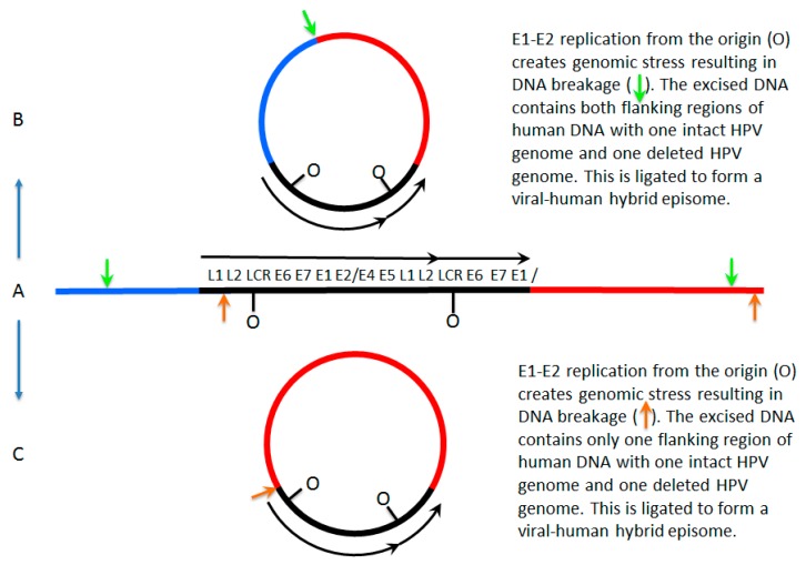
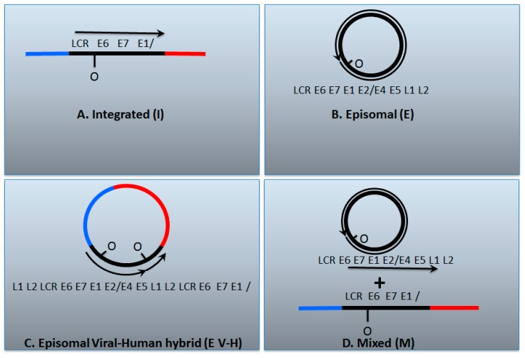
Human papillomaviruses (HPV) are detected in 70–80% of oropharyngeal cancers in the developed world, the incidence of which has reached epidemic proportions. The current paradigm regarding the status of the viral genome in these cancers is that there are three situations: one where the viral genome remains episomal, one where the viral genome integrates into the host genome and a third where there is a mixture of both integrated and episomal HPV genomes. Our recent work suggests that this third category has been mischaracterized as having integrated HPV genomes; evidence indicates that this category consists of virus–human hybrid episomes. Most of these hybrid episomes are consistent with being maintained by replication from HPV origin. We discuss our evidence to support this new paradigm, how such genomes can arise, and more importantly the implications for the clinical management of HPV positive head and neck cancers following accurate determination of the viral genome status.
Overview
The claims and statistics of this paper haven’t been checked yet.
Run the check — free, no account needed. It reads the open-access text and checks the sources it cites.
Integrity signals
Registry
Cites
Statistics
Figures
Verification
Claims the paper makes
The claims and statistics of this paper haven’t been checked yet.
Run the check — free, no account needed. It reads the open-access text and checks the sources it cites.
Claims other papers make about this one
Each is checked against this paper’s own text: does it say what the citing paper says it does?
In the citing paper
“in the case of HPV-18-positive cancers, the HPV-18 genome is integrated in 100% of cases, whilst in the case of HPV-16, the virus can remain episomal in up to 25% of cancers [13,14,15]”
From The Not-So-Good, the Bad and the Ugly: HPV E5, E6 and E7 Oncoproteins in the Orchestration of Carcinogenesis (2021) · cites this paper as [15]
In the citing paper
“More than 70% of HPV-mediated cancers are caused by high-risk types 16 and 18 alone”
From The Not-So-Good, the Bad and the Ugly: HPV E5, E6 and E7 Oncoproteins in the Orchestration of Carcinogenesis (2021) · cites this paper as [15]
Self-check
Percentages, sums and reported differences re-computed from the paper’s own numbers.
The self-check needs the paper’s full text.
Citations
Cites
No references could be resolved yet.
Research works printed but not linked: 105
- Nulton, 2017, Analysis of the Cancer Genome Atlas Sequencing Data Reveals Novel Properties of the Human Papillomavirus 16 Genome in Head and Neck Squamous Cell Carcinoma, Oncotarget, 8, 17684, 10.18632/oncotarget.15179 · DOI
- Hebner, 2006, Human Papillomaviruses: Basic Mechanisms of Pathogenesis and Oncogenicity, Rev. Med. Virol., 16, 83, 10.1002/rmv.488 · DOI
- Doorbar, 2012, The Biology and Life-Cycle of Human Papillomaviruses, Vaccine, 30, F55, 10.1016/j.vaccine.2012.06.083 · DOI
- Calton, C.M., Bronnimann, M.P., Manson, A.R., Li, S., Chapman, J.A., Suarez-Berumen, M., Williamson, T.R., Molugu, S.K., Bernal, R.A., and Campos, S.K. (2017). Translocation of the Papillomavirus L2/vDNA Complex Across the Limiting Membrane Requires the Onset of Mitosis. PLoS Pathog., 13. · DOI
- Aydin, I., Weber, S., Snijder, B., Samperio Ventayol, P., Kuhbacher, A., Becker, M., Day, P.M., Schiller, J.T., Kann, M., and Pelkmans, L. (2014). Large Scale RNAi Reveals the Requirement of Nuclear Envelope Breakdown for Nuclear Import of Human Papillomaviruses. PLoS Pathog., 10. · DOI
- Thierry, 2009, Transcriptional Regulation of the Papillomavirus Oncogenes by Cellular and Viral Transcription Factors in Cervical Carcinoma, Virology, 384, 375, 10.1016/j.virol.2008.11.014 · DOI
- Moody, 2010, Human Papillomavirus Oncoproteins: Pathways to Transformation, Nat. Rev. Cancer, 10, 550, 10.1038/nrc2886 · DOI
- McBride, 2013, The Papillomavirus E2 Proteins, Virology, 445, 57, 10.1016/j.virol.2013.06.006 · DOI
- Bergvall, 2013, The E1 Proteins, Virology, 445, 35, 10.1016/j.virol.2013.07.020 · DOI
- Masterson, 1998, A C-Terminal Helicase Domain of the Human Papillomavirus E1 Protein Binds E2 and the DNA Polymerase Alpha-Primase p68 Subunit, J. Virol., 72, 7407, 10.1128/JVI.72.9.7407-7419.1998 · DOI
- Melendy, 1995, Cellular Factors Required for Papillomavirus DNA Replication, J. Virol., 69, 7857, 10.1128/jvi.69.12.7857-7867.1995 · DOI
- Hu, 2006, Cellular Topoisomerase I Modulates Origin Binding by Bovine Papillomavirus Type 1 E1, J. Virol., 80, 4363, 10.1128/JVI.80.9.4363-4371.2006 · DOI
- Clower, 2006, Papillomavirus E1 Protein Binds to and Stimulates Human Topoisomerase I, J. Virol., 80, 1584, 10.1128/JVI.80.3.1584-1587.2006 · DOI
- Loo, 2004, Recruitment of Replication Protein A by the Papillomavirus E1 Protein and Modulation by Single-Stranded DNA, J. Virol., 78, 1605, 10.1128/JVI.78.4.1605-1615.2004 · DOI
- Boner, 2002, A Functional Interaction between the Human Papillomavirus 16 Transcription/Replication Factor E2 and the DNA Damage Response Protein TopBP1, J. Biol. Chem., 277, 22297, 10.1074/jbc.M202163200 · DOI
- Donaldson, 2012, An Interaction between Human Papillomavirus 16 E2 and TopBP1 is Required for Optimum Viral DNA Replication and Episomal Genome Establishment, J. Virol., 86, 12806, 10.1128/JVI.01002-12 · DOI
- Gauson, 2015, Evidence Supporting a Role for TopBP1 and Brd4 in the Initiation but Not Continuation of Human Papillomavirus 16 E1/E2-Mediated DNA Replication, J. Virol., 89, 17684, 10.1128/JVI.00335-15 · DOI
- Donaldson, 2007, TopBP1 Regulates Human Papillomavirus Type 16 E2 Interaction with Chromatin, J. Virol., 81, 4338, 10.1128/JVI.02353-06 · DOI
- You, 2004, Interaction of the Bovine Papillomavirus E2 Protein with Brd4 Tethers the Viral DNA to Host Mitotic Chromosomes, Cell, 117, 349, 10.1016/S0092-8674(04)00402-7 · DOI
- Parish, 2006, ChlR1 is Required for Loading Papillomavirus E2 Onto Mitotic Chromosomes and Viral Genome Maintenance, Mol. Cell, 24, 867, 10.1016/j.molcel.2006.11.005 · DOI
- Chappell, 2015, Homologous Recombination Repair Factors, Rad51 and BRCA1, are Necessary for Productive Replication of Human Papillomavirus 31, J. Virol., 90, 2639, 10.1128/JVI.02495-15 · DOI
- Moody, C.A., and Laimins, L.A. (2009). Human Papillomaviruses Activate the ATM DNA Damage Pathway for Viral Genome Amplification upon Differentiation. PLoS Pathog., 5. · DOI · Paper page
- Gillespie, 2012, Human Papillomaviruses Recruit Cellular DNA Repair and Homologous Recombination Factors to Viral Replication Centers, J. Virol., 86, 9520, 10.1128/JVI.00247-12 · DOI
- Gautam, D., Moody, C.A., Weitzman, M.D., Weitzman, J.B., Moody, C.A., Laimins, L.A., Hong, S., Dutta, A., Laimins, L.A., and Gillespie, K.A. (2016). Impact of the DNA Damage Response on Human Papillomavirus Chromatin. PLoS Pathog., 12. · DOI
- Moody, 2011, Nuclear Accumulation of the Papillomavirus E1 Helicase Blocks S-Phase Progression and Triggers an ATM-Dependent DNA Damage Response, J. Virol., 85, 8996, 10.1128/JVI.00542-11 · DOI
- Anacker, 2014, Productive Replication of Human Papillomavirus 31 Requires DNA Repair Factor Nbs1, J. Virol., 88, 8528, 10.1128/JVI.00517-14 · DOI
- Anacker, 2016, HPV31 Utilizes the ATR-Chk1 Pathway to Maintain Elevated RRM2 Levels and a Replication-Competent Environment in Differentiating Keratinocytes, Virology, 499, 383, 10.1016/j.virol.2016.09.028 · DOI
- Johnson, 2017, The Rb Binding Domain of HPV31 E7 is Required to Maintain High Levels of DNA Repair Factors in Infected Cells, Virology, 500, 22, 10.1016/j.virol.2016.09.029 · DOI
- Bristol, M.L., Wang, X., Smith, N.W., Son, M.P., Evans, M.R., and Morgan, I.M. (2016). DNA Damage Reduces the Quality, but Not the Quantity of Human Papillomavirus 16 E1 and E2 DNA Replication. Viruses, 8. · DOI
- Reinson, 2013, Engagement of the ATR-Dependent DNA Damage Response at the HPV18 Replication Centers during the Initial Amplification, J. Virol., 87, 951, 10.1128/JVI.01943-12 · DOI
- Sakakibara, 2011, The Papillomavirus E1 Helicase Activates a Cellular DNA Damage Response in Viral Replication Foci, J. Virol., 85, 8981, 10.1128/JVI.00541-11 · DOI
- Sakakibara, N., Chen, D., Jang, M.K., Kang, D.W., Luecke, H.F., Wu, S.Y., Chiang, C.M., and McBride, A.A. (2013). Brd4 is Displaced from HPV Replication Factories as they Expand and Amplify Viral DNA. PLoS Pathog., 9. · DOI
- Sakakibara, N., Chen, D., and McBride, A.A. (2013). Papillomaviruses use Recombination-Dependent Replication to Vegetatively Amplify their Genomes in Differentiated Cells. PLoS Pathog., 9. · DOI
- King, 2010, Human Papillomavirus E1 and E2 Mediated DNA Replication is Not Arrested by DNA Damage Signalling, Virology, 406, 95, 10.1016/j.virol.2010.06.033 · DOI
- 2009, Papillomaviruses in the Causation of Human Cancers—A Brief Historical Account, Virology, 384, 260, 10.1016/j.virol.2008.11.046 · DOI · Paper page
- Cullen, 1991, Analysis of the Physical State of Different Human Papillomavirus DNAs in Intraepithelial and Invasive Cervical Neoplasm, J. Virol., 65, 606, 10.1128/jvi.65.2.606-612.1991 · DOI · Paper page
- Durst, 1985, The Physical State of Human Papillomavirus Type 16 DNA in Benign and Malignant Genital Tumours, J. Gen. Virol., 66, 1515, 10.1099/0022-1317-66-7-1515 · DOI
- Kristiansen, 1994, Coexistence of Episomal and Integrated HPV16 DNA in Squamous Cell Carcinoma of the Cervix, J. Clin. Pathol., 47, 253, 10.1136/jcp.47.3.253 · DOI
- Jeon, 1995, Integration of Human Papillomavirus Type 16 into the Human Genome Correlates with a Selective Growth Advantage of Cells, J. Virol., 69, 2989, 10.1128/jvi.69.5.2989-2997.1995 · DOI
- Cooper, 1992, Integration of Human Papillomavirus Types 16 and 18 in Cervical Adenocarcinoma, J. Clin. Pathol., 45, 382, 10.1136/jcp.45.5.382 · DOI
- Couturier, 1991, Integration of Papillomavirus DNA near Myc Genes in Genital Carcinomas and its Consequences for Proto-Oncogene Expression, J. Virol., 65, 4534, 10.1128/jvi.65.8.4534-4538.1991 · DOI
- Cooper, 1991, In Situ Human Papillomavirus (HPV) Genotyping of Cervical Intraepithelial Neoplasia in South African and British Patients: Evidence for Putative HPV Integration in Vivo, J. Clin. Pathol., 44, 400, 10.1136/jcp.44.5.400 · DOI
- Fukushima, 1990, The Physical State of Human Papillomavirus 16 DNA in Cervical Carcinoma and Cervical Intraepithelial Neoplasia, Cancer, 66, 2155, 10.1002/1097-0142(19901115)66:10<2155::AID-CNCR2820661019>3.0.CO;2-Q · DOI
- Wagatsuma, 1990, Analysis of Integrated Human Papillomavirus Type 16 DNA in Cervical Cancers: Amplification of Viral Sequences Together with Cellular Flanking Sequences, J. Virol., 64, 813, 10.1128/jvi.64.2.813-821.1990 · DOI
- Woodworth, 1988, Characterization of Normal Human Exocervical Epithelial Cells Immortalized in Vitro by Papillomavirus Types 16 and 18 DNA, Cancer Res., 48, 4620
- Lehn, 1988, Physical State and Biological Activity of Human Papillomavirus Genomes in Precancerous Lesions of the Female Genital Tract, J. Gen. Virol., 69, 187, 10.1099/0022-1317-69-1-187 · DOI
- Choo, 1987, Integration of Human Papillomavirus Type 16 into Cellular DNA of Cervical Carcinoma: Preferential Deletion of the E2 Gene and Invariable Retention of the Long Control Region and the E6/E7 Open Reading Frames, Virology, 161, 259, 10.1016/0042-6822(87)90195-4 · DOI
- Baker, 1987, Structural and Transcriptional Analysis of Human Papillomavirus Type 16 Sequences in Cervical Carcinoma Cell Lines, J. Virol., 61, 962, 10.1128/jvi.61.4.962-971.1987 · DOI
- Durst, 1987, Papillomavirus Sequences Integrate Near Cellular Oncogenes in some Cervical Carcinomas, Proc. Natl. Acad. Sci. USA, 84, 1070, 10.1073/pnas.84.4.1070 · DOI · Paper page
- Choo, 1987, Presence of Episomal and Integrated Human Papillomavirus DNA Sequences in Cervical Carcinoma, J. Med. Virol., 21, 101, 10.1002/jmv.1890210202 · DOI
- Shirasawa, 1987, Integration and Transcription of Human Papillomavirus Type 16 and 18 Sequences in Cell Lines Derived from Cervical Carcinomas, J. Gen. Virol., 68, 583, 10.1099/0022-1317-68-2-583 · DOI
- Mincheva, 1987, Chromosomal Integration Sites of Human Papillomavirus DNA in Three Cervical Cancer Cell Lines Mapped by in Situ Hybridization, Med. Microbiol. Immunol., 176, 245, 10.1007/BF00190531 · DOI
- Romanczuk, 1992, Disruption of either the E1 or the E2 Regulatory Gene of Human Papillomavirus Type 16 Increases Viral Immortalization Capacity, Proc. Natl. Acad. Sci. USA, 89, 3159, 10.1073/pnas.89.7.3159 · DOI
- Daniel, 1995, Changes in the Physical State and Expression of Human Papillomavirus Type 16 in the Progression of Cervical Intraepithelial Neoplasia Lesions Analysed by PCR, J. Gen. Virol., 76, 2589, 10.1099/0022-1317-76-10-2589 · DOI
- Vernon, 1997, Association of Human Papillomavirus Type 16 Integration in the E2 Gene with Poor Disease-Free Survival from Cervical Cancer, Int. J. Cancer, 74, 50, 10.1002/(SICI)1097-0215(19970220)74:1<50::AID-IJC9>3.0.CO;2-# · DOI
- Park, 1997, Physical Status and Expression of HPV Genes in Cervical Cancers, Gynecol. Oncol., 65, 121, 10.1006/gyno.1996.4596 · DOI
- Yoshinouchi, 1999, Analysis by Multiplex PCR of the Physical Status of Human Papillomavirus Type 16 DNA in Cervical Cancers, J. Clin. Microbiol., 37, 3514, 10.1128/JCM.37.11.3514-3517.1999 · DOI
- Gray, 2010, In Vitro Progression of Human Papillomavirus 16 Episome-Associated Cervical Neoplasia Displays Fundamental Similarities to Integrant-Associated Carcinogenesis, Cancer Res., 70, 4081, 10.1158/0008-5472.CAN-09-3335 · DOI
- Herdman, 2006, Interferon-Beta Treatment of Cervical Keratinocytes Naturally Infected with Human Papillomavirus 16 Episomes Promotes Rapid Reduction in Episome Numbers and Emergence of Latent Integrants, Carcinogenesis, 27, 2341, 10.1093/carcin/bgl172 · DOI
- Bouvard, 1994, Characterization of the Human Papillomavirus E2 Protein: Evidence of Trans-Activation and Trans-Repression in Cervical Keratinocytes, EMBO J., 13, 5451, 10.1002/j.1460-2075.1994.tb06880.x · DOI
- Demeret, 1997, Different Mechanisms Contribute to the E2-Mediated Transcriptional Repression of Human Papillomavirus Type 18 Viral Oncogenes, J. Virol., 71, 9343, 10.1128/jvi.71.12.9343-9349.1997 · DOI
- Romanczuk, 1990, Mutational Analysis of cis Elements Involved in E2 Modulation of Human Papillomavirus Type 16 P97 and Type 18 P105 Promoters, J. Virol., 64, 2849, 10.1128/jvi.64.6.2849-2859.1990 · DOI
- Bechtold, 2003, Human Papillomavirus Type 16 E2 Protein has no Effect on Transcription from Episomal Viral DNA, J. Virol., 77, 2021, 10.1128/JVI.77.3.2021-2028.2003 · DOI
- Hwang, 1996, Activation of the Endogenous p53 Growth Inhibitory Pathway in HeLa Cervical Carcinoma Cells by Expression of the Bovine Papillomavirus E2 Gene, Oncogene, 12, 795
- DeFilippis, 2003, Endogenous Human Papillomavirus E6 and E7 Proteins Differentially Regulate Proliferation, Senescence, and Apoptosis in HeLa Cervical Carcinoma Cells, J. Virol., 77, 1551, 10.1128/JVI.77.2.1551-1563.2003 · DOI
- Goodwin, 2001, Induced Senescence in HeLa Cervical Carcinoma Cells Containing Elevated Telomerase Activity and Extended Telomeres, Cell Growth Differ., 12, 525
- Parish, 2006, E2 Proteins from High- and Low-Risk Human Papillomavirus Types Differ in their Ability to Bind p53 and Induce Apoptotic Cell Death E2 Proteins from High- and Low-Risk Human Papillomavirus Types Differ in their Ability to Bind p53 and Induce Apoptotic Cell, J. Virol., 80, 4580, 10.1128/JVI.80.9.4580-4590.2006 · DOI
- Kalantari, 2015, Assessment of the HPV DNA Methylation Status in Cervical Lesions, Methods Mol. Biol., 1249, 267, 10.1007/978-1-4939-2013-6_20 · DOI
- Kalantari, 2004, Conserved Methylation Patterns of Human Papillomavirus Type 16 DNA in Asymptomatic Infection and Cervical Neoplasia, J. Virol., 78, 12762, 10.1128/JVI.78.23.12762-12772.2004 · DOI
- Das, 2010, Association of Viral Load with HPV16 Positive Cervical Cancer Pathogenesis: Causal Relevance in Isolates Harboring Intact Viral E2 Gene, Virology, 402, 197, 10.1016/j.virol.2010.03.030 · DOI
- Bhattacharjee, 2006, CpG Methylation of HPV 16 LCR at E2 Binding Site Proximal to P97 is Associated with Cervical Cancer in Presence of Intact E2, Virology, 354, 280, 10.1016/j.virol.2006.06.018 · DOI
- Fernandez, 2009, The Dynamic DNA Methylomes of Double-Stranded DNA Viruses Associated with Human Cancer, Genome Res., 19, 438, 10.1101/gr.083550.108 · DOI
- Wentzensen, 2012, Methylation of HPV18, HPV31, and HPV45 Genomes and Cervical Intraepithelial Neoplasia Grade 3, J. Natl. Cancer Inst., 104, 1738, 10.1093/jnci/djs425 · DOI
- Badal, 2003, CpG Methylation of Human Papillomavirus Type 16 DNA in Cervical Cancer Cell Lines and in Clinical Specimens: Genomic Hypomethylation Correlates with Carcinogenic Progression, J. Virol., 77, 6227, 10.1128/JVI.77.11.6227-6234.2003 · DOI
- Kalantari, 2008, Effects of Cellular Differentiation, Chromosomal Integration and 5-aza-2′-Deoxycytidine Treatment on Human Papillomavirus-16 DNA Methylation in Cultured Cell Lines, Virology, 374, 292, 10.1016/j.virol.2007.12.016 · DOI
- Gillison, 2000, Evidence for a Causal Association between Human Papillomavirus and a Subset of Head and Neck Cancers, J. Natl. Cancer Inst., 92, 709, 10.1093/jnci/92.9.709 · DOI
- Gillison, 2004, Human Papillomavirus-Associated Head and Neck Cancer is a Distinct Epidemiologic, Clinical, and Molecular Entity, Semin. Oncol., 31, 744, 10.1053/j.seminoncol.2004.09.011 · DOI
- Gillison, 2001, Human Papillomavirus-Associated Head and Neck Squamous Cell Carcinoma: Mounting Evidence for an Etiologic Role for Human Papillomavirus in a Subset of Head and Neck Cancers, Curr. Opin. Oncol., 13, 183, 10.1097/00001622-200105000-00009 · DOI
- Garnaes, 2015, Increasing Incidence of Base of Tongue Cancers from 2000 to 2010 due to HPV: The Largest Demographic Study of 210 Danish Patients, Br. J. Cancer, 113, 131, 10.1038/bjc.2015.198 · DOI
- Garnaes, 2015, A High and Increasing HPV Prevalence in Tonsillar Cancers in Eastern Denmark, 2000–2010: The Largest Registry-Based Study to Date, Int. J. Cancer, 136, 2196, 10.1002/ijc.29254 · DOI
- Carlander, 2017, Continuing Rise in Oropharyngeal Cancer in a High HPV Prevalence Area: A Danish Population-Based Study from 2011 to 2014, Eur. J. Cancer, 70, 75, 10.1016/j.ejca.2016.10.015 · DOI
- Ang, 2010, Human Papillomavirus and Survival of Patients with Oropharyngeal Cancer, N. Engl. J. Med., 363, 24, 10.1056/NEJMoa0912217 · DOI
- Garnaes, 2016, Double Positivity for HPV DNA/p16 in Tonsillar and Base of Tongue Cancer Improves Prognostication: Insights from a Large Population-Based Study, Int. J. Cancer, 139, 2598, 10.1002/ijc.30389 · DOI
- Larsen, 2016, Novel Nomograms for Survival and Progression in HPV+ and HPV− Oropharyngeal Cancer: A Population-Based Study of 1542 Consecutive Patients, Oncotarget, 7, 71761, 10.18632/oncotarget.12335 · DOI
- Hafkamp, 2003, A Subset of Head and Neck Squamous Cell Carcinomas Exhibits Integration of HPV 16/18 DNA and Overexpression of p16INK4A and p53 in the Absence of Mutations in p53 Exons 5–8, Int. J. Cancer, 107, 394, 10.1002/ijc.11389 · DOI
- Begum, 2005, Tissue Distribution of Human Papillomavirus 16 DNA Integration in Patients with Tonsillar Carcinoma, Clin. Cancer Res., 11, 5694, 10.1158/1078-0432.CCR-05-0587 · DOI
- Samama, 2002, HPV DNA Detection by in Situ Hybridization with Catalyzed Signal Amplification on Thin-Layer Cervical Smears, J. Histochem. Cytochem., 50, 1417, 10.1177/002215540205001014 · DOI
- Kim, 2007, HPV Integration Begins in the Tonsillar Crypt and Leads to the Alteration of p16, EGFR and c-Myc during Tumor Formation, Int. J. Cancer, 120, 1418, 10.1002/ijc.22464 · DOI
- Deng, 2013, Viral Load, Physical Status, and E6/E7 mRNA Expression of Human Papillomavirus in Head and Neck Squamous Cell Carcinoma, Head Neck, 35, 800, 10.1002/hed.23034 · DOI
- Olthof, N.C., Speel, E.M., Kolligs, J., Haesevoets, A., Henfling, M., Ramaekers, F.C.S., Preuss, S.F., Drebber, U., Wieland, U., and Silling, S. (2014). Comprehensive Analysis of HPV16 Integration in OSCC Reveals no Significant Impact of Physical Status on Viral Oncogene and Virally Disrupted Human Gene Expression. PLoS ONE, 9. · DOI
- Cancer Genome Atlas Network (2015). Comprehensive Genomic Characterization of Head and Neck Squamous Cell Carcinomas. Nature, 517, 576–582. · DOI · Paper page
- Parfenov, 2014, Characterization of HPV and Host Genome Interactions in Primary Head and Neck Cancers, Proc. Natl. Acad. Sci. USA, 111, 15544, 10.1073/pnas.1416074111 · DOI
- Vojtechova, 2016, Analysis of the Integration of Human Papillomaviruses in Head and Neck Tumours in Relation to Patients’ Prognosis, Int. J. Cancer, 138, 386, 10.1002/ijc.29712 · DOI
- Lim, 2016, Human Papillomavirus Integration Pattern and Demographic, Clinical, and Survival Characteristics of Patients with Oropharyngeal Squamous Cell Carcinoma, Head Neck, 38, 1139, 10.1002/hed.24429 · DOI
- Kadaja, 2007, Genomic Instability of the Host Cell Induced by the Human Papillomavirus Replication Machinery, EMBO J., 26, 2180, 10.1038/sj.emboj.7601665 · DOI
- Akagi, 2014, Genome-Wide Analysis of HPV Integration in Human Cancers Reveals Recurrent, Focal Genomic Instability, Genome Res., 24, 185, 10.1101/gr.164806.113 · DOI
- Evans, M.R., James, C.D., Loughran, O., Nulton, T.J., Wang, X., Bristol, M.L., Windle, B., and Morgan, I.M. (2017). An Oral Keratinocyte Life Cycle Model Identifies Novel Host Genome Regulation by Human Papillomavirus 16 Relevant to HPV Positive Head and Neck Cancer. Oncotarget. · DOI
- Kelly, 2016, Treatment De-Intensification Strategies for Head and Neck Cancer, Eur. J. Cancer, 68, 125, 10.1016/j.ejca.2016.09.006 · DOI
- Fakhry, 2008, Improved Survival of Patients with Human Papillomavirus-Positive Head and Neck Squamous Cell Carcinoma in a Prospective Clinical Trial, J. Natl. Cancer Inst., 100, 261, 10.1093/jnci/djn011 · DOI
- Fischer, 2010, P16 Expression in Oropharyngeal Cancer: Its Impact on Staging and Prognosis Compared with the Conventional Clinical Staging Parameters, Ann. Oncol., 21, 1961, 10.1093/annonc/mdq210 · DOI
- Haughey, 2016, Pathology-Based Staging for HPV-Positive Squamous Carcinoma of the Oropharynx, Oral Oncol., 62, 11, 10.1016/j.oraloncology.2016.09.004 · DOI
- Lydiatt, 2017, Head and Neck Cancers-Major Changes in the American Joint Committee on Cancer Eighth Edition Cancer Staging Manual, CA Cancer. J. Clin., 67, 122, 10.3322/caac.21389 · DOI
- Malm, 2017, Evaluation of Proposed Staging Systems for Human Papillomavirus-Related Oropharyngeal Squamous Cell Carcinoma, Cancer, 123, 1768, 10.1002/cncr.30512 · DOI
- Porceddu, 2017, Validation of the ICON-S Staging for HPV-Associated Oropharyngeal Carcinoma using a Pre-Defined Treatment Policy, Oral Oncol., 66, 81, 10.1016/j.oraloncology.2017.01.002 · DOI
- Ramqvist, 2015, Studies on Human Papillomavirus (HPV) 16 E2, E5 and E7 mRNA in HPV-Positive Tonsillar and Base of Tongue Cancer in Relation to Clinical Outcome and Immunological Parameters, Oral Oncol., 51, 1126, 10.1016/j.oraloncology.2015.09.007 · DOI
Cited by
Basukala, O., & Banks, L. (2021) · Viruses · doi:10.3390/v13101892 · Cited by 137
Metadata
- Authors
- Morgan, I., DiNardo, L., & Windle, B.
- Venue
- Viruses
- Publisher
- MDPI AG
- Published
- 2017-08-03
- Volume
- 9
- Issue
- 8
- Pages
- 208
- ISSN
- 1999-4915
- PMC
- PMC5580465
- Cited by
- 65
- References
- 0
- Language
- en
- Funders
- National Institute for Dental and Craniofacial Research (R03DE026230-01)
- National Cancer Institute (P30 CA016059)
About these checks
Registry, retraction, Cites and statistics results are computed, not judged, with one exception: a printed reference that no index lists is read once by a model before it counts as a citation error. Claims are extracted by a model and kept only when their quote appears verbatim in the text; each cited source is then checked, with a model’s help, for whether it says what the claim relies on. Those verdicts can be wrong — read the claim’s quote and the cited source before relying on one.
Statistics and arithmetic are recomputed, not judged.
Confidence bands are shown only for fields calibrated on our annotated test set. None is calibrated yet, so no band is shown.
What counts as a claim ›How we measure confidence ›How we check this ›
Are you an author, or do you see a problem with this page? Request a review
How to cite
Cite CoChat for the checks on this page, and the paper for its own findings.
- This page
- CoChat. (2026). Citation check: Integration of Human Papillomavirus Genomes in Head and Neck Cancer: Is It Time to Consider a Paradigm Shift? https://app.cochat.ai/papers/10.3390/v9080208
- The paper
- Morgan, I., DiNardo, L., & Windle, B. (2017). Integration of Human Papillomavirus Genomes in Head and Neck Cancer: Is It Time to Consider a Paradigm Shift? Viruses. https://doi.org/10.3390/v9080208
