

The DOI matches its Crossref record; 55 of the 55 references it cites were found in a registry or an index.
Isolation and characterization of platelet-derived extracellular vesicles
Open accessAbstract
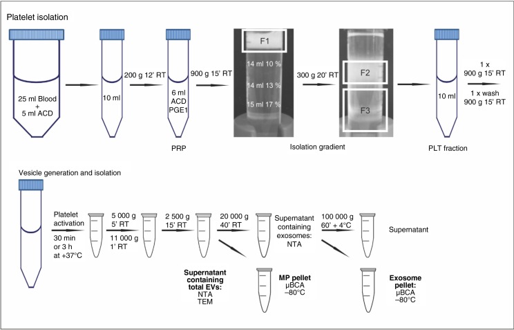
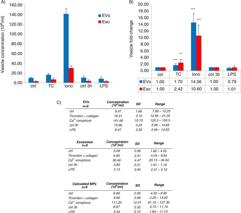
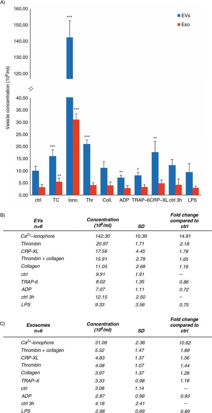
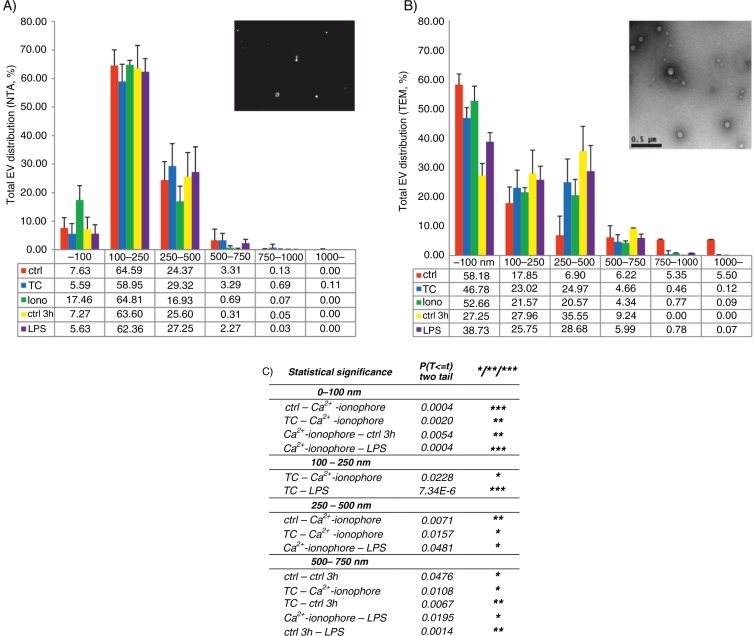
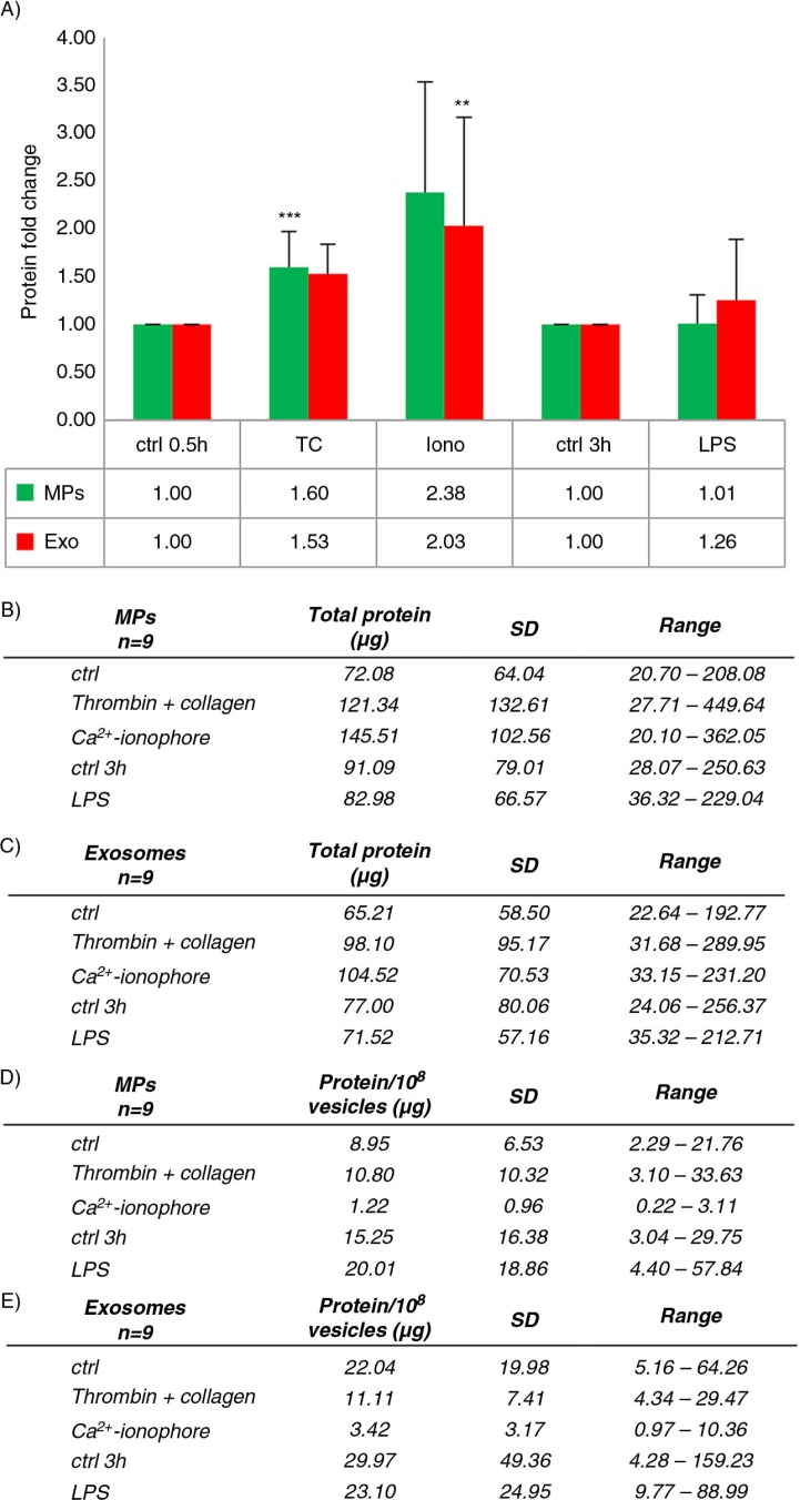
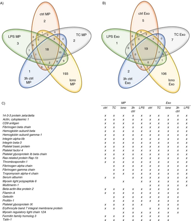
BACKGROUND: Platelet-derived extracellular vesicles (EVs) participate, for example, in haemostasis, immunity and development. Most studies of platelet EVs have targeted microparticles, whereas exosomes and EV characterization under various conditions have been less analyzed. Studies have been hampered by the difficulty in obtaining EVs free from contaminating cells and platelet remnants. Therefore, we optimized an EV isolation protocol and compared the quantity and protein content of EVs induced by different agonists. METHODS: Platelets isolated with iodixanol gradient were activated by thrombin and collagen, lipopolysaccharide (LPS) or Ca(2+) ionophore. Microparticles and exosomes were isolated by differential centrifugations. EVs were quantitated by nanoparticle tracking analysis (NTA) and total protein. Size distributions were determined by NTA and electron microscopy. Proteomics was used to characterize the differentially induced EVs. RESULTS: The main EV populations were 100-250 nm and over 90% were <500 nm irrespective of the activation. However, activation pathways differentially regulated the quantity and the quality of EVs, which also formed constitutively. Thrombogenic activation was the most potent physiological EV-generator. LPS was a weak inducer of EVs, which had a selective protein content from the thrombogenic EVs. Ca(2+) ionophore generated a large population of protein-poor and unselectively packed EVs. By proteomic analysis, EVs were highly heterogeneous after the different activations and between the vesicle subpopulations. CONCLUSIONS: Although platelets constitutively release EVs, vesiculation can be increased, and the activation pathway determines the number and the cargo of the formed EVs. These activation-dependent variations render the use of protein content in sample normalization invalid. Since most platelet EVs are 100-250 nm, only a fraction has been analyzed by previously used methods, for example, flow cytometry. As the EV subpopulations could not be distinguished and large vesicle populations may be lost by differential centrifugation, novel methods are required for the isolation and the differentiation of all EVs.
Overview
The claims and statistics of this paper haven’t been checked yet.
Run the check — free, no account needed. It reads the open-access text and checks the sources it cites.
Integrity signals
Registry
Cites
Statistics
Figures
Verification
Claims the paper makes
The claims and statistics of this paper haven’t been checked yet.
Run the check — free, no account needed. It reads the open-access text and checks the sources it cites.
Claims other papers make about this one
Each is checked against this paper’s own text: does it say what the citing paper says it does?
The check wasn’t confident either way, so it gives no verdict — never disputed.
In the citing paper
“Despite the extensive literature regarding antibodies targeting the cardiolipin (also known as the mitochondrial antigen M1) in SLE, the anti-mitochondrial autoantibody repertoire and their antigenic targets remains mostly uncharacterized (12, 34–37).”
From Autoantibodies in Systemic Lupus Erythematosus Target Mitochondrial RNA (2019) · cites this paper as [37]
Experimental — accuracy not yet measured
Self-check
Percentages, sums and reported differences re-computed from the paper’s own numbers.
The self-check needs the paper’s full text.
Citations
Cites
Primer3 on the WWW for General Users and for Biologist Programmers
Rozen, S., & Skaletsky, H. (2003) · Humana Press eBooks · doi:10.1385/1-59259-192-2:365 · Cited by 15,068
Membrane vesicles as conveyors of immune responses
Théry, C., Ostrowski, M., & Segura, É. (2009) · Nature reviews. Immunology · doi:10.1038/nri2567 · Cited by 3,996
Witwer, K. W., Buzás, E. I., Bemis, L., Bora, A., Lässer, C., Lötvall, J. O., Hoen, E. N. M. N., Piper, M. G., Sivaraman, S., Skog, J., et al. (2013) · Journal of Extracellular Vesicles · doi:10.3402/jev.v2i0.20360 · Cited by 2,513
Filipe, V., Hawe, A., & Jiskoot, W. (2010) · Pharmaceutical Research · doi:10.1007/s11095-010-0073-2 · Cited by 1,885
Heijnen, H. F. G., Schiel, A. E., Fijnheer, R., Geuze, H. J., & Sixma, J. J. (1999) · Blood · doi:10.1182/blood.v94.11.3791 · Cited by 1,488
Sizing and phenotyping of cellular vesicles using Nanoparticle Tracking Analysis
Dragovic, R., Gardiner, C., Brooks, A. S., Tannetta, D., Ferguson, D. J. P., Hole, P., Carr, B., Redman, C. W. G., Harris, A. L., Dobson, P. J., et al. (2011) · Nanomedicine Nanotechnology Biology and Medicine · doi:10.1016/j.nano.2011.04.003 · Cited by 1,368
Platelet α-granules: Basic biology and clinical correlates
Blair, P., & Flaumenhaft, R. (2009) · Blood Reviews · doi:10.1016/j.blre.2009.04.001 · Cited by 1,157
Exosomes: proteomic insights and diagnostic potential
Simpson, R. J., Lim, J. W., Moritz, R. L., & Mathivanan, S. (2009) · Expert Review of Proteomics · doi:10.1586/epr.09.17 · Cited by 1,119
Arraud, N., Linares, R., Tan, S., Gounou, C., Pasquet, J., Mornet, S., & Brisson, A. R. (2014) · Journal of Thrombosis and Haemostasis · doi:10.1111/jth.12554 · Cited by 776
Sims, P. J., Wiedmer, T., Esmon, C. T., Weiss, H. J., & Shattil, S. J. (1989) · Journal of Biological Chemistry · doi:10.1016/s0021-9258(18)71457-9 · Cited by 595
Optical and non‐optical methods for detection and characterization of microparticles and exosomes
van der Pol, E., Hoekstra, A. G., Sturk, A., Otto, C., van Leeuwen, T. G. J. M., & Nieuwland, R. (2010) · Journal of Thrombosis and Haemostasis · doi:10.1111/j.1538-7836.2010.04074.x · Cited by 562
Jimenez, J. J., Jy, W., Mauro, L. M., Soderland, C., Horstman, L. L., & Ahn, Y. S. (2003) · Thrombosis Research · doi:10.1016/s0049-3848(03)00064-1 · Cited by 515
Holme, P. A., Sølvik, U. Ø., Hamers, M. J. A. G., Solum, N. O., Brosstad, F. R., Barstad, R. M., & Sakaríassen, K. S. (1997) · Arteriosclerosis Thrombosis and Vascular Biology · doi:10.1161/01.atv.17.4.646 · Cited by 442
Single vs. swarm detection of microparticles and exosomes by flow cytometry
van der Pol, E., van Gemert, M. J. C., Sturk, A., Nieuwland, R., & van Leeuwen, T. G. J. M. (2012) · Journal of Thrombosis and Haemostasis · doi:10.1111/j.1538-7836.2012.04683.x · Cited by 441
Platelets regulate lymphatic vascular development through CLEC-2–SLP-76 signaling
Bertozzi, C. C., Schmaier, A. A., Mericko, P. A., Hess, P. R., Zou, Z., Chen, M., Chen, C., Xu, B., Lu, M., Zhou, D. D., et al. (2010) · Blood · doi:10.1182/blood-2010-02-270876 · Cited by 437
Lacroix, R., Judicone, C., PONCELET, P., Robert, S., ARNAUD, L., Sampol, J., & Dignat‐George, F. (2011) · Journal of Thrombosis and Haemostasis · doi:10.1111/j.1538-7836.2011.04610.x · Cited by 401
Platelets: bridging hemostasis, inflammation, and immunity
Jenne, C. N., Urrutia, R., & Kubes, P. (2013) · International Journal of Laboratory Hematology · doi:10.1111/ijlh.12084 · Cited by 382
Reininger, A. J. (2006) · Blood · doi:10.1182/blood-2005-02-0618 · Cited by 344
Flaumenhaft, R. C., Dilks, J. R., Richardson, J., Alden, E. C., Patel-Hett, S. R., Battinelli, E. M., Klement, G. L., Sola-Visner, M. C., & Italiano, J. E. (2008) · Blood · doi:10.1182/blood-2008-06-163832 · Cited by 318
Clinical relevance of microparticles from platelets and megakaryocytes
Italiano, J. E., Mairuhu, A. T. A., & Flaumenhaft, R. C. (2010) · Current Opinion in Hematology · doi:10.1097/moh.0b013e32833e77ee · Cited by 299
Pre-analytical and analytical issues in the analysis of blood microparticles
Yuana, Y., Bertina, R. M., & Osanto, S. (2010) · Thrombosis and Haemostasis · doi:10.1160/th10-09-0595 · Cited by 268
REVIEW ARTICLE: Coated-platelets: an emerging component of the procoagulant response
Dale, G. L. (2005) · Journal of Thrombosis and Haemostasis · doi:10.1111/j.1538-7836.2005.01274.x · Cited by 266
Atomic force microscopy: a novel approach to the detection of nanosized blood microparticles
Yuana, Y., Oosterkamp, T. H., Bahatyrova, S., Ashcroft, B. A., RODRIGUEZ, P. G., Bertina, R. M., & Osanto, S. (2009) · Journal of Thrombosis and Haemostasis · doi:10.1111/j.1538-7836.2009.03654.x · Cited by 262
Modulation of B-cell exosome proteins by gamma herpesvirus infection
Meckes, D. G., Gunawardena, H. P., DeKroon, R. M., Heaton, P. R., Edwards, R. H., Özgür, S., Griffith, J. D., Damania, B., & Raab‐Traub, N. (2013) · Proceedings of the National Academy of Sciences · doi:10.1073/pnas.1303906110 · Cited by 248
Brown, G. T., & McIntyre, T. M. (2011) · The Journal of Immunology · doi:10.4049/jimmunol.1001623 · Cited by 222
Apoptosis in the anucleate platelet
Leytin, V. (2011) · Blood Reviews · doi:10.1016/j.blre.2011.10.002 · Cited by 207
Proteomic and functional characterisation of platelet microparticle size classes
Dean, W. L., Lee, M. J., Cummins, T. D., Schultz, D. J., & Powell, D. W. (2009) · Thrombosis and Haemostasis · doi:10.1160/th09-04-243 · Cited by 199
Platelet-Derived Microvesicles: Multitalented Participants in Intercellular Communication
Aatonen, M., Grönholm, M., & Siljander, P. R. M. (2012) · Seminars in Thrombosis and Hemostasis · doi:10.1055/s-0031-1300956 · Cited by 194
Self-citationGambim, M. H., Carmo, A. O., Marti, L. C., Veríssimo-Filho, S., Lopes, L. R., & Janiszewski, M. (2007) · Critical Care · doi:10.1186/cc6133 · Cited by 186
Procoagulant platelets: are they necrotic?
Jackson, S. P., & Schoenwaelder, S. M. (2010) · Blood · doi:10.1182/blood-2010-01-261669 · Cited by 171
The emerging role of platelets in adaptive immunity
Elzey, B. D., Sprague, D. L., & Ratliff, T. L. (2005) · Cellular Immunology · doi:10.1016/j.cellimm.2005.12.005 · Cited by 170
Kilpinen, L., Impola, U., Sankkila, L., Ritamo, I., Aatonen, M., Kilpinen, S. K., Tuimala, J., Valmu, L., Levijoki, J. M., Finckenberg, P., et al. (2013) · Journal of Extracellular Vesicles · doi:10.3402/jev.v2i0.21927 · Cited by 169
Self-citationCauwenberghs, S., Feijge, M. A. H., Harper, A. G. S., Sage, S. O., Curvers, J., & Heemskerk, J. W. M. (2006) · FEBS Letters · doi:10.1016/j.febslet.2006.08.082 · Cited by 160
Platelet-derived microparticles associate with fibrin during thrombosis
Siljander, P. R. M., Carpén, O., & Lassila, R. (1996) · Blood · doi:10.1182/blood.v87.11.4651.bloodjournal87114651 · Cited by 155
Self-citationCellular prion protein is released on exosomes from activated platelets
Robertson, C., Booth, S. A., Beniac, D. R., Coulthart, M. B., Booth, T. F., & McNicol, A. (2006) · Blood · doi:10.1182/blood-2005-02-0802 · Cited by 137
Platelet Adhesion Enhances the Glycoprotein VI–Dependent Procoagulant Response
Siljander, P. R. M., Farndale, R. W., Feijge, M. A. H., Comfurius, P., Kos, S., Bevers, E. M., & Heemskerk, J. W. M. (2001) · Arteriosclerosis Thrombosis and Vascular Biology · doi:10.1161/01.atv.21.4.618 · Cited by 117
Self-citationSpinelli, S. L., Pollock, S. J., Murant, T. I., O'Brien, J. J., Blumberg, N., Francis, C. W., Taubman, M. B., Ray, D. M., & Phipps, R. P. (2007) · Thrombosis and Haemostasis · doi:10.1160/th07-05-0328 · Cited by 100
Gender-specific and menstrual cycle dependent differences in circulating microparticles
Tóth, B., Nikolajek, K., Rank, A., Nieuwland, R., Lohse, P., Pihusch, V., Friese, K., & Thaler, C. (2007) · Platelets · doi:10.1080/09537100701525843 · Cited by 84
Gennebäck, N., Hellman, U., Malm, L., Larsson, G., Ronquist, G., Waldenström, A. P., & Mörner, S. (2013) · Journal of Extracellular Vesicles · doi:10.3402/jev.v2i0.20167 · Cited by 83
Endocytosis by human platelets: metabolic and freeze-fracture studies.
Zucker‐Franklin, D. (1981) · The Journal of Cell Biology · doi:10.1083/jcb.91.3.706 · Cited by 78
Cell-Derived Microparticles Promote Coagulation after Moderate Exercise
Soßdorf, M., Otto, G. P., Claus, R. A., Gabriel, H. H. W., & Lösche, W. (2010) · Medicine & Science in Sports & Exercise · doi:10.1249/mss.0b013e3182068645 · Cited by 74
Siljander, P. R. M., Vuist, W. M. J., Breikers, G., Reutelingsperger, C. P. M., Barnes, M. J., Knight, C. G., Lassila, R., Farndale, R. W., & Heemskerk, J. W. M. (1999) · Thrombosis and Haemostasis · doi:10.1055/s-0037-1614571 · Cited by 71
Self-citationFurie, B., & Furie, B. C. (2006) · Blood Cells Molecules and Diseases · doi:10.1016/j.bcmd.2005.12.018 · Cited by 69
Hoen, E. N. M. N., van der Vlist, E. J., de Boer-Brouwer, M., Arkesteijn, G. J. A., Stoorvogel, W., & Wauben, M. H. M. (2012) · Journal of Leukocyte Biology · doi:10.1189/jlb.0911480 · Cited by 57
Cypryk, W., Öhman, T., Eskelinen, E., Matikainen, S., & Nyman, T. A. (2014) · Journal of Proteome Research · doi:10.1021/pr4012552 · Cited by 54
Self-citationVasina, E. M., Cauwenberghs, S., Staudt, M., Feijge, M. A. H., Weber, C., Koenen, R. R., & Heemskerk, J. W. M. (2013) · Europe PMC (PubMed Central) · Cited by 53
Soop, A., Hållström, L., FROSTELL, C. G., Wallén, H., Mobarrez, F., & Pisetsky, D. S. (2013) · Scandinavian Journal of Immunology · doi:10.1111/sji.12076 · Cited by 41
Vince, R. V., Sandström, M., Taylor, L. K., McNaughton, L. R., Laden, G., & Madden, L. A. (2008) · Thrombosis and Haemostasis · doi:10.1160/th08-01-0030 · Cited by 40
Contact- and agonist-regulated microvesiculation of human platelets
Liu, X., Liu, L., Zaske, A. M., Zhou, Z., Fu, Y., Yang, X., Conyers, J. L., Li, M., Zhang, Y., Dong, J.-F., et al. (2013) · Thrombosis and Haemostasis · doi:10.1160/th12-11-0853 · Cited by 38
Procoagulant platelet balloons: evidence from cryopreparation and electron microscopy
Hess, M. W., & Siljander, P. R. M. (2001) · Histochemistry and Cell Biology · doi:10.1007/s004180100272 · Cited by 32
Self-citationShrinkage in preparatory steps for SEM
Jensen, O. A., Prause, J. U., & Laursen, H. (1981) · Graefe s Archive for Clinical and Experimental Ophthalmology · doi:10.1007/bf00407662 · Cited by 30
Birschmann, I., Mietner, S., Dittrich, M. T., Pfrang, J., Dandekar, T., & Walter, U. (2007) · Thrombosis Research · doi:10.1016/j.thromres.2007.08.022 · Cited by 23
Pook, M., Tamming, L., Padari, K., Tiido, T., Maimets, T., Patarroyo, M. E., Juronen, E., Jaks, V., & Ingerpuu, S. (2014) · Journal of Thrombosis and Haemostasis · doi:10.1111/jth.12513 · Cited by 18
Shrinkage in Preparatory Steps for SEM
Jensen, O. A., Prause, J. U., & Laursen, H. (1981) · Current research in ophthalmic electron microscopy · doi:10.1007/978-3-642-81614-7_3 · Cited by 6
Microparticle Sizing and Counting Using Light Scattering Methods
Gabriel, D. A., & Giordano, K. (2010) · Seminars in Thrombosis and Hemostasis · doi:10.1055/s-0030-1267036 · Cited by 4
Cited by
Sorted by citations
Biological properties of extracellular vesicles and their physiological functions
Yáñez‐Mó, M., Siljander, P. R. ‐., Andreu, Z., Bedina Zavec, A., Borràs, F. E., Buzas, E. I., Buzas, K., Casal, E., Cappello, F., Carvalho, J., et al. (2015) · Journal of Extracellular Vesicles · doi:10.3402/jev.v4.27066 · Cited by 5,926
Self-citationKowal, J., Arras, G., Colombo, M., Jouve, M., Morath, J. P., Primdal-Bengtson, B., Dingli, F., Loew, D., Tkach, M., & Théry, C. (2016) · Proceedings of the National Academy of Sciences · doi:10.1073/pnas.1521230113 · Cited by 3,426
Extracellular vesicles in cancer — implications for future improvements in cancer care
Xu, R., Rai, A., Chen, M., Suwakulsiri, W., Greening, D. W., & Simpson, R. J. (2018) · Nature Reviews Clinical Oncology · doi:10.1038/s41571-018-0036-9 · Cited by 1,472
Methodological Guidelines to Study Extracellular Vesicles
Coumans, F. A. W., Brisson, A. R., Buzás, E. I., Dignat‐George, F., Drees, E. E. E., Andaloussi, S. E., Emanueli, C., Gąsecka, A., Hendrix, A., Hill, A. F., et al. (2017) · Circulation Research · doi:10.1161/circresaha.117.309417 · Cited by 1,052
Self-citationExtracellular Vesicles in Cancer: Exosomes, Microvesicles and the Emerging Role of Large Oncosomes
Minciacchi, V. R., Freeman, M. R., & Vizio, D. D. (2015) · Seminars in Cell and Developmental Biology · doi:10.1016/j.semcdb.2015.02.010 · Cited by 1,014
Metadata
- Authors
- Aatonen, M. T., Öhman, T., Nyman, T. A., Laitinen, S., Grönholm, M., & Siljander, P. R. ‐.
- Affiliations
- University of Helsinki (FI)
- Finnish Red Cross (FI)
- Corresponding author
- Siljander, Pia R.‐M.
- ORCID
- Venue
- Journal of Extracellular Vesicles
- Publisher
- Wiley
- Published
- 2014-01
- Volume
- 3
- Issue
- 1
- ISSN
- 2001-3078 (ISSN-L)
- PMC
- PMC4125723
- OpenAlex
- W1991525001
- Cited by
- 333
- References
- 55
- Citations by year
- 2026: 20 · 2025: 25 · 2024: 40 · 2023: 27 · 2022: 40 · 2021: 28 · 2020: 20 · 2019: 35 · 2018: 16 · 2017: 25 · 2016: 41 · 2015: 15 · 2014: 1
- FWCI
- 11.20
- Type
- journal article
- Language
- en
- Open access
- gold
- doi.org
- Licence
- Locations
- Journal of Extracellular Vesicles (publishedVersion, cc-by-nc)
- Europe PMC (PubMed Central) (publishedVersion, cc-by-nc-nd)
- PubMed Central (publishedVersion, cc-by-nc-nd)
- PubMed (publishedVersion)
- DOAJ (DOAJ: Directory of Open Access Journals) (submittedVersion, cc-by-sa)
- Funders
- Magnus Ehrnroothin Säätiö
- Otto A. Malm Lahjoitusrahasto
- Topics
- Life Sciences › Biochemistry, Genetics and Molecular Biology › Molecular Biology › Extracellular vesicles in disease
- Health Sciences › Medicine › Hematology › Platelet Disorders and Treatments
- Life Sciences › Biochemistry, Genetics and Molecular Biology › Cell Biology › Caveolin-1 and cellular processes
- Keywords
- platelet-derived extracellular vesicles, exosomes, microparticles, EV isolation, platelet activation, thrombin, collagen, lipopolysaccharide, calcium ionophore, vesicle size distribution, EV proteome, vesicle heterogeneity, sample normalization
About these checks
Registry, retraction, Cites and statistics results are computed, not judged, with one exception: a printed reference that no index lists is read once by a model before it counts as a citation error. Claims are extracted by a model and kept only when their quote appears verbatim in the text; each cited source is then checked, with a model’s help, for whether it says what the claim relies on. Those verdicts can be wrong — read the claim’s quote and the cited source before relying on one.
Statistics and arithmetic are recomputed, not judged.
Confidence bands are shown only for fields calibrated on our annotated test set. None is calibrated yet, so no band is shown.
What counts as a claim ›How we measure confidence ›How we check this ›
Are you an author, or do you see a problem with this page? Request a review
