

The DOI matches its Crossref record; 3 of the 3 references it cites were found in a registry or an index.
Genetic Diversity, Predictive Protein Structures, and Interaction Networks of Cysteine-Rich Receptor-Like Kinases in Arabidopsis thaliana
Open accessPublished version of a preprint
Abstract
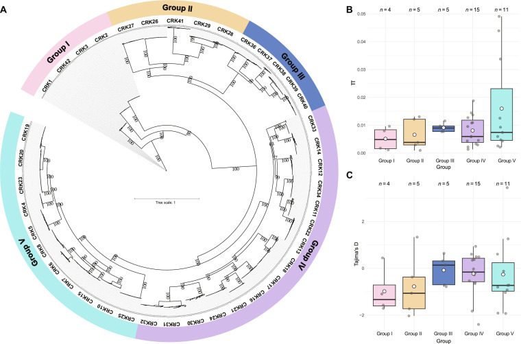
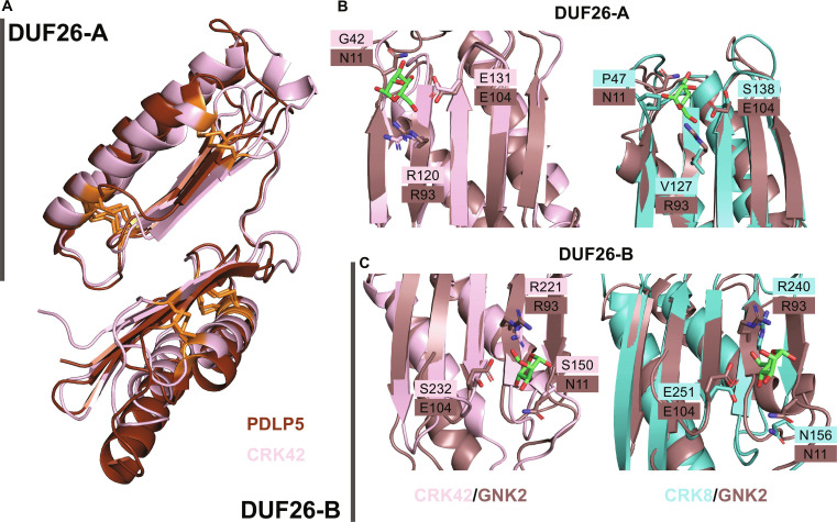
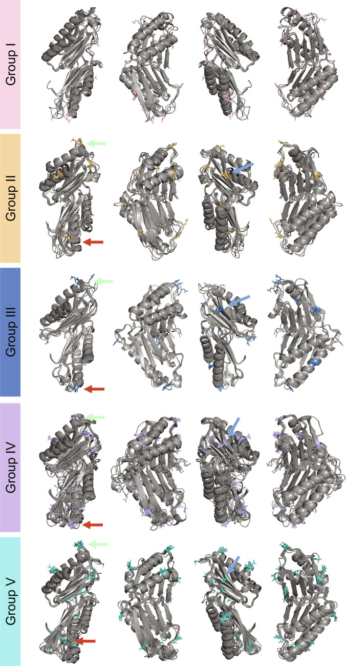
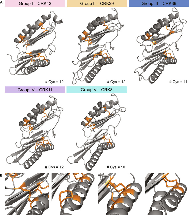
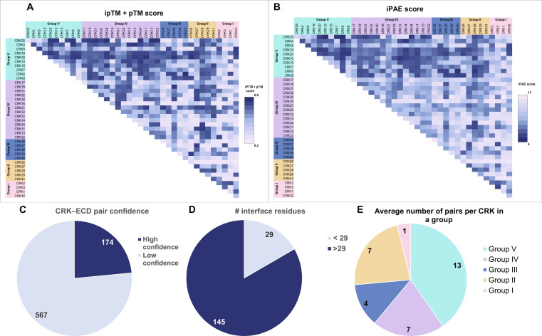
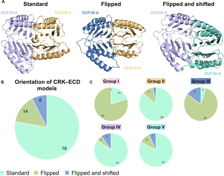
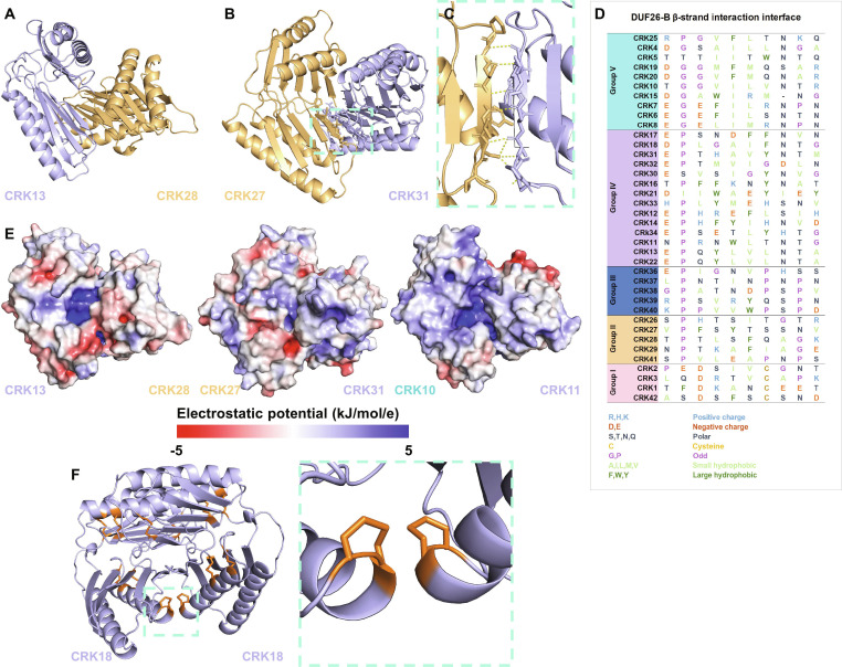
Cysteine-rich receptor-like kinases (CRKs) are a large subfamily of plant receptor-like kinases implicated in immunity and development, yet their ligands, interaction partners, and mechanistic roles remain poorly defined. We combined population-genetic analyses and AlphaFold-based structural prediction to characterize the Arabidopsis thaliana CRK family. Phylogenetic reconstruction from 69 natural accessions resolved 5 well-supported CRK clades. Nucleotide diversity (π) and neutrality tests revealed heterogeneous diversity across loci, with evidence of both positive and negative selection pressure acting on different CRKs. AlphaFold models of CRK extracellular domains (ECDs) recapitulate the DUF26 structure observed in plasmodesmata localizing protein (PDLP)5/PDLP8 and ginkbilobin-2 but display distinct biochemical properties and disulfide-bond topologies. Pairwise AlphaFold dimer modeling of all 780 CRK–ECD combinations produced 145 high-confidence interaction models; ~78% of these adopt a shared dimer conformation characterized by an extended intermolecular β-sheet at the interface. Integrating evolutionary and structural approaches reveal clade-specific selective regimes and conserved structural features of CRK–ECDs that likely underpin receptor–receptor interactions. Predicted high-confidence dimer interfaces suggest a general mode of CRK–ECD association that can guide targeted biochemical and genetic validation, accelerating functional dissection of this important receptor family.
Overview
The claims and statistics of this paper haven’t been checked yet.
Run the check — free, no account needed. It reads the open-access text and checks the sources it cites.
Integrity signals
Registry
Cites
Statistics
Figures
Verification
Claims the paper makes
The claims and statistics of this paper haven’t been checked yet.
Run the check — free, no account needed. It reads the open-access text and checks the sources it cites.
Self-check
Percentages, sums and reported differences re-computed from the paper’s own numbers.
The self-check needs the paper’s full text.
Citations
Cites
Highly accurate protein structure prediction with AlphaFold
Jumper, J., Evans, R., Pritzel, A., Green, T., Figurnov, M., Ronneberger, O., Tunyasuvunakool, K., Bates, R., Žídek, A., Potapenko, A., et al. (2021) · Nature · doi:10.1038/s41586-021-03819-2 · Cited by 43,764
Accurate structure prediction of biomolecular interactions with AlphaFold 3
Abramson, J., Adler, J., Dunger, J., Evans, R., Green, T., Pritzel, A., Ronneberger, O., Willmore, L., Ballard, A. J., Bambrick, J., et al. (2024) · Nature · doi:10.1038/s41586-024-07487-w · Cited by 15,861 · cited 2×
“The recently released AF3 is a step closer to including these conditions, allowing for the prediction of glycosylation and small lipids [ 71 ].”
Varadi, M., Bertoni, D., Magana, P., Paramval, U., Pidruchna, I., Radhakrishnan, M., Tsenkov, M., Nair, S., Mirdita, M., Yeo, J., et al. (2024) · Nucleic Acids Research · doi:10.1093/nar/gkad1011 · Cited by 2,052 · cited 1×
“Full-length CRK models were extracted from the AF database [ 13 , 94 ].”
Research works printed but not linked: 93
- 10.1104/pp.126.2.473 · DOI
- Martin-Ramirez S, Stouthamer J, Smakowska-Luzan E. More questions than answers: Insights into potential cysteine-rich receptor-like kinases redox signalling in Arabidopsis. Plant J. 2025;122(2): Article e70176. · DOI
- 10.1146/annurev-arplant-042916-040957 · DOI · Paper page
- Shiu SH, Bleecker AB. Plant receptor-like kinase gene family: Diversity, function, and signaling. Sci STKE. 2001;2001(113):re22.
- Bourdais G, Burdiak P, Gauthier A, Nitsch L, Salojärvi J, Rayapuram C, Idänheimo N, Hunter K, Kimura S, et al. Large-scale phenomics identifies primary and fine-tuning roles for CRKs in responses related to oxidative stress. PLOS Genet. 2015;11(7): Article e1005373. · DOI
- 10.1073/pnas.181141598 · DOI · Paper page
- 10.1105/tpc.020834 · DOI · Paper page
- 10.1016/j.tplants.2023.03.028 · DOI
- Vaattovaara A, Brandt B, Rajaraman S, Safronov O, Veidenberg A, Luklová M, Kangasjärvi J, Löytynoja A, Hothorn M, Salojärvi J, et al. Mechanistic insights into the evolution of DUF26-containing proteins in land plants. Commun. Biol. 2019;2:56. · DOI
- Yadeta KA, Elmore JM, Creer AY, Feng B, Franco JY, Rufian JS, He P, Phinney B, Coaker G. A cysteine-rich protein kinase associates with a membrane immune complex and the cysteine residues are required for cell death. Plant Physiol. 2017;173(1):771–787. · DOI
- Restrepo-Montoya D, Brueggeman R, McClean PE, Osorno JM. Computational identification of receptor-like kinases ‘rLK’ and receptor-like proteins ‘rLP’ in legumes. BMC Genomics. 2020;21(1):459. · DOI
- del Hierro I, Mélida H, Broyart C, Santiago J, Molina A. Computational prediction method to decipher receptor–glycoligand interactions in plant immunity. Plant J. 2021;105(6):1710. · DOI
- Ali F. Patterns of change in nucleotide diversity over gene length. Genome Biol Evol. 2024;16(4):evae078. · DOI
- Tatarinova TV, Chekalin E, Nikolsky Y, Bruskin S, Chebotarov D, McNally KL, Alexandrov N. Nucleotide diversity analysis highlights functionally important genomic regions. Sci Rep. 2016;6:35730. · DOI
- 10.1038/s44319-024-00122-9 · DOI
- 10.1016/j.cub.2024.07.061 · DOI
- Lomize AL, Schnitzer KA, Tood SC, Cherepanov S, Outerial C, Deane CM, Pogozheva ID. Membranome 3.0: Database of single-pass membrane proteins with AlphaFold models. Protein Sci. 2022;31(5): Article e4318. · DOI
- Dobson L, Szekeres LI, Gerdán C, Langó T, Zeke A, Tusnády GE. TmAlphaFold database: Membrane localization and evaluation of AlphaFold2 predicted alpha-helical transmembrane protein structures. Nucleic Acids Res. 2023;51(D1):D517–D522. · DOI
- 10.1104/pp.114.242636 · DOI
- Cui W, Lee JY. Arabidopsis callose synthases CalS1/8 regulate plasmodesmal permeability during stress. Nat Plants. 2016;2(5):16034. · DOI
- Sawano Y, Miyakawa T, Yamazaki H, Tanokura M, Hatano KI. Purification, characterization, and molecular gene cloning of an antifungal protein from Ginkgo biloba seeds. Biol Chem. 2007;388(3):273–280. · DOI
- Miyakawa T, Miyazono KI, Sawano Y, Hatano K-I, Tanokura M. Crystal structure of ginkbilobin-2 with homology to the extracellular domain of plant cysteine-rich receptor-like kinases. Proteins. 2009;77(1):247–251. · DOI
- 10.1074/jbc.M109.063073 · DOI
- 10.1038/s41477-024-01841-6 · DOI
- 10.1016/j.plantsci.2018.05.007 · DOI
- de Oliveira MVV, Xu G, Li B, de Souza Vespoli L, Meng X, Chen X, Yu X, de Souza SA, Intorne A, de Manhães AM, et al. Specific control of Arabidopsis BAK1/SERK4-regulated cell death by protein glycosylation. Nat Plants. 2016;2:15218. · DOI
- 10.1110/ps.03234503 · DOI
- 10.1016/j.jmb.2017.03.017 · DOI
- Evans R O’Neill M Pritzel A Antropova N Senior A Green T Žídek A Bates R Blackwell S Yim J et al. Protein complex prediction with AlphaFold-Multimer. bioRxiv. 2021. https://doi.org/10.1101/2021.10.04.463034 · DOI · Paper page
- Yan C, Wu F, Jernigan RL, Dobbs D, Honavar V. Characterization of protein-protein interfaces. Protein J. 2008;27(1):59–70. · DOI
- Wei X, Liu X, Zhang X, Guo S, Shi J. Structural insights into ligand recognition and receptor activation of plant leucine-rich repeat (LRR) transmembrane receptors. N Crops. 2024;1: Article 100022. · DOI
- Liu W, Pellegrini M, Wang X. Detecting communities based on network topology. Sci Rep. 2014;4:5739. · DOI
- 10.1038/s41477-021-00874-5 · DOI · Paper page
- 10.1111/tpj.13312 · DOI
- 10.1111/nph.18305 · DOI
- Pasion J, Hind SR. Utilizing Tajima’s D to identify potential microbe-associated molecular patterns in Xanthomonas euvesicatoria and X. perforans. Physiol Mol Plant Pathol. 2021;116: Article 101744. · DOI
- Steidele CE, Stam R. Multi-omics approach highlights differences between RLP classes in Arabidopsis thaliana. BMC Genomics. 2021;22(1):557. · DOI
- Kileeg Z, Mott GA. A species-wide inventory of receptor-like kinases in Arabidopsis thaliana. BMC Biol. 2025;23(1):266. · DOI
- Ngou BPM, Wyler M, Schmid MW, Kadota Y, Shirasu K. Evolutionary trajectory of pattern recognition receptors in plants. Nat Commun. 2024;15(1):308. · DOI
- Fischer I, Diévart A, Droc G, Dufayard JF, Chantret N. Evolutionary dynamics of the leucine-rich repeat receptor-like kinase (LRR-RLK) subfamily in angiosperms. Plant Physiol. 2016;170(3):1595–1610. · DOI
- Stouthamer J, Martin-Ramirez S, Smakowska-Luzan E. An interactome assay for detecting interactions between extracellular domains of receptor kinases. Methods Mol Biol. 2023;2690:193–204. · DOI
- Butenko MA, Wildhagen M, Albert M, Jehle A, Kalbacher H, Aalen RB, Felix G. Tools and strategies to match peptide-ligand receptor pairs. Plant Cell. 2014;26(5):1838–1847. · DOI
- Miyakawa T, Sawano Y, Miyazono K-I, Hatano K-I, Tanokura M. Crystallization and preliminary X-ray analysis of ginkbilobin-2 from Ginkgo biloba seeds: A novel antifungal protein with homology to the extracellular domain of plant cysteine-rich receptor-like kinases. Acta Crystallogr Sect F Struct Biol Cryst Commun. 2007;63(Pt 9):737–739. · DOI
- Man J, Harrington TA, Lally K, Bartlett ME. Asymmetric evolution of protein domains in the leucine-rich repeat receptor-like kinase family of plant signaling proteins. Mol Biol Evol. 2023;40(10):msad220. · DOI
- Hurst CH, Wright KM, Turnbull D, Leslie K, Jones S, Hemsley PA. Juxta-membrane S-acylation of plant receptor-like kinases is likely fortuitous and does not necessarily impact upon function. Sci Rep. 2019;9(1):12818. · DOI
- Li C, Wu HM, Cheung AY. FERONIA and her pals: Functions and mechanisms. Plant Physiol. 2016;171(4):2379. · DOI
- Li C, Yeh F-L, Cheung AY, Duan Q, Kita D, Liu M-C, Maman J, Luu EJ, Wu BW, Gates L, et al. Glycosylphosphatidylinositol-anchored proteins as chaperones and co-receptors for FERONIA receptor kinase signaling in Arabidopsis. ELife. 2015;4:1–21.
- Trempel F, Kajiura H, Ranf S, Grimmer J, Westphal L, Zipfel C, Scheel D, Fujiyama K, Lee J. Altered glycosylation of exported proteins, including surface immune receptors, compromises calcium and downstream signaling responses to microbe-associated molecular patterns in Arabidopsis thaliana. BMC Plant Biol. 2016;16:31. · DOI
- Czernic P, Visser B, Sun W, Savouré A, Deslandes L, Marco Y, Montagu MV, Verbruggen N. Characterization of an Arabidopsis thaliana receptor-like protein kinase gene activated by oxidative stress and pathogen attack. Plant J. 1999;18(3):321–327. · DOI
- Hung C-W, Koudelka T, Anastasi C, Becker A, Moali C, Tholey A. Characterization of post-translational modifications in full-length human BMP-1 confirms the presence of a rare vicinal disulfide linkage in the catalytic domain and highlights novel features of the EGF domain. J Proteome. 2016;138:136–145. · DOI
- Butera D, Passam F, Ju L, Cook KM, Woon H, Aponte-Santamaría C, Gardiner E, Davis AK, Murphy DA, Bronowska A, et al. Autoregulation of von Willebrand factor function by a disulfide bond switch. Sci Adv. 2018;4(2). · DOI
- 10.1073/pnas.052706099 · DOI · Paper page
- Levy ED, Teichmann S. Structural, evolutionary, and assembly principles of protein oligomerization. Prog Mol Biol Transl Sci. 2013;117:25–51. · DOI
- 10.1016/j.bbrc.2014.02.013 · DOI
- 10.1093/jxb/erad080 · DOI
- Imai S, Hirozawa H, Sugahara S, Ishizaki C, Higuchi M, Matsushita Y, Suzuki T, Mochizuki N, Nagatani A, Ueguchi C. The CRK14 gene encoding a cysteine-rich receptor-like kinase is implicated in the regulation of global proliferative arrest in Arabidopsis thaliana. Genes Cells. 2024;29(9):735–745. · DOI
- Kimura S, Waszczak C, Hunter K, Wrzaczek M. Bound by fate: The role of reactive oxygen species in receptor-like kinase signaling. Plant Cell. 2017;29(4):638–654. · DOI
- 10.1038/s41586-020-2032-3 · DOI · Paper page
- Wrzaczek M, Brosché M, Salojärvi J, Kangasjärvi S, Idänheimo N, Mersmann S, Robatzek S, Karpiński S, Karpińska B, Kangasjärvi J. Transcriptional regulation of the CRK/DUF26 group of receptor-like protein kinases by ozone and plant hormones in Arabidopsis. BMC Plant Biol. 2010;10:95. · DOI · Paper page
- 10.1093/jxb/erad236 · DOI
- 10.1016/j.jmb.2010.09.027 · DOI
- 10.1016/j.freeradbiomed.2022.12.001 · DOI
- 10.1093/jxb/eraa414 · DOI
- Molina A, Sánchez-Vallet A, Jordá L, Carrasco-López C, Rodríguez-Herva JJ, López-Solanilla E. Plant cell walls: Source of carbohydrate-based signals in plant-pathogen interactions. Curr Opin Plant Biol. 2024;82: Article 102630. · DOI
- Ma L-S, Wang L, Trippel C, Mendoza-Mendoza A, Ullmann S, Moretti M, Carsten A, Kahnt J, Reissmann S, Zechmann B, et al. The Ustilago maydis repetitive effector Rsp3 blocks the antifungal activity of mannose-binding maize proteins. Nat Commun. 2018;9(1):1711. · DOI
- 10.1093/jxb/erab328 · DOI
- Pierdzig L Seinsche C Kalla S Armiento S Schulz L.M. Rismondo J Molinaro A De Castro C Petutschnig EK Lipka V. Cysteine-rich receptor-like kinases mediate wall teichoic acid perception in Arabidopsis. bioRxiv. 2025. https://doi.org/10.1101/2025.09.18.677107 · DOI
- 10.1038/s41477-024-01836-3 · DOI
- 10.1016/j.cell.2022.07.012 · DOI
- Terwilliger TC, Liebschner D, Croll TI, Williams CJ, McCoy AJ, Poon BK, Afonine PV, Oeffner RD, Richardson JS, Read RJ, et al. AlphaFold predictions are valuable hypotheses and accelerate but do not replace experimental structure determination. Nat Methods. 2023;21(1):110–116. · DOI · Paper page
- 10.1016/j.csbj.2024.09.010 · DOI
- 10.1016/j.csbj.2025.07.021 · DOI
- Lian Q, Huettel B, Walkemeier B, Mayjonade B, Lopez-Roques C, Gil L, Roux F, Schneeberger K, Mercier R. A pan-genome of 69 Arabidopsis thaliana accessions reveals a conserved genome structure throughout the global species range. Nat Genet. 2024;56(6):982–991. · DOI
- 10.1186/1471-2105-10-421 · DOI · Paper page
- Danecek P, Bonfield JK, Liddle J, Marshall J, Ohan V, Pollard MO, Withwham A, Keane T, McCarthy SA, Davies RM, et al. Twelve years of SAMtools and BCFtools. Gigascience. 2021;10(2):giab008. · DOI · Paper page
- Katoh K, Standley DM. MAFFT multiple sequence alignment software version 7: Improvements in performance and usability. Mol Biol Evol. 2013;30(4):772–780. · DOI · Paper page
- Capella-Gutiérrez S, Silla-Martínez JM, Gabaldón T. trimAl: A tool for automated alignment trimming in large-scale phylogenetic analyses. Bioinformatics. 2009;25(15):1972–1973. · DOI · Paper page
- 10.1093/molbev/msaa015 · DOI · Paper page
- Letunic I, Bork P. Interactive tree of life (iTOL) v6: Recent updates to the phylogenetic tree display and annotation tool. Nucleic Acids Res. 2024;52(W1):W78–W82. · DOI · Paper page
- Leigh JW, Bryant D. Popart: Full-feature software for haplotype network construction. Methods Ecol Evol. 2015;6(9):1110–1116. · DOI
- Clement M Snell Q Walke P Posada D Crandall K. TCS: Estimating gene genealogies. In: Proceedings - International Parallel and Distributed Processing Symposium IPDPS. IEEE; 2002. · DOI
- Pond SLK, Frost SDW, Muse SV. HyPhy: Hypothesis testing using phylogenies. Bioinformatics. 2005;21(5):676–679. · DOI · Paper page
- Murrell B, Wertheim JO, Moola S, Weighill T, Scheffler K, Pond SLK. Detecting individual sites subject to episodic diversifying selection. PLoS Genet. 2012;8(7): Article e1002764. · DOI · Paper page
- 10.1093/molbev/mst030 · DOI · Paper page
- Pond SLK, Frost SDW. Not so different after all: A comparison of methods for detecting amino acid sites under selection. Mol Biol Evol. 2005;22(5):1208–1222. · DOI · Paper page
- Larsson A. AliView: A fast and lightweight alignment viewer and editor for large datasets. Bioinformatics. 2014;30(22):3276–3278. · DOI
- Tareen A, Kinney JB. Logomaker: Beautiful sequence logos in python. Bioinformatics. 2020;36(7):2272–2274. · DOI
- 10.1038/msb.2011.75 · DOI · Paper page
- 10.1002/pro.3290 · DOI · Paper page
- Edgar RC. MUSCLE: Multiple sequence alignment with high accuracy and high throughput. Nucleic Acids Res. 2004;32(5):1792–1797. · DOI · Paper page
- Madeira F, Madhusoodanan N, Lee J, Eusebi A, Niewielska A, Tivey AR, Lopez R, Butcher S. The EMBL-EBI job dispatcher sequence analysis tools framework in 2024. Nucleic Acids Res. 2024;52(W1):W521–W525. · DOI · Paper page
- Krissinel E, Henrick K. Inference of macromolecular assemblies from crystalline state. J Mol Biol. 2007;372(3):774–797. · DOI · Paper page
- 10.1002/pro.3280 · DOI
Metadata
- Authors
- Stouthamer, J., Pereira, D., Martin-Ramirez, S., Mutte, S., Mott, G. A., & Smakowska-Luzan, E.
- Affiliations
- Wageningen University & Research (NL)
- Laboratory of Biochemistry, Wageningen University and Research, 6708 WE Wageningen, The Netherlands.
- University of Toronto Scarborough (CA)
- ORCID
- Venue
- Computational and Structural Biotechnology Journal
- Published
- 2026-03-01
- Volume
- 35
- Issue
- 1
- Pages
- 0043-0043
- ISSN
- 2001-0370 (ISSN-L)
- PMC
- PMC13058244
- OpenAlex
- W7137929205
- Cited by
- 2
- References
- 3
- Citations by year
- 2026: 2
- FWCI
- 7.36
- Type
- journal article
- Language
- en
- Open access
- gold
- doi.org
- Licence
- cc-by
- Locations
- Computational and Structural Biotechnology Journal (publishedVersion, cc-by)
- bioRxiv (Cold Spring Harbor Laboratory) (acceptedVersion, public-domain)
- PubMed (publishedVersion)
- PubMed Central (publishedVersion, cc-by)
- DOAJ (DOAJ: Directory of Open Access Journals) (submittedVersion)
- Socio-Environmental Systems Modeling (submittedVersion, cc-by)
- Funders
- NWO Talent Programme Vidi (VI.Vidi.193.074)
- Natural Sciences and Engineering Research Council of Canada through a Discovery Grant Award (RGPIN-2019-06395)
- Topics
- Life Sciences › Agricultural and Biological Sciences › Plant Science › Plant Molecular Biology Research
- Life Sciences › Agricultural and Biological Sciences › Plant Science › Plant-Microbe Interactions and Immunity
- Life Sciences › Biochemistry, Genetics and Molecular Biology › Molecular Biology › Protein Structure and Dynamics
- Keywords
- cysteine-rich receptor-like kinases, Arabidopsis thaliana, genetic diversity, extracellular domain structure, protein dimerization, receptor-receptor interactions, positive and negative selection, phylogenetic clades, plant immunity, population genetics
About these checks
Registry, retraction, Cites and statistics results are computed, not judged, with one exception: a printed reference that no index lists is read once by a model before it counts as a citation error. Claims are extracted by a model and kept only when their quote appears verbatim in the text; each cited source is then checked, with a model’s help, for whether it says what the claim relies on. Those verdicts can be wrong — read the claim’s quote and the cited source before relying on one.
Statistics and arithmetic are recomputed, not judged.
Confidence bands are shown only for fields calibrated on our annotated test set. None is calibrated yet, so no band is shown.
What counts as a claim ›How we measure confidence ›How we check this ›
Are you an author, or do you see a problem with this page? Request a review
Data sources
Bibliographic data: Semantic Scholar, OpenAlex, Crossref.
Contains information from the Semantic Scholar Academic Graph Datasets, made available under the ODC Attribution License (ODC-BY 1.0).
Semantic Scholar’s data is described in: Kinney, R., et al. (2023). The Semantic Scholar Open Data Platform. arXiv:2301.10140.
