

The DOI matches its Crossref record; 113 of the 114 references it cites were found in a registry or an index.
Genomic Basis for Methicillin Resistance in Staphylococcus aureus
Open accessAbstract
Since the discovery of the first strain in 1961 in England, MRSA, the most notorious multidrug-resistant hospital pathogen, has spread all over the world. MRSA repeatedly turned down the challenges by number of chemotherapeutics, the fruits of modern organic chemistry. Now, we are in short of effective therapeutic agents against MRSA prevailing among immuno-compromised patients in the hospital. On top of this, we recently became aware of the rise of diverse clones of MRSA, some of which have increased pathogenic potential compared to the classical hospital-associated MRSA, and the others from veterinary sources. They increased rapidly in the community, and started menacing otherwise healthy individuals by causing unexpected acute infection. This review is intended to provide a whole picture of MRSA based on its genetic makeup as a versatile pathogen and our tenacious colonizer.
Overview
The claims and statistics of this paper haven’t been checked yet.
Run the check — free, no account needed. It reads the open-access text and checks the sources it cites.
Integrity signals
Registry
Cites
Statistics
Figures
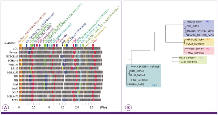
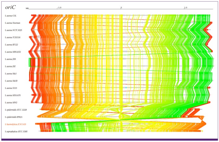
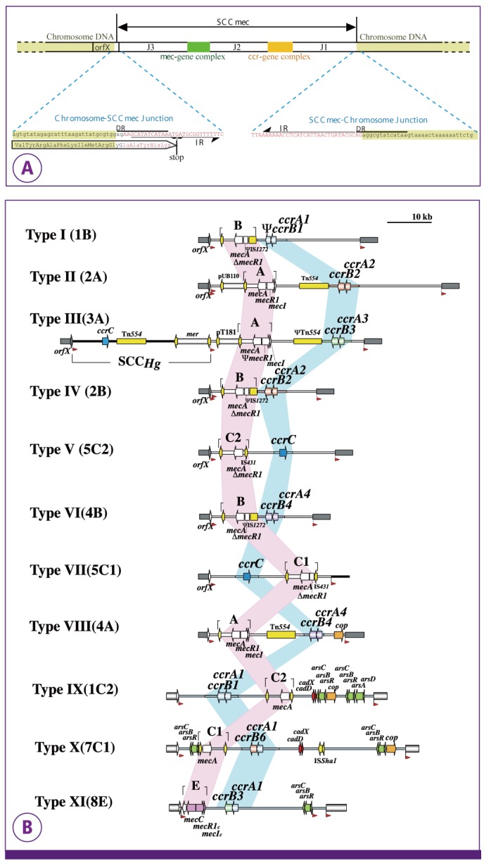
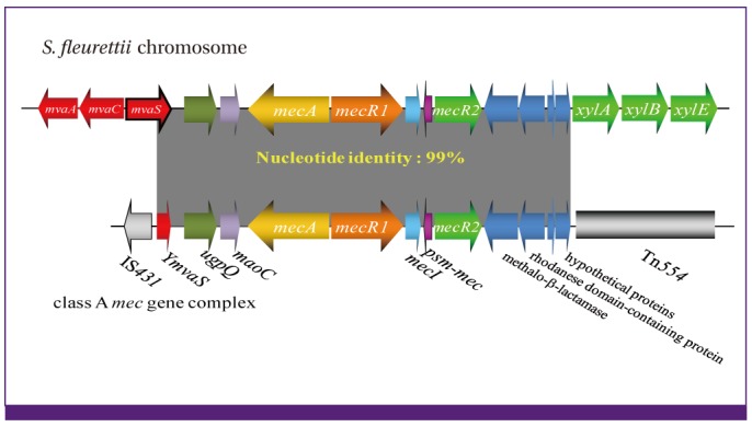
Verification
Claims the paper makes
The claims and statistics of this paper haven’t been checked yet.
Run the check — free, no account needed. It reads the open-access text and checks the sources it cites.
Claims other papers make about this one
Each is checked against this paper’s own text: does it say what the citing paper says it does?
In the citing paper
“To date, 11 different SCCmec allotypes have been described with varying degrees of genetic homology and different sizes, insertion sequences and accompanying resistance genes (68, 69).”
From Mechanisms of Antibiotic Resistance (2016) · cites this paper as [68]
Self-check
Percentages, sums and reported differences re-computed from the paper’s own numbers.
The self-check needs the paper’s full text.
Citations
Cites
Methicillin-resistant Staphylococcus aureus clinical strain with reduced vancomycin susceptibility
Hiramatsu, K. (1997) · Journal of Antimicrobial Chemotherapy · doi:10.1093/jac/40.1.135 · Cited by 1,984
Self-citationWhole genome sequencing of meticillin-resistant Staphylococcus aureus
Kuroda, M., Ohta, T., Uchiyama, I., Baba, T., Yuzawa, H., Kobayashi, I., Cui, L., Oguchi, A., Aoki, K.-I., Nagai, Y., et al. (2001) · The Lancet · doi:10.1016/s0140-6736(00)04403-2 · Cited by 1,950
Self-citation"Celbenin" - resistant Staphylococci
Jevons, M. P. (1961) · BMJ · doi:10.1136/bmj.1.5219.124-a · Cited by 1,730
Diep, B. A., Gill, S. R., Chang, R. F., Phan, T. H., Chen, J. H., Davidson, M. G., Lin, F., Lin, J., Carleton, H. A., Mongodin, E. F., et al. (2006) · The Lancet · doi:10.1016/s0140-6736(06)68231-7 · Cited by 1,633
The evolutionary history of methicillin-resistant Staphylococcus aureus (MRSA)
Enright, M. C., Robinson, D. A., Randle, G. A., Feil, E. J., Grundmann, H., & Spratt, B. G. (2002) · Proceedings of the National Academy of Sciences · doi:10.1073/pnas.122108599 · Cited by 1,591
The Changing Epidemiology of Staphylococcus aureus ?
Chambers, H. F. “. (2001) · Emerging infectious diseases · doi:10.3201/eid0702.010204 · Cited by 1,419
Genome and virulence determinants of high virulence community-acquired MRSA
Baba, T., Takeuchi, F., Kuroda, M., Yuzawa, H., Aoki, K.-I., Oguchi, A., Nagai, Y., Iwama, N., Asano, K., Naimi, T. S., et al. (2002) · The Lancet · doi:10.1016/s0140-6736(02)08713-5 · Cited by 1,356
Self-citationInfection with Vancomycin-Resistant Staphylococcus aureus Containing the vanA Resistance Gene
Chang, S., Sievert, D. M., Hageman, J. C., Boulton, M. L., Tenover, F. C., Downes, F. P., Shah, S., Rudrik, J. T., Pupp, G. R., Brown, W. J., et al. (2003) · New England Journal of Medicine · doi:10.1056/nejmoa025025 · Cited by 1,095
Katayama, Y., Ito, T., & Hiramatsu, K. (2000) · Antimicrobial Agents and Chemotherapy · doi:10.1128/aac.44.6.1549-1555.2000 · Cited by 1,042
Self-citationGill, S. R., Fouts, D. E., Archer, G. L., Mongodin, E. F., DeBoy, R. T., Ravel, J., Paulsen, I. T., Kolonay, J. F., Brinkac, L. M., Beanan, M., et al. (2005) · Journal of Bacteriology · doi:10.1128/jb.187.7.2426-2438.2005 · Cited by 1,026
Ito, T., Katayama, Y., Asada, K., Mori, N., Tsutsumimoto, K., Tiensasitorn, C., & Hiramatsu, K. (2001) · Antimicrobial Agents and Chemotherapy · doi:10.1128/aac.45.5.1323-1336.2001 · Cited by 964
Self-citationKondo, Y., Ito, T., Xue, X., Watanabe, S., Kreiswirth, B. N., Étienne, J., & Hiramatsu, K. (2006) · Antimicrobial Agents and Chemotherapy · doi:10.1128/aac.00165-06 · Cited by 958
Self-citationGarcía-Álvarez, L., Holden, M. T. G., Lindsay, H., Webb, C. R., Brown, D. F. J., Curran, M. D., Walpole, E., Brooks, K. L., Pickard, D., Teale, C. J., et al. (2011) · The Lancet Infectious Diseases · doi:10.1016/s1473-3099(11)70126-8 · Cited by 941
Hartman, B. J., & Tomasz, A. (1984) · Journal of Bacteriology · doi:10.1128/jb.158.2.513-516.1984 · Cited by 893
Stokes, H. W., & Hall, R. M. (1989) · Molecular Microbiology · doi:10.1111/j.1365-2958.1989.tb00153.x · Cited by 889
Dissemination of New Methicillin-Resistant Staphylococcus aureus Clones in the Community
Okuma, K., Iwakawa, K., Turnidge, J. D., Grubb, W. B., Bell, J. M., O’Brien, F. G., Coombs, G. W., Pearman, J. W., Tenover, F. C., Kapi, M., et al. (2002) · Journal of Clinical Microbiology · doi:10.1128/jcm.40.11.4289-4294.2002 · Cited by 888
Self-citationHolden, M. T. G., Feil, E. J., Lindsay, J. A., Peacock, S. J., Day, N., Enright, M. C., Foster, T. J., Moore, C. E., Hurst, L. D., Atkin, R. J., et al. (2004) · Proceedings of the National Academy of Sciences · doi:10.1073/pnas.0402521101 · Cited by 885
The emergence and evolution of methicillin-resistant Staphylococcus aureus
Hiramatsu, K., Cui, L., Kuroda, M., & Ito, T. (2001) · Trends in Microbiology · doi:10.1016/s0966-842x(01)02175-8 · Cited by 825
Self-citation(2009) · Antimicrobial Agents and Chemotherapy · doi:10.1128/aac.00579-09 · Cited by 804
Ito, T., Xue, X., Takeuchi, F., Okuma, K., Yuzawa, H., & Hiramatsu, K. (2004) · Antimicrobial Agents and Chemotherapy · doi:10.1128/aac.48.7.2637-2651.2004 · Cited by 677
Self-citationMa, X. X., Ito, T., Tiensasitorn, C., Jamklang, M., Chongtrakool, P., Boyle-Vavra, S., Daum, R. S., & Hiramatsu, K. (2002) · Antimicrobial Agents and Chemotherapy · doi:10.1128/aac.46.4.1147-1152.2002 · Cited by 587
Self-citationBaba, T., Bae, T., Schneewind, O., Takeuchi, F., & Hiramatsu, K. (2007) · Journal of Bacteriology · doi:10.1128/jb.01000-07 · Cited by 564
Self-citationCellbenin-resistant staphylococci.
Jevons, M. P. (1961) · Medical Entomology and Zoology · Cited by 553
Kuroda, M., Kuroda, H., Oshima, T., Takeuchi, F., Mori, H., & Hiramatsu, K. (2003) · Molecular Microbiology · doi:10.1046/j.1365-2958.2003.03599.x · Cited by 515
Self-citationIto, T., Katayama, Y., & Hiramatsu, K. (1999) · Antimicrobial Agents and Chemotherapy · doi:10.1128/aac.43.6.1449 · Cited by 451
Self-citationLindsay, J. A., Ruzin, A. V., Ross, H. F., Kurepina, N., & Novick, R. P. (1998) · Molecular Microbiology · doi:10.1046/j.1365-2958.1998.00947.x · Cited by 414
Zhang, Y., Ren, S., Li, H., Wang, Y., Fu, G., Yang, J., Qin, Z., Miao, Y., Wang, W., Chen, R., et al. (2003) · Molecular Microbiology · doi:10.1046/j.1365-2958.2003.03671.x · Cited by 387
Shore, A. C., Deasy, E. C., Slickers, P., Brennan, G. I., O'Connell, B. C., Monecke, S., Ehricht, R., & Coleman, D. C. (2011) · Antimicrobial Agents and Chemotherapy · doi:10.1128/aac.00187-11 · Cited by 383
Chongtrakool, P., Ito, T., Xue, X., Kondo, Y., Trakulsomboon, S., Tiensasitorn, C., Jamklang, M., Chavalit, T., Song, J., & Hiramatsu, K. (2006) · Antimicrobial Agents and Chemotherapy · doi:10.1128/aac.50.3.1001-1012.2006 · Cited by 378
Self-citationUtsui, Y., & Yokota, T. (1985) · Antimicrobial Agents and Chemotherapy · doi:10.1128/aac.28.3.397 · Cited by 367
Udo, E. E. (1993) · Journal of Hospital Infection · doi:10.1016/0195-6701(93)90100-e · Cited by 358
The complete genomes and proteomes of 27 Staphylococcus aureus bacteriophages
Kwan, T., Liu, J., DuBow, M. S., Gros, P., & Pelletier, J. (2005) · Proceedings of the National Academy of Sciences · doi:10.1073/pnas.0501140102 · Cited by 334
Takeuchi, F., Watanabe, S., Baba, T., Yuzawa, H., Ito, T., Morimoto, Y., Kuroda, M., Cui, L., Takahashi, M., Ankai, A., et al. (2005) · Journal of Bacteriology · doi:10.1128/jb.187.21.7292-7308.2005 · Cited by 331
Self-citationMatsuhashi, M., Song, M., Ishino, F., Wachi, M., Doi, M., Inoue, M., Ubukata, K., Yamashita, N., & Konno, M. (1986) · Journal of Bacteriology · doi:10.1128/jb.167.3.975-980.1986 · Cited by 316
Phage-Mediated Intergeneric Transfer of Toxin Genes
Chen, J. Y.-S., & Novick, R. P. (2009) · Science · doi:10.1126/science.1164783 · Cited by 314
Standard Nomenclature for the Superantigens Expressed byStaphylococcus
Lina, G., Bohach, G. A., Nair, S. P., Hiramatsu, K., Jouvin‐Marche, E., & Mariuzza, R. A. (2004) · The Journal of Infectious Diseases · doi:10.1086/420852 · Cited by 310
Self-citationSong, M. D., Wachi, M., Doi, M., Ishino, F., & Matsuhashi, M. (1987) · FEBS Letters · doi:10.1016/0014-5793(87)80373-3 · Cited by 301
Fitzgerald, J. R., Monday, S. R., Foster, T. J., Bohach, G. A., Hartigan, P. J., Meaney, W. J., & Smyth, C. J. (2001) · Journal of Bacteriology · doi:10.1128/jb.183.1.63-70.2001 · Cited by 280
Highlander, S. K., Hultén, K. G., Qin, X., Jiang, H., Yerrapragada, S., Mason, E. O., Shang, Y., Williams, T. M., Fortunov, R. M., Liu, Y., et al. (2007) · BMC Microbiology · doi:10.1186/1471-2180-7-99 · Cited by 261
A Proteolytic Transmembrane Signaling Pathway and Resistance to β-Lactams in Staphylococci
Zhang, H. Z., Hackbarth, C. J., Chansky, K., & Chambers, H. F. “. (2001) · Science · doi:10.1126/science.1055144 · Cited by 253
Molecular Correlates of Host Specialization in Staphylococcus aureus
Herron‐Olson, L., Fitzgerald, J. R., Musser, J. M., & Kapur, V. (2007) · PLoS ONE · doi:10.1371/journal.pone.0001120 · Cited by 245
Methicillin-Resistant Staphylococcus aureus as a Community Organism
Moreno, F., Crisp, C., Jorgensen, J. H., & PATTERSON, J. E. (1995) · Clinical Infectious Diseases · doi:10.1093/clinids/21.5.1308 · Cited by 239
Langley, R. J., Wines, B. D., Willoughby, N., Basu, I., Proft, T., & Fraser, J. D. (2005) · The Journal of Immunology · doi:10.4049/jimmunol.174.5.2926 · Cited by 237
Ubukata, K., Yamashita, N., & Konno, M. (1985) · Antimicrobial Agents and Chemotherapy · doi:10.1128/aac.27.5.851 · Cited by 234
Yamaguchi, T., Nishifuji, K., Sasaki, M., Fudaba, Y., Aepfelbacher, M., Takata, T., Ohara, M., Komatsuzawa, H., Amagai, M., & Sugai, M. (2002) · Infection and Immunity · doi:10.1128/iai.70.10.5835-5845.2002 · Cited by 230
Vancomycin resistance in staphylococci
Hiramatsu, K. (1998) · Drug Resistance Updates · doi:10.1016/s1368-7646(98)80029-0 · Cited by 226
Self-citationOrigin and Molecular Evolution of the Determinant of Methicillin Resistance in Staphylococci
Tsubakishita, S., Kuwahara‐Arai, K., Sasaki, T., & Hiramatsu, K. (2010) · Antimicrobial Agents and Chemotherapy · doi:10.1128/aac.00356-10 · Cited by 212
Self-citationHiramatsu, K. (1995) · Microbiology and Immunology · doi:10.1111/j.1348-0421.1995.tb02239.x · Cited by 206
Self-citationKuroda, M., Kuwahara‐Arai, K., & Hiramatsu, K. (2000) · Biochemical and Biophysical Research Communications · doi:10.1006/bbrc.2000.2277 · Cited by 190
Self-citationIntrinsic resistance to β‐lactam antibiotics in staphylococcus aureus
Brown, D. F. J., & Reynolds, P. E. (1980) · FEBS Letters · doi:10.1016/0014-5793(80)80455-8 · Cited by 188
Kozitskaya, S., Cho, S.-H., Dietrich, K., Marre, R., Naber, K. G., & Ziebuhr, W. (2004) · Infection and Immunity · doi:10.1128/iai.72.2.1210-1215.2004 · Cited by 187
Wu, S., C, P., de LENCASTRE, H., & Tomasz, A. (1996) · Microbial Drug Resistance · doi:10.1089/mdr.1996.2.435 · Cited by 185
Hiramatsu, K., Asada, K., Suzuki, E., Okonogi, K., & Yokota, T. (1992) · FEBS Letters · doi:10.1016/0014-5793(92)80039-j · Cited by 182
Self-citationSchnellmann, C., Gerber, V., Rossano, A., Jaquier, V., Panchaud, Y., Doherr, M. G., Thomann, A., Straub, R., & Perreten, V. (2006) · Journal of Clinical Microbiology · doi:10.1128/jcm.00868-06 · Cited by 178
Molecular genetics of methicillin-resistant Staphylococcus aureus
Hiramatsu, K., Katayama, Y., Yuzawa, H., & Ito, T. (2002) · International Journal of Medical Microbiology · doi:10.1078/1438-4221-00192 · Cited by 168
Self-citationWilliamson, R., Bouguénec, C. L., Gutmann, L., & Horaud, T. (1985) · Microbiology · doi:10.1099/00221287-131-8-1933 · Cited by 157
Ryffel, C., Kayser, F. H., & Berger‐Bächi, B. (1992) · Antimicrobial Agents and Chemotherapy · doi:10.1128/aac.36.1.25 · Cited by 151
Dissemination of Methicillin-Resistant Staphylococci among Healthy Japanese Children
Hisata, K., Kuwahara‐Arai, K., Yamanoto, M., Ito, T., Nakatomi, Y., Cui, L., Baba, T., Terasawa, M., Sotozono, C., Kinoshita, S., et al. (2005) · Journal of Clinical Microbiology · doi:10.1128/jcm.43.7.3364-3372.2005 · Cited by 147
Self-citationCharacterization and Expression Analysis of Staphylococcus aureus Pathogenicity Island 3
Yarwood, J. M., McCormick, J. K., Paustian, M. L., Orwin, P. M., Kapur, V., & Schlievert, P. M. (2002) · Journal of Biological Chemistry · doi:10.1074/jbc.m111661200 · Cited by 146
Distribution of mec regulator genes in methicillin-resistant Staphylococcus clinical strains
Suzuki, E., Kuwahara‐Arai, K., Richardson, J. F., & Hiramatsu, K. (1993) · Antimicrobial Agents and Chemotherapy · doi:10.1128/aac.37.6.1219 · Cited by 144
Self-citationMechanisms of heteroresistance in methicillin-resistant Staphylococcus aureus
Ryffel, C., Strässle, A., Kayser, F. H., & Berger‐Bächi, B. (1994) · Antimicrobial Agents and Chemotherapy · doi:10.1128/aac.38.4.724 · Cited by 140
Colony Spreading in Staphylococcus aureus
Kaito, C., & Sekimizu, K. (2006) · Journal of Bacteriology · doi:10.1128/jb.01635-06 · Cited by 139
Kuwahara‐Arai, K., Kondo, N., Hori, S., Tateda-Suzuki, E., & Hiramatsu, K. (1996) · Antimicrobial Agents and Chemotherapy · doi:10.1128/aac.40.12.2680 · Cited by 136
Self-citationA Role in vivo for Penicillin‐Binding Protein‐4 of Staphylococcus aureus
Wyke, A. W., Ward, J. B., HAYES, M. V., & Curtis, N. A. C. (1981) · European Journal of Biochemistry · doi:10.1111/j.1432-1033.1981.tb05620.x · Cited by 136
Clarke, S. R., Harris, L. G., Richards, R. G., & Foster, S. J. (2002) · Infection and Immunity · doi:10.1128/iai.70.12.6680-6687.2002 · Cited by 133
Genome Analysis of the Meat Starter Culture BacteriumStaphylococcus carnosusTM300
Rosenstein, R., Nerz, C., Biswas, L., Resch, A., Raddatz, G., Schuster, S. C., & Götz, F. (2008) · Applied and Environmental Microbiology · doi:10.1128/aem.01982-08 · Cited by 130
Kaito, C., Saito, Y., Nagano, G., Ikuo, M., Omae, Y., Hanada, Y., Han, X., Kuwahara‐Arai, K., Hishinuma, T., Baba, T., et al. (2011) · PLoS Pathogens · doi:10.1371/journal.ppat.1001267 · Cited by 130
Self-citationÚbeda, C., Tormo‐Más, M. Á., Cucarella, C., Trotonda, M. P., Foster, T. J., Lasa, Í., & Penadés, J. R. (2003) · Molecular Microbiology · doi:10.1046/j.1365-2958.2003.03577.x · Cited by 126
Baba, T., Kuwahara‐Arai, K., Uchiyama, I., Takeuchi, F., Ito, T., & Hiramatsu, K. (2008) · Journal of Bacteriology · doi:10.1128/jb.01058-08 · Cited by 126
Self-citationMongkolrattanothai, K., Boyle, S., Murphy, T. V., & Daum, R. S. (2004) · Antimicrobial Agents and Chemotherapy · doi:10.1128/aac.48.5.1823-1836.2004 · Cited by 123
Ruppé, É., Barbier, F., Mesli, Y., Maïga, A., Cojocaru, R., Benkhalfat, M., Benchouk, S., Hassaïne, H., Maïga, I., Diallo, A., et al. (2008) · Antimicrobial Agents and Chemotherapy · doi:10.1128/aac.00724-08 · Cited by 122
The SaPIs: Mobile Pathogenicity Islands of Staphylococcus
Novick, R. P., & Subedi, A. (2007) · KARGER eBooks · doi:10.1159/000100857 · Cited by 121
Lee, S. M., Ender, M., Adhikari, R. P., Smith, J. M. B., Berger‐Bächi, B., & Cook, G. M. (2007) · Antimicrobial Agents and Chemotherapy · doi:10.1128/aac.01239-06 · Cited by 119
Kaito, C., Saito, Y., Ikuo, M., Omae, Y., Mao, H., Nagano, G., Fujiyuki, T., Numata, S., Han, X., Obata, K., et al. (2013) · PLoS Pathogens · doi:10.1371/journal.ppat.1003269 · Cited by 118
Self-citationGustafson, J. E., Strässle, A., Hächler, H., Kayser, F. H., & Berger‐Bächi, B. (1994) · Journal of Bacteriology · doi:10.1128/jb.176.5.1460-1467.1994 · Cited by 111
Chromosomal map location of the methicillin resistance determinant in Staphylococcus aureus
Kuhl, S. A., Pattee, P. A., & Baldwin, J. N. (1978) · Journal of Bacteriology · doi:10.1128/jb.135.2.460-465.1978 · Cited by 104
sarU , a sarA Homolog, Is Repressed by SarT and Regulates Virulence Genes in Staphylococcus aureus
Manna, A. C., & Cheung, A. L. (2002) · Infection and Immunity · doi:10.1128/iai.71.1.343-353.2003 · Cited by 103
Matsuo, M., Hishinuma, T., Katayama, Y., Cui, L., Kapi, M., & Hiramatsu, K. (2011) · Antimicrobial Agents and Chemotherapy · doi:10.1128/aac.00398-11 · Cited by 100
Self-citationLuong, T. T., Ouyang, S., Bush, K., & Lee, C. Y. (2002) · Journal of Bacteriology · doi:10.1128/jb.184.13.3623-3629.2002 · Cited by 99
Staphylococcus aureus Pathogenicity Island DNA Is Packaged in Particles Composed of Phage Proteins
Tormo‐Más, M. Á., Ferrer, M. D., Maiques, E., Úbeda, C., Selva, L., Lasa, Í., Calvete, J. J., Novick, R. P., & Penadés, J. R. (2008) · Journal of Bacteriology · doi:10.1128/jb.01349-07 · Cited by 98
Staphylococcal Cassette Chromosome mec -Like Element in Macrococcus caseolyticus
Tsubakishita, S., Kuwahara‐Arai, K., Baba, T., & Hiramatsu, K. (2010) · Antimicrobial Agents and Chemotherapy · doi:10.1128/aac.00575-09 · Cited by 97
Self-citationO’Neill, A. J., Larsen, A. R., Skov, R. L., Henriksen, A. S., & Chopra, I. (2007) · Journal of Clinical Microbiology · doi:10.1128/jcm.01984-06 · Cited by 96
Chatterjee, S. S., Chen, L., Joo, H., Cheung, G. Y. C., Kreiswirth, B. N., & Otto, M. J. (2011) · PLoS ONE · doi:10.1371/journal.pone.0028781 · Cited by 94
Jamaluddin, T. Z. M. T., Kuwahara‐Arai, K., Hisata, K., Terasawa, M., Cui, L., Baba, T., Sotozono, C., Kinoshita, S., Ito, T., & Hiramatsu, K. (2008) · Journal of Clinical Microbiology · doi:10.1128/jcm.02262-07 · Cited by 86
Self-citationTesch, W., Ryffel, C., Strässle, A., Kayser, F. H., & Berger‐Bächi, B. (1990) · Antimicrobial Agents and Chemotherapy · doi:10.1128/aac.34.9.1703 · Cited by 83
SaPI operon I is required for SaPI packaging and is controlled by LexA
Úbeda, C., Maiques, E., Tormo‐Más, M. Á., Campoy, S., Lasa, Í., Barbé, J., Novick, R. P., & Penadés, J. R. (2007) · Molecular Microbiology · doi:10.1111/j.1365-2958.2007.05758.x · Cited by 83
Aubry-Damon, H., Legrand, P., Brun‐Buisson, C., Astier, A., Soussy, C., & Leclercq, R. (1997) · Clinical Infectious Diseases · doi:10.1086/513749 · Cited by 80
Hongo, I., Baba, T., Oishi, K., Morimoto, Y., Ito, T., & Hiramatsu, K. (2009) · The Journal of Infectious Diseases · doi:10.1086/605332 · Cited by 80
Self-citationHürlimann-Dalel, R. L., Ryffel, C., Kayser, F. H., & Berger‐Bächi, B. (1992) · Antimicrobial Agents and Chemotherapy · doi:10.1128/aac.36.12.2617 · Cited by 79
A Staphylococcus xylosus Isolate with a New mecC Allotype
Harrison, E. M., Paterson, G. K., Holden, M. T. G., Morgan, F. J. E., Larsen, A. R., Petersen, A., Léroy, S., Vliegher, S. D., Perreten, V., Fox, L. K., et al. (2012) · Antimicrobial Agents and Chemotherapy · doi:10.1128/aac.01882-12 · Cited by 78
Uchiyama, I. (2006) · Nucleic Acids Research · doi:10.1093/nar/gkl978 · Cited by 76
Uchiyama, I. (2006) · Nucleic Acids Research · doi:10.1093/nar/gkj448 · Cited by 75
Sjöström, J.-E., Löfdahl, S., & Philipson, L. (1975) · Journal of Bacteriology · doi:10.1128/jb.123.3.905-915.1975 · Cited by 72
Intrinsic Penicillin Resistance in Enterococci
Fontana, R., Ligozzi, M., Pittaluga, F., & Satta, G. (1996) · Microbial Drug Resistance · doi:10.1089/mdr.1996.2.209 · Cited by 71
Hiramatsu, K., Suzuki, E., Takayama, H., Katayama, Y., & Yokota, T. (1990) · Antimicrobial Agents and Chemotherapy · doi:10.1128/aac.34.4.600 · Cited by 70
Self-citationKondo, N., Kuwahara‐Arai, K., Murakami-Kuroda, H., Tateda-Suzuki, E., & Hiramatsu, K. (2001) · Antimicrobial Agents and Chemotherapy · doi:10.1128/aac.45.3.815-824.2001 · Cited by 68
Self-citationKharroubi, A. E., Jacques, P., Piras, G., BEEUMEN, J. V., Coyette, J., & Ghuysen, J. (1991) · Biochemical Journal · doi:10.1042/bj2800463 · Cited by 67
Changing epidemiology of methicillin‐resistant Staphylococcus aureus in Western Australia
Riley, T. V., Pearman, J. W., & Rouse, I. L. (1995) · The Medical Journal of Australia · doi:10.5694/j.1326-5377.1995.tb124656.x · Cited by 67
Hiramatsu, K., Igarashi, M., Morimoto, Y., Baba, T., Umekita, M., & Akamatsu, Y. (2012) · International Journal of Antimicrobial Agents · doi:10.1016/j.ijantimicag.2012.02.007 · Cited by 62
Self-citationMaki, H., & Murakami, K. (1997) · Journal of Bacteriology · doi:10.1128/jb.179.22.6944-6948.1997 · Cited by 62
Genetic basis for molecular epidemiology of MRSA
Hiramatsu, K., Kondo, N., & Ito, T. (1996) · Journal of Infection and Chemotherapy · doi:10.1007/bf02351563 · Cited by 60
Self-citationGenetic behavior of the methicillin resistance determinant in Staphylococcus aureus
Stewart, G. C., & Rosenblum, E. D. (1980) · Journal of Bacteriology · doi:10.1128/jb.144.3.1200-1202.1980 · Cited by 52
Subedi, A., Úbeda, C., Adhikari, R. P., Penadés, J. R., & Novick, R. P. (2007) · Microbiology · doi:10.1099/mic.0.2007/006932-0 · Cited by 52
Wada, A., Katayama, Y., Hiramatsu, K., & Yokota, T. (1991) · Biochemical and Biophysical Research Communications · doi:10.1016/0006-291x(91)90430-f · Cited by 50
Self-citationAiba, Y., Katayama, Y., Hishinuma, T., Murakami-Kuroda, H., Cui, L., & Hiramatsu, K. (2013) · Antimicrobial Agents and Chemotherapy · doi:10.1128/aac.00720-13 · Cited by 48
Self-citationKaito, C., Omae, Y., Matsumoto, Y., Nagata, M., Yamaguchi, H., Aoto, T., Ito, T., Hiramatsu, K., & Sekimizu, K. (2008) · PLoS ONE · doi:10.1371/journal.pone.0003921 · Cited by 47
Self-citationPiras, G., Raze, D., Kharroubi, A. E., Hastir, D., Englebert, S., Coyette, J., & Ghuysen, J. (1993) · Journal of Bacteriology · doi:10.1128/jb.175.10.2844-2852.1993 · Cited by 46
Yamakawa, J., Kurosawa, H., Kaneko, K., Yamakawa, J., AMINAKA, M., Hori, S., Cui, L., Ito, T., Jin, J., Hiramatsu, K., et al. (2011) · Journal of Infection and Chemotherapy · doi:10.1007/s10156-011-0330-2 · Cited by 35
Self-citationUchiyama, I. (2008) · BMC Genomics · doi:10.1186/1471-2164-9-515 · Cited by 33
Genetic Basis for Molecular Epidemiology of MRSA.
Kondo, N., Ito, T., & Hiramatsu, K. (1997) · Nippon Saikingaku Zasshi · doi:10.3412/jsb.52.417 · Cited by 29
Self-citationWatanabe, S., Ito, T., Morimoto, Y., Takeuchi, F., & Hiramatsu, K. (2007) · Journal of Bacteriology · doi:10.1128/jb.01485-06 · Cited by 22
Self-citationEffects of Penicillin-Binding Protein 4 Overproduction in Staphylococcus aureus
Henze, U., Roos, M. A., & Berger‐Bächi, B. (1996) · Microbial Drug Resistance · doi:10.1089/mdr.1996.2.193 · Cited by 21
The Staphylococcus aureus Genome
Baba, T., Takeuchi, F., Kuroda, M., Ito, T., Yuzawa, H., & Hiramatsu, K. (2004) · Elsevier eBooks · doi:10.1016/b978-1-898563-96-9.50008-7 · Cited by 12
Self-citation
Research works printed but not linked: 4
- Baba, 2004, Molecular and Clinical Aspects, 66
- Hiramatsu, Molecular Evolution of MRSA 2010, 11
- Williamson, 1985, J Gen Microbiol, 131, 1933
- Aiba, Antimicrob Agents Chemother
Cited by
Sorted by citations
Mechanisms of Antibiotic Resistance
Munita, J. M., & Arias, C. A. (2016) · Microbiology Spectrum · doi:10.1128/microbiolspec.vmbf-0016-2015 · Cited by 2,782
What is a resistance gene? Ranking risk in resistomes
Martínez, J. L., Coque, T. M., & Baquero, F. (2014) · Nature Reviews Microbiology · doi:10.1038/nrmicro3399 · Cited by 1,033
Antimicrobial Resistance in Bacteria: Mechanisms, Evolution, and Persistence
Christaki, E., Marcou, M., & Tofarides, A. G. (2019) · Journal of Molecular Evolution · doi:10.1007/s00239-019-09914-3 · Cited by 875
Antibiotic Resistance and the MRSA Problem
Vestergaard, M. R., Frees, D., & Ingmer, H. (2019) · Microbiology Spectrum · doi:10.1128/microbiolspec.gpp3-0057-2018 · Cited by 540
Mechanisms of antibiotic resistance
Munita, J. M., & Arias, C. A. (2016) · Virulence Mechanisms of Bacterial Pathogens · doi:10.1128/9781555819286.ch17 · Cited by 421
Metadata
- Authors
- Hiramatsu, K., Ito, T., Tsubakishita, S., Sasaki, T., Takeuchi, F., Morimoto, Y., Katayama, Y., Matsuo, M., Kuwahara-Arai, K., Hishinuma, T., & Baba, T.
- Affiliations
- Juntendo University (JP)
- Rakuno Gakuen University (JP)
- National Institute of Infectious Diseases (JP)
- Department of Bacteriology, Juntendo University, Tokyo, Japan.
- Corresponding author
- Hiramatsu, Keiichi
- ORCID
- Venue
- Infection & Chemotherapy
- Publisher
- XMLink
- Published
- 2013
- Volume
- 45
- Issue
- 2
- Pages
- 117
- ISSN
- 1598-8112 (ISSN-L)
- 2093-2340
- 2092-6448
- PMC
- PMC3780952
- OpenAlex
- W1969730343
- Cited by
- 189
- References
- 114
- Citations by year
- 2026: 6 · 2025: 14 · 2024: 14 · 2023: 17 · 2022: 16 · 2021: 17 · 2020: 20 · 2019: 25 · 2018: 16 · 2017: 9 · 2016: 15 · 2015: 12 · 2014: 8
- FWCI
- 1.48
- Type
- journal article
- Language
- en
- Open access
- gold
- doi.org
- Licence
- Locations
- Infection and Chemotherapy (publishedVersion, cc-by-nc)
- Europe PMC (PubMed Central) (publishedVersion, cc-by-nc)
- PubMed Central (publishedVersion, cc-by-nc)
- PubMed (publishedVersion)
- Institutional Repositories DataBase (IRDB) (publishedVersion, other-oa)
- Funders
- Ministry of Education, Culture, Sports, Science and Technology (S1201013)
- Japan Society for the Promotion of Science (24780298)
- Topics
- Health Sciences › Medicine › Infectious Diseases › Antimicrobial Resistance in Staphylococcus
- Health Sciences › Medicine › Microbiology (medical) › Antibiotic Resistance in Bacteria
- Life Sciences › Biochemistry, Genetics and Molecular Biology › Genetics › Bacterial Genetics and Biotechnology
- Keywords
- methicillin resistance, Staphylococcus aureus, methicillin-resistant Staphylococcus aureus, multidrug resistance, antimicrobial resistance, MRSA clones, community-acquired MRSA, hospital-acquired MRSA, bacterial genomics, pathogenicity, immunocompromised patients
About these checks
Registry, retraction, Cites and statistics results are computed, not judged, with one exception: a printed reference that no index lists is read once by a model before it counts as a citation error. Claims are extracted by a model and kept only when their quote appears verbatim in the text; each cited source is then checked, with a model’s help, for whether it says what the claim relies on. Those verdicts can be wrong — read the claim’s quote and the cited source before relying on one.
Statistics and arithmetic are recomputed, not judged.
Confidence bands are shown only for fields calibrated on our annotated test set. None is calibrated yet, so no band is shown.
What counts as a claim ›How we measure confidence ›How we check this ›
Are you an author, or do you see a problem with this page? Request a review
