

The DOI matches its Crossref record; 62 of the 62 references it cites were found in a registry or an index; 13 of the 39 sources its claims cite were checked for support.
Ectodermal Dysplasia – An Overview and Update
Open accessAbstract
Ectodermal dysplasias are a heterogeneous group of disorders characterized by abnormal development of ectodermal structures.
Overview
CoChat Summary
## TL;DR
This review summarizes ectodermal dysplasias as a heterogeneous group of inherited disorders affecting ectodermal structures such as hair, teeth, nails, and sweat glands, and notes that classification has shifted from phenotype-based schemes to pathway/gene-based groupings. It emphasizes careful clinical assessment, genetic diagnosis, counseling, and multidisciplinary management across several major ED subtypes.
## Key findings
- Ectodermal dysplasias are described as a heterogeneous group affecting “hair, teeth, nails, and sweat glands,” and advances in genetics have identified causative genes and pathways in at least 80 EDs.
- The paper says current classification groups disorders into four pathway-based categories: EDA/NFKB, WNT, TP63, and structural disorders.
- The older Freire-Maia 1-2-3-4 system eventually included around 154 entities, but the paper notes that newer schemes shift toward genetic/pathway classification.
- For hypohidrotic ectodermal dysplasia (HED), the classic triad is hypohidrosis/anhidrosis, hypotrichosis, and hypodontia; the text also states atopic eczema is seen in two-thirds of patients.
- For HED with immunodeficiency, the paper reports 74% survival with allogenic hematopoietic stem cell transplantation (HSCT).
- For incontinentia pigmenti, the paper states the common deletion of exons 4 to 10 in IKBKG is present in about 70–80% of cases, and “de novo” mutations account for 65% of cases.
## Methods
- This is a narrative review/overview of ectodermal dysplasia, organized by pathway and syndrome subtype, with sections on definitions, classification, diagnosis, and management.
- The paper draws on molecular genetics, clinical features, diagnostic criteria, and management approaches across multiple ED entities; no original patient cohort or new dataset is described.
- Its main analytical approach is comparative clinical and genetic synthesis of subtype-specific signs, inheritance patterns, confirmatory tests, and treatment options.
## Limitations
- The paper does not report a systematic search strategy, study selection criteria, or evidence-graded review process; this is evident from the text and not explicitly discussed.
- It does not present original outcome data or comparative statistics across patient groups; this is evident from the text.
- The review itself states that there is considerable overlap in clinical features among ED conditions, which limits purely clinical classification and diagnosis.
Model-written summary. Check it against the paper before relying on it.
Integrity signals
Registry
Cites
Statistics
Figures
Verification
Claims the paper makes
11 Valid · 3 Not valid · 23 Not checked · 7 Own results and interpretations
The cited sources support only part of this claim — read each source’s verdict below.
In the paper
“They are currently divided into four groups of disorders based on the pathway involved, which includes the ectodysplasin/nuclear factor-kappa B (NFKB) pathway, wingless-type MMTV integration site family, member 10 ([wingless related integration site] WNT10), tumor protein p63 (TP63), and the structural group.”
§ Ectodermal Dysplasia – An Overview and Update > Abstract
What the sources say
- Partly supportedcites [1] sourceExperimental — accuracy not yet measured
In “Classification Scheme and Clustering” (page unavailable), the paper groups ectodermal dysplasias by genotype, molecular pathway and phenotype, identifying EDA-, WNT- and TP63-associated genes alongside genes involved in cellular structure. It does not state that the disorders are divided into exactly four groups, and it names the broader WNT pathway rather than a WNT10 group. (LLM, conf=0.85)
plain- Asserted: passes — The authors put it forward as true.
- Checkable: passes — It can be checked against evidence.
- Load-bearing: unclear — It is unclear whether the paper’s conclusions depend on it.
- Based: passes — The paper gives a reason why it holds.
- Partly supported
Every source the paper cites for this claim was checked and supports it.
In the paper
“The most recent classification was proposed by Peschel et al.[5] as a part of the five-yearly update on EDs. This system adhered to the previous genetic scheme of classification while adding 15 new entities or genes that have been recently studied.”
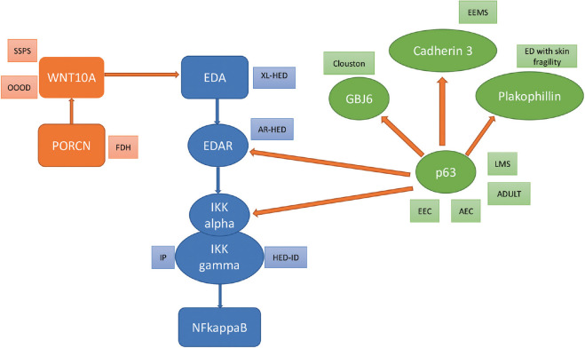
§ Ectodermal Dysplasia – An Overview and Update > Definitions and Classification ‘‘Pure ectodermal dysplasias’’ are entities with only ectodermal signs without disturbances derivatives of other embryonic layers. On the other hand, ‘‘syndromes of ectodermal dysplasia and malformation’’ present ectodermal signs and also defects of another embryonic origin, such as cleft lip/palate. Freire-Maia offered the most common major classification scheme in 1971, with primary emphasis on grouping the disorders based on the involvement of hair, teeth, nails, or eccrine sweat glands, which were termed as 1,2, 3, and 4, respectively. This was referred to as the 1-2-3-4 system, where the nomenclature of the subgroup depended on the structures involved. For example, if hair and teeth are involved, the subgroup would be named 1-2.[2] This classification finally came to include around 154 entities,[3] amongst which multiple entities belong to other classifications like pachyonychia congenita, dyskeratosis congenita, and Rothmund-Thompson syndrome. Continuous revision of the classification systems has been attempted, and a shift has occurred to classifying these disorders based on the genetic defect or the developmental pathway involved which helps in offering a better understanding of these disorders.[4] The classification described by Wright et al.[1] groups these disorders broadly into defects of the ectodysplasin A (EDA)/nuclear factor kappa B (NFKB) pathway, wingless related integration site (WNT) pathway, tumor protein p63 (TP63) pathway, other structural defects, and unknown causes [Table 1]. Figure 1 demonstrates the inter-relationships between the various molecules and pathways and the associated disorders. This system, however, excludes entities that have been included in other classification systems, like palmoplantar keratodermas or poikilodermas. The most recent classification was proposed by Peschel et al.[5] as a part of the five-yearly update on EDs. This system adhered to the previous genetic scheme of classification while adding 15 new entities or genes that have been recently studied.
What the sources say
- Supportedcites [5] source
Section 2 says the five-yearly update retained the molecular pathway-based classification and added 15 entities not listed in the previous classification paper. This supports the citing sentence’s description of the updated genetic scheme and the number of additions. (LLM, conf=0.93)
plain- Asserted: passes — The authors put it forward as true.
- Checkable: passes — It can be checked against evidence.
- Load-bearing: unclear — It is unclear whether the paper’s conclusions depend on it.
- Based: passes — The paper gives a reason why it holds.
- Supported
The cited sources support only part of this claim — read each source’s verdict below.
In the paper
“It is classified further based on the mutated gene and the type of inheritance[6]: (a) X-linked HED-EDA1 mutation (also known as Christ–Siemens Touraine syndrome) [X-linked recessive] (b) EDAR (EDA receptor) mutation- autosomal dominant and recessive (c) EDARADD (EDAR associated death domain encoding gene) mutation- autosomal dominant and recessive (d) X-linked HED with immunodeficiency- NEMO (NFK”
§ Ectodermal Dysplasia – An Overview and Update > (A) Disorders of the EDA/NFKB pathway > Hypohidrotic ectodermal dysplasia (HED)
What the sources say
- Partly supportedcites [6] sourceExperimental — accuracy not yet measured
The Abstract (page not provided) identifies EDA1 with X-linked HED and EDAR and EDARADD with autosomal dominant and recessive forms; the Results also document both inheritance patterns for EDAR. The cited paper does not identify a NEMO/IKBKG immunodeficiency category or use the name Christ–Siemens–Touraine syndrome. It also identifies WNT10A as a cause of HED/EDA, which the cited classification omits. (LLM, conf=0.87)
plain- Asserted: passes — The authors put it forward as true.
- Checkable: passes — It can be checked against evidence.
- Load-bearing: unclear — It is unclear whether the paper’s conclusions depend on it.
- Based: passes — The paper gives a reason why it holds.
- Partly supported
Every source the paper cites for this claim was checked and supports it.
In the paper
“Apart from this, PRIMA-1MET, a p53 reactivator, when applied topically, has been shown to augment epidermal differentiation and improve wound healing in patients with AEC syndrome.[57]”
§ Ectodermal Dysplasia – An Overview and Update > (C) Disorders of the TP63 pathway > Ankyloblepharon-ectodermal defects-cleft lip/palate (AEC) syndrome
What the sources say
- Supportedcites [57] source
The Abstract identifies PRIMA-1MET as a p53-reactivating compound and reports re-epithelialization after topical treatment of two AEC patients. The Results and discussion report that it rescued differentiation in patient-derived keratinocytes and suggest that the treated scalp’s new epidermal covering reflects enhanced early differentiation. The differentiation finding is based chiefly on cultured cells, rather than a direct measurement of differentiation after topical application. (LLM, conf=0.89)
plainstates a cause- Asserted: passes — The authors put it forward as true.
- Checkable: passes — It can be checked against evidence.
- Load-bearing: unclear — It is unclear whether the paper’s conclusions depend on it.
- Based: passes — The paper gives a reason why it holds.
- Supported
Every source the paper cites for this claim was checked and supports it.
In the paper
“The characteristic feature is ankyloblepharon filiforme adnatum (fusion of the eyelids).[54]”
§ Ectodermal Dysplasia – An Overview and Update > (C) Disorders of the TP63 pathway > Ankyloblepharon-ectodermal defects-cleft lip/palate (AEC) syndrome
What the sources say
- Supportedcites [54] source
The Overview of AEC Syndrome describes characteristic eyelid fusion as part of the syndrome. The Collaborative Roundtable Discussion names ankyloblepharon filiforme adnatum in its discussion of eyelid findings, supporting the citing sentence. (LLM, conf=0.92)
plain- Asserted: passes — The authors put it forward as true.
- Checkable: passes — It can be checked against evidence.
- Load-bearing: unclear — It is unclear whether the paper’s conclusions depend on it.
- Based: passes — The paper gives a reason why it holds.
- Supported
10 more claims
Create a free account to see all 44 claims and to verify the 3 references still open.
- It may state two separate claims as one. 24
- Probably not a substantial claim. 8
Claims other papers make about this one
Each is checked against this paper’s own text: does it say what the citing paper says it does?
The source backs part of the claim, not all of it — or backs it for a different population, condition or comparison.
In the citing paper
“Tooth agenesis with insufficient bone volume presents substantial challenges for implant rehabilitation [4, 5].”
From Implant-supported fixed prosthodontic treatment for ectodermal dysplasia: a case report (2026) · cites this paper as [5]
Experimental — accuracy not yet measured
Citations
Cites
Zonana, J., Elder, M. E., Schneider, L. C., Orlow, S. J., Moss, C., Golabi, M., Shapira, S. K., Farndon, P. A., Wara, D. W., Emmal, S. A., et al. (2000) · The American Journal of Human Genetics · doi:10.1086/316914 · Cited by 521 · cited 1×
“[22] Incontinentia pigmenti (IP) is allelic to this disorder and is caused by a deletion, in IKBK .”
BackgroundEctodermal dysplasias: A clinical classification and a causal review
Pinheiro, M., & Freire‐Maia, N. (1994) · American Journal of Medical Genetics · doi:10.1002/ajmg.1320530207 · Cited by 413 · cited 1×
“[2] This classification finally came to include around 154 entities, [3] amongst which multiple entities belong to other classifications like pachyonychia congenita, dyskeratosis congenita, and Rothmund‑Thompson syndrome.”
BackgroundIncontinentia pigmenti (Bloch-Sulzberger syndrome)
Landy, S. J., & Donnai, D. (1993) · Journal of Medical Genetics · doi:10.1136/jmg.30.1.53 · Cited by 404 · cited 2×
“[25] Null mutations are lethal in utero in males, whereas females survive because of lyonization, and hence, most cases of IP occur in females.”
BackgroundGOLTZ, R. W. (1962) · Archives of Dermatology · doi:10.1001/archderm.1962.01590120006002 · Cited by 342 · cited 1×
“Also known as Goltz syndrome or Goltz–Gorlin syndrome, [37] FDH is an X‑linked dominant disorder that is caused by deletions in the porcupine O‑acyltransferase gene ( PORCN ), which is required for the secretion of WNT proteins.”
BackgroundCluzeau, C., Hadj-Rabia, S., Jambou, M., Mansour, S., Guigue, P., Masmoudi, S., Bal, E., Chassaing, N., Vincent, M.-C., Viot, G., et al. (2011) · Human Mutation · doi:10.1002/humu.21384 · Cited by 311 · cited 1×
“It is classified further based on the mutated gene and the type of inheritance [6] : (a) X‑linked HED‑EDA1 mutation (also known as Christ–Siemens Touraine syndrome) [X‑linked recessive] (b) EDAR (EDA receptor) mutation‑ autosomal dominant and recessive (c) EDARADD (EDAR associated death domain…”
BackgroundSmahi, A. (2002) · Human Molecular Genetics · doi:10.1093/hmg/11.20.2371 · Cited by 300 · cited 1×
“[24,25] It is inherited in an X‑linked dominant fashion (“de novo” in 65% of cases).”
BackgroundMutations in GJB6 cause hidrotic ectodermal dysplasia
Lamartine, J., Munhoz Essenfelder, G., Kibar, Z., Lanneluc, I., Callouet, E., Laoudj, D., Lemaître, G., Hand, C., Hayflick, S. J., Zonana, J., et al. (2000) · Nature Genetics · doi:10.1038/79851 · Cited by 284 · cited 1×
“Also known as Clouston syndrome, it is caused by mutations in the connexin‑30 or GJB6 gene , on chromosome 13 [58] and is inherited in an autosomal dominant fashion.”
BackgroundAdaimy, L., Chouery, E., Mégarbané, H., Mroueh, S., Delague, V., Nicolas, E., Belguith, H., de Mazancourt, P., & Mégarbané, A. (2007) · The American Journal of Human Genetics · doi:10.1086/520064 · Cited by 248 · cited 1×
“[44] It is characterized by severe oligodontia, onychodysplasia, palmoplantar hyperkeratosis, dry skin, hypotrichosis, and hyperhidrosis of the palms and soles.”
BackgroundBohring, A., Stamm, T., Spaich, C., Haase, C., Spree, K., Hehr, U., Hoffmann, M., Ledig, S., Sel, S., Wieacker, P., et al. (2009) · The American Journal of Human Genetics · doi:10.1016/j.ajhg.2009.06.001 · Cited by 232 · cited 1×
“[47] Clinical features include diffuse palmoplantar keratoderma, hypodontia, hypotrichosis, nail dystrophy, and multiple periocular and eyelid apocrine hidrocystomas.”
BackgroundEctodermal dysplasias: Classification and organization by phenotype, genotype and molecular pathway.
Wright, J. T., Fete, M., Schneider, H., Zinser, M., Koster, M. I., Clarke, A. J., Hadj‐Rabia, S., Tadini, G., Pagnan, N., Visinoni, A. F., et al. (2019) · American Journal of Medical Genetics Part A · doi:10.1002/ajmg.a.61045 · Cited by 185 · cited 2×
“Ectodermal dysplasias (ED) are a group of inherited disorders affecting the development or homeostasis of two or more ectodermal derivatives, [1] including hair, teeth, nails, and other structures like glands, retina, cochlea, central nervous system (CNS), and adrenal medulla.”
Background
Cited by
Morandini, A. C., Adeogun, O., Black, M., Holman, E., Collins, K., James, W., Lally, L., Fordyce, A., Dobbs, R., McDaniel, E., et al. (2025) · Frontiers in Pediatrics · doi:10.3389/fped.2025.1523313 · Cited by 8
“It is also common to see peg shaped or conical teeth and hypodontia or absence of teeth (18).”
InfluentialWang, B., Zhao, J.-L., Wang, G.-Y., Cai, W.-Y., Xiao, Y.-T., Wang, J.-S., Wang, C., Li, Y., Peng, X., Yao, T.-Y., et al. (2025) · Stem Cell Research & Therapy · doi:10.1186/s13287-024-04129-w · Cited by 5
“Mutations in the p63 gene result in severe abnormalities, leading to syndromes of ectodermal dysplasia that affect the cornea and other ectodermal tissues [36, 37], highlighting the critical role of this transcription factor in normal corneal development.”
Expanding the Mutational Spectrum of TSPEAR in Ectodermal Dysplasia Type 14: A Familial Case Study
Sirica, R., Ottaiano, A., Brasi, D. D., Marcella, S., Acquaviva, F., Ianniello, M., Petrillo, N., Angelis, V. D., Ruggiero, R., D’Angelo, R., et al. (2025) · Genes · doi:10.3390/genes16050519 · Cited by 3
“Anhidrotic ectodermal dysplasia (AED) involves the same genetic alterations but represents a more severe variant, characterized by a near-complete absence of sweating, leading to significant thermoregulatory challenges [3,4].”
Implant-supported fixed prosthodontic treatment for ectodermal dysplasia: a case report
Wang, Y., Chen, S., & Zhou, Y. (2026) · BMC Oral Health · doi:10.1186/s12903-026-08326-2
“Tooth agenesis with insufficient bone volume presents substantial challenges for implant rehabilitation [4, 5].”
Multidisciplinary interventions in ectodermal dysplasia: A clinical case report
Chapoñan-Lavalle, A., Garcia, L. G. A., Randich, K. H., Chaponan-Lavalle, Y., Ordaya-Gonzales, K., & Arriola‐Montenegro, J. (2024) · Journal of Family Medicine and Primary Care · doi:10.4103/jfmpc.jfmpc_844_24
“[7,8] Non‑cutaneous manifestations are rare but may include bone abnormalities.”
Metadata
- Authors
- Dev, A., Malhi, K., & Mahajan, R.
- Affiliations
- Post Graduate Institute of Medical Education and Research (IN)
- Corresponding author
- Mahajan, Rahul
- Venue
- Indian Dermatology Online Journal
- Published
- 2024-04-23
- Volume
- 15
- Issue
- 3
- Pages
- 405-414
- ISSN
- 2229-5178 (ISSN-L)
- 2249-5673
- PMC
- PMC11152464
- OpenAlex
- W4396216437
- Semantic Scholar
- 44d16cc8d339d5f6a0d39b53bc459bdf3c0e216a
- Cited by
- 19
- References
- 62
- Citations by year
- 2026: 7 · 2025: 11 · 2024: 1
- FWCI
- 0.77
- Type
- journal article
- Language
- en
- Open access
- diamond
- doi.org
- Licence
- cc-by-nc-sa
- Locations
- Indian Dermatology Online Journal (publishedVersion, cc-by-nc-sa)
- PubMed Central (publishedVersion, cc-by-nc-sa)
- PubMed (publishedVersion)
- DOAJ (DOAJ: Directory of Open Access Journals) (submittedVersion)
- Topics
- Health Sciences › Dentistry › General Dentistry › dental development and anomalies
- Health Sciences › Medicine › Dermatology › Genetic and rare skin diseases.
- Health Sciences › Medicine › Dermatology › Hair Growth and Disorders
- Keywords
- ectodermal dysplasia, genetic classification, clinical features, multidisciplinary management, genetic counseling, family screening, prosthesis, rehabilitation
About these checks
Registry, retraction, Cites and statistics results are computed, not judged, with one exception: a printed reference that no index lists is read once by a model before it counts as a citation error. Claims are extracted by a model and kept only when their quote appears verbatim in the text; each cited source is then checked, with a model’s help, for whether it says what the claim relies on. Those verdicts can be wrong — read the claim’s quote and the cited source before relying on one.
The claim verdicts this report counts are experimental: their accuracy is not yet measured.
A language model drew the claims from the paper on 2026-10-10, under version 3 of the claim definition (model deepseek-v4.1-flash, prompt extract-v3).
A second model labelled each claim on 2026-10-10 (model jev-1.13-20260917, definition revision gold-guideline-v0.2).
Cited sources are checked with a language model’s help (gpt-6-sol).
Statistics and arithmetic are recomputed, not judged.
Confidence bands are shown only for fields calibrated on our annotated test set. None is calibrated yet, so no band is shown.
What counts as a claim ›How we measure confidence ›How we check this ›
Are you an author, or do you see a problem with this page? Request a review
How to cite
Cite CoChat for the checks on this page, and the paper for its own findings.
- This page
- CoChat. (2026). Citation check: Ectodermal Dysplasia – An Overview and Update. https://app.cochat.ai/papers/10.4103/idoj.idoj_599_23
- The paper
- Dev, A., Malhi, K., & Mahajan, R. (2024). Ectodermal Dysplasia – An Overview and Update. Indian Dermatology Online Journal. https://doi.org/10.4103/idoj.idoj_599_23
Data sources
Bibliographic data: Semantic Scholar, OpenAlex, Crossref.
Contains information from the Semantic Scholar Academic Graph Datasets, made available under the ODC Attribution License (ODC-BY 1.0).
Semantic Scholar’s data is described in: Kinney, R., et al. (2023). The Semantic Scholar Open Data Platform. arXiv:2301.10140.
