Cited by 58

The DOI matches its Crossref record; 2 of the 2 references it cites were found in a registry or an index.

Ubiquitination and proteasomal degradation of ATG12 regulates its proapoptotic activity

Haller, Martina, Hock, Andreas K, Giampazolias, Evangelos, Oberst, Andrew, Green, Douglas R, Debnath, Jayanta, Ryan, Kevin M, Vousden, Karen H, Tait, Stephen W G·2014·Autophagy

Overview

The claims and statistics of this paper haven’t been checked yet.

Run the check — free, no account needed. It reads the open-access text and checks the sources it cites.

A few free checks a day · papers already verified open instantly · results are public

Integrity signals

Registry

Crossref · 10.4161/15548627.2014.981914

Cites

2 of 2 references verified0 citation errors · 0 not checkable

Statistics

Figures

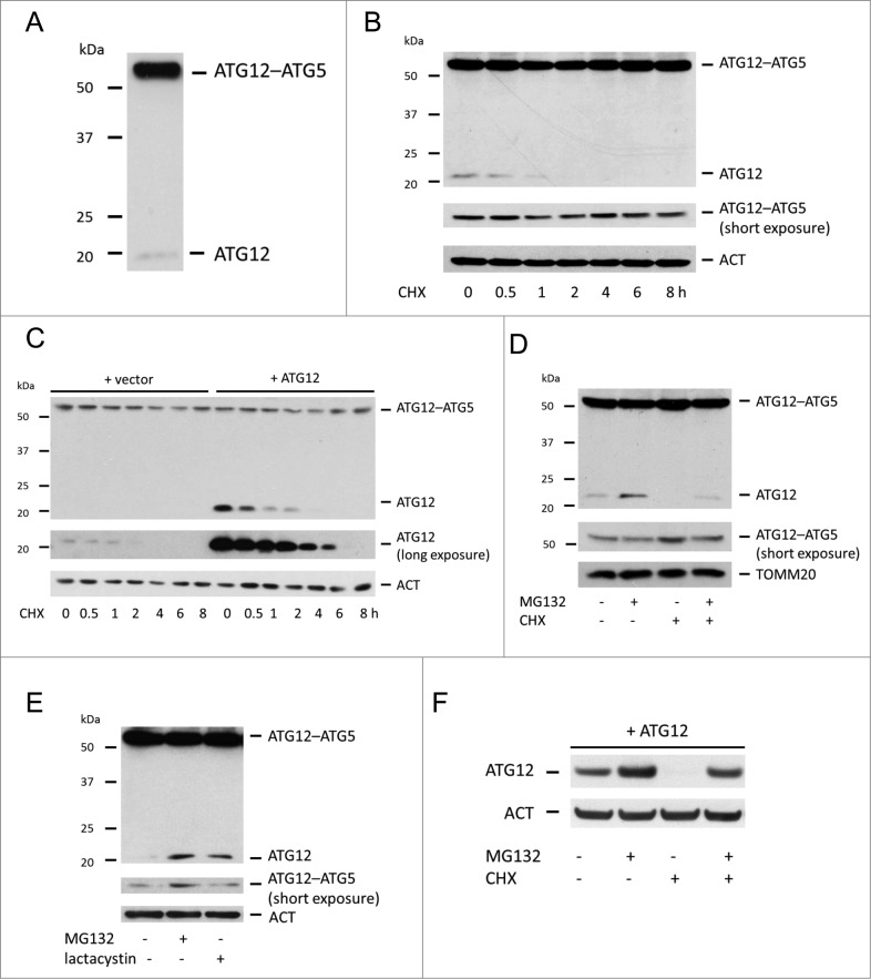
Figure 1. Free ATG12 is rapidly degraded in a proteasome-dependent manner (A) Western blot detection of ATG12 in U2OS cells. (B) Endogenous ATG12 expression in U2OS cells following CHX treatment. (C) U2OS expressing empty vector or ATG12 were treated with CHX for various times and probed for ATG12 expression. (D) U2OS cells were treated were treated for 8 h as indicated with MG132 and/or CHX and probed for ATG12 expression. (E) U2OS were treated for 8 h with MG132 or lactacystin and examined for ATG12 expression. (F) U2OS expressing ATG12 were treated for 8 h with MG132 and/or CHX as indicated and cell lysates were probed for ATG12 expression. ACT or TOMM20 were used as a loading control.
Figure 1. Free ATG12 is rapidly degraded in a proteasome-dependent manner (A) Western blot detection of ATG12 in U2OS cells. (B) Endogenous ATG12 expression in U2OS cells following CHX treatment. (C) U2OS expressing empty vector or ATG12 were treated with CHX for various times and probed for ATG12 expression. (D) U2OS cells were treated were treated for 8 h as indicated with MG132 and/or CHX and probed for ATG12 expression. (E) U2OS were treated for 8 h with MG132 or lactacystin and examined for ATG12 expression. (F) U2OS expressing ATG12 were treated for 8 h with MG132 and/or CHX as indicated and cell lysates were probed for ATG12 expression. ACT or TOMM20 were used as a loading control.CC BY-NC · PubMed Central
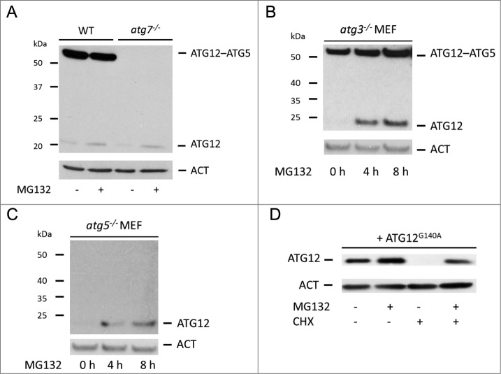
Figure 2. Proteasomal degradation of free ATG12 protein occurs independent of autophagy (A) E1A and Ras-transformed WT or Atg7 knockout MEF were treated for 8 h with MG132 and cell lysates were probed for ATG12 expression. (B) Atg3 or (C) Atg5 knockout MEFs were treated with MG132 for 4 h and 8 h and analyzed for ATG12 expression. (D) U2OS cells expressing ATG12G140A were treated for 8 h with MG132 and/or CHX as indicated and lysates were examined for ATG12 expression. In all immunoblots, ACT was used as a loading control.
Figure 2. Proteasomal degradation of free ATG12 protein occurs independent of autophagy (A) E1A and Ras-transformed WT or Atg7 knockout MEF were treated for 8 h with MG132 and cell lysates were probed for ATG12 expression. (B) Atg3 or (C) Atg5 knockout MEFs were treated with MG132 for 4 h and 8 h and analyzed for ATG12 expression. (D) U2OS cells expressing ATG12G140A were treated for 8 h with MG132 and/or CHX as indicated and lysates were examined for ATG12 expression. In all immunoblots, ACT was used as a loading control.CC BY-NC · PubMed Central
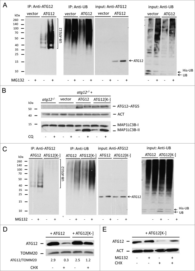
Figure 3. Direct ubiquitination of free ATG12 regulates its proteasomal degradation (A) 293T cells expressing vector or ATG12 and His-ubiquitin were treated with MG132 as indicated. Cell lysates were subject to His-tag affinity isolation and immunoblotted for ATG12 and ubiquitin expression (B) Atg12 knockout MEFs, as well as MEFs stably expressing Atg12, were treated for 4 h with chloroquine, then examined for ATG12 and LC3B expression. (C) 293T cells expressing ATG12 or ATG12[K-] together with His-ubiquitin, were treated as indicated, subject to His-tag affinity isolation and probed for ATG12 and ubiquitin expression. (D) U2OS cells expressing ATG12 or ATG12[K-] and treated for 6 h with CHX and analyzed for ATG12 and TOMM20 expression, densitometric analysis was performed using ImageJ software, normalizing to TOMM20 levels. (E) U2OS cells expressing ATG12[K-] were treated with MG132 and/or CHX for 8 h as indicated and examined for ATG12 expression. ACT or TOMM20 were used as loading controls.
Figure 3. Direct ubiquitination of free ATG12 regulates its proteasomal degradation (A) 293T cells expressing vector or ATG12 and His-ubiquitin were treated with MG132 as indicated. Cell lysates were subject to His-tag affinity isolation and immunoblotted for ATG12 and ubiquitin expression (B) Atg12 knockout MEFs, as well as MEFs stably expressing Atg12, were treated for 4 h with chloroquine, then examined for ATG12 and LC3B expression. (C) 293T cells expressing ATG12 or ATG12[K-] together with His-ubiquitin, were treated as indicated, subject to His-tag affinity isolation and probed for ATG12 and ubiquitin expression. (D) U2OS cells expressing ATG12 or ATG12[K-] and treated for 6 h with CHX and analyzed for ATG12 and TOMM20 expression, densitometric analysis was performed using ImageJ software, normalizing to TOMM20 levels. (E) U2OS cells expressing ATG12[K-] were treated with MG132 and/or CHX for 8 h as indicated and examined for ATG12 expression. ACT or TOMM20 were used as loading controls.CC BY-NC · PubMed Central
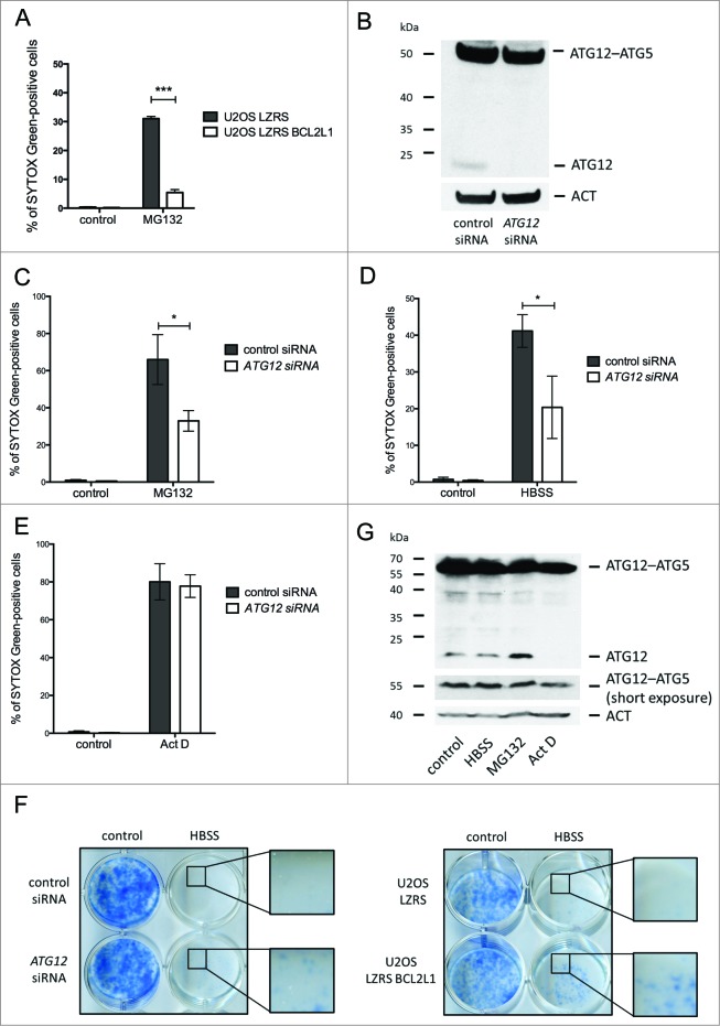
Figure 4. Free ATG12 promotes proteasome inhibitor-mediated cell death (A) U2OS cells stably expressing empty vector or BCL2L1 were treated with MG132 and cell viability was determined by SYTOX Green staining using an Incucyte Imager; data represents the mean +/− standard error of the mean (SEM) of 3 experiments at a representative time-point (24 h). (B) U2OS cells were assessed for ATG12 expression 2 d post-transfection with control or ATG12 siRNA. Cell viability of control or ATG12 siRNA-transfected U2OS cells, treated with MG132 (C) HBSS (D) or Act D (E) was determined by SYTOX Green staining using an Incucyte Imager; representative time-points shown (24 h MG132, 30 h HBSS, 24 h Act D). Graphs represent the mean +/− SEM of 4 experiments. (F) U2OS, transfected with control or ATG12 siRNA, or stably expressing vector or BCL2L1 were starved in HBSS. After 48 h of starvation, cells were washed, cultivated for 7 d in DMEM and colonies were stained with methylene blue. (G) U2OS cells were treated with HBSS (24 h), MG132 (16 h) or Act D (16 h) and examined for ATG12 expression. In all immunoblots, ACT was used as a loading control.
Figure 4. Free ATG12 promotes proteasome inhibitor-mediated cell death (A) U2OS cells stably expressing empty vector or BCL2L1 were treated with MG132 and cell viability was determined by SYTOX Green staining using an Incucyte Imager; data represents the mean +/− standard error of the mean (SEM) of 3 experiments at a representative time-point (24 h). (B) U2OS cells were assessed for ATG12 expression 2 d post-transfection with control or ATG12 siRNA. Cell viability of control or ATG12 siRNA-transfected U2OS cells, treated with MG132 (C) HBSS (D) or Act D (E) was determined by SYTOX Green staining using an Incucyte Imager; representative time-points shown (24 h MG132, 30 h HBSS, 24 h Act D). Graphs represent the mean +/− SEM of 4 experiments. (F) U2OS, transfected with control or ATG12 siRNA, or stably expressing vector or BCL2L1 were starved in HBSS. After 48 h of starvation, cells were washed, cultivated for 7 d in DMEM and colonies were stained with methylene blue. (G) U2OS cells were treated with HBSS (24 h), MG132 (16 h) or Act D (16 h) and examined for ATG12 expression. In all immunoblots, ACT was used as a loading control.CC BY-NC · PubMed Central
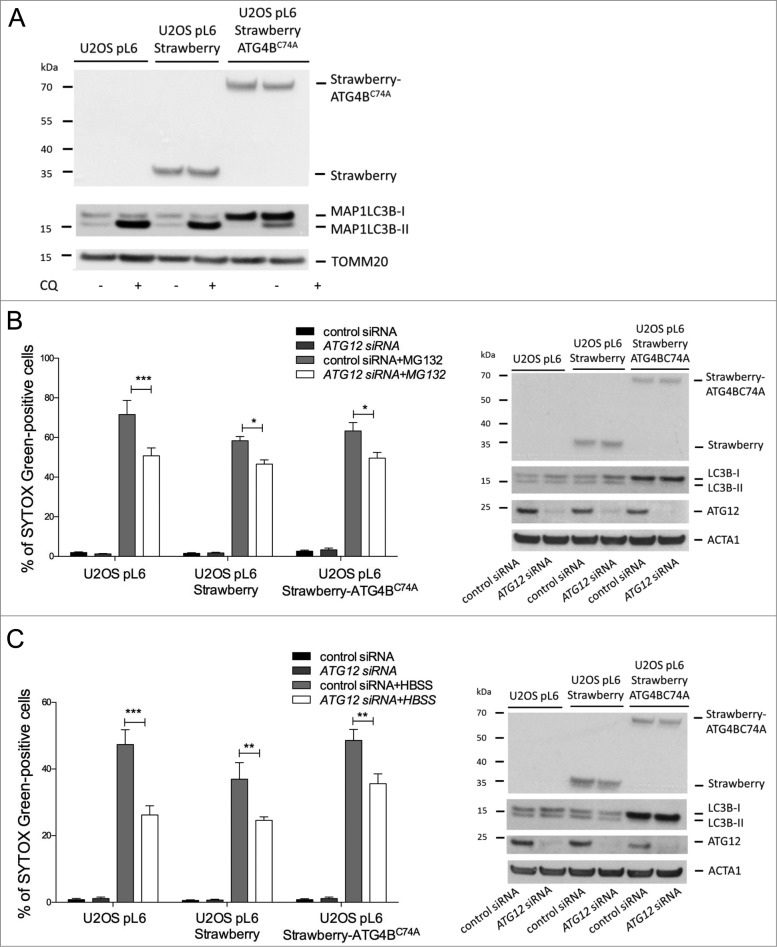
Figure 5. Free ATG12 promotes cell death independent of autophagy (A) U2OS cells stably expressing vector, Strawberry or Strawberry-ATG4BC74A were treated for 4 h with chloroquine and cell lysates were blotted for RFP and LC3B. U2OS cells stably expressing vector, Strawberry or Strawberry-ATG4BC74A were transfected with control or ATG12 siRNA. (B) Following MG132 treatment (24 h) or (C) HBSS starvation (48 h), cell viability was determined by SYTOX Green exclusion in an Incuyte Imager. Graphs show the mean +/− SEM of 3 (B) or 5 (C) experiments at representative time-points (24h MG132, 48 h HBSS). Western blots show cell lysates of U2OS cells stably expressing vector, Strawberry or Strawberry-ATG4BC74A, transfected with control or ATG12 siRNA, treated for 8 h with MG132 (B) or HBSS (C) and probed for RFP, LC3B, and ATG12. In all immunoblots, ACT or TOMM20 were used as a loading control.
Figure 5. Free ATG12 promotes cell death independent of autophagy (A) U2OS cells stably expressing vector, Strawberry or Strawberry-ATG4BC74A were treated for 4 h with chloroquine and cell lysates were blotted for RFP and LC3B. U2OS cells stably expressing vector, Strawberry or Strawberry-ATG4BC74A were transfected with control or ATG12 siRNA. (B) Following MG132 treatment (24 h) or (C) HBSS starvation (48 h), cell viability was determined by SYTOX Green exclusion in an Incuyte Imager. Graphs show the mean +/− SEM of 3 (B) or 5 (C) experiments at representative time-points (24h MG132, 48 h HBSS). Western blots show cell lysates of U2OS cells stably expressing vector, Strawberry or Strawberry-ATG4BC74A, transfected with control or ATG12 siRNA, treated for 8 h with MG132 (B) or HBSS (C) and probed for RFP, LC3B, and ATG12. In all immunoblots, ACT or TOMM20 were used as a loading control.CC BY-NC · PubMed Central

Verification

Claims the paper makes

The claims and statistics of this paper haven’t been checked yet.

Run the check — free, no account needed. It reads the open-access text and checks the sources it cites.

A few free checks a day · papers already verified open instantly · results are public

Claims other papers make about this one

Each is checked against this paper’s own text: does it say what the citing paper says it does?

Self-check

Percentages, sums and reported differences re-computed from the paper’s own numbers.

The self-check needs the paper’s full text.

Citations

Cites

Show all 2 references

Research works printed but not linked: 23

  1. 10.1101/sqb.2011.76.011023 · DOI
  2. 10.1016/j.tcb.2013.12.003 · DOI
  3. 10.2353/ajpath.2007.070188 · DOI · Paper page
  4. 10.1016/j.molcel.2009.01.021 · DOI · Paper page
  5. 10.1016/j.neulet.2009.03.085 · DOI
  6. 10.1038/nrneurol.2011.200 · DOI
  7. 10.1038/nrm2673 · DOI
  8. 10.1038/embor.2008.163 · DOI
  9. 10.1016/j.cell.2007.03.045 · DOI · Paper page
  10. 10.1016/j.cell.2010.07.018 · DOI · Paper page
  11. 10.1016/j.molcel.2011.10.014 · DOI
  12. 10.1091/mbc.E08-03-0309 · DOI
  13. 10.1038/26506 · DOI
  14. 10.1038/nsmb.2431 · DOI
  15. 10.1091/mbc.E07-12-1257 · DOI
  16. 10.1091/mbc.E08-03-0312 · DOI
  17. 10.1146/annurev-biochem-093010-153308 · DOI
  18. 10.1016/j.molcel.2011.08.025 · DOI
  19. 10.1128/MCB.25.9.3483-3491.2005 · DOI
  20. 10.1091/mbc.E11-07-0609 · DOI
  21. 10.1038/embor.2012.208 · DOI
  22. 10.1016/j.molcel.2011.08.034 · DOI
  23. 10.1038/emboj.2011.419 · DOI

Cited by

Sorted by citations

Metadata

Authors
Haller, M., Hock, A. K., Giampazolias, E., Oberst, A., Green, D. R., Debnath, J., Ryan, K. M., Vousden, K. H., & Tait, S. W. G.
Venue
Autophagy
Published
2014-12-02
Volume
10
Issue
12
Pages
2269-2278
ISSN
  • 1554-8627
  • 1554-8635
Cited by
58
References
2
Language
en

About these checks

Registry, retraction, Cites and statistics results are computed, not judged, with one exception: a printed reference that no index lists is read once by a model before it counts as a citation error. Claims are extracted by a model and kept only when their quote appears verbatim in the text; each cited source is then checked, with a model’s help, for whether it says what the claim relies on. Those verdicts can be wrong — read the claim’s quote and the cited source before relying on one.

Statistics and arithmetic are recomputed, not judged.

Confidence bands are shown only for fields calibrated on our annotated test set. None is calibrated yet, so no band is shown.

What counts as a claim ›How we measure confidence ›How we check this ›

Are you an author, or do you see a problem with this page? Request a review