

The DOI matches its Crossref record; 48 of the 48 references it cites were found in a registry or an index.
Hippocampal activation is associated with longitudinal amyloid accumulation and cognitive decline
Open accessAbstract
The amyloid hypothesis suggests that beta-amyloid (Aβ) deposition leads to alterations in neural function and ultimately to cognitive decline in Alzheimer’s disease. However, factors that underlie Aβ deposition are incompletely understood. One proposed model suggests that synaptic activity leads to increased Aβ deposition. More specifically, hyperactivity in the hippocampus may be detrimental and could be one factor that drives Aβ deposition. To test this model, we examined the relationship between hippocampal activity during a memory task using fMRI and subsequent longitudinal change in Aβ using PIB-PET imaging in cognitively normal older adults. We found that greater hippocampal activation at baseline was associated with increased Aβ accumulation. Furthermore, increasing Aβ accumulation mediated the influence of hippocampal activation on declining memory performance, demonstrating a crucial role of Aβ in linking hippocampal activation and memory. These findings support a model linking increased hippocampal activation to subsequent Aβ deposition and cognitive decline.
Overview
The claims and statistics of this paper haven’t been checked yet.
Run the check — free, no account needed. It reads the open-access text and checks the sources it cites.
Integrity signals
Registry
Cites
Statistics
Figures
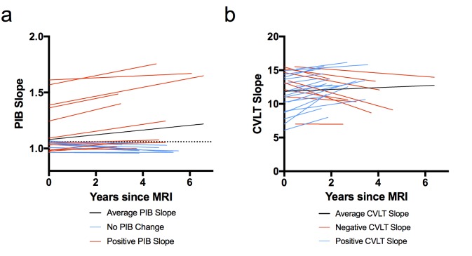
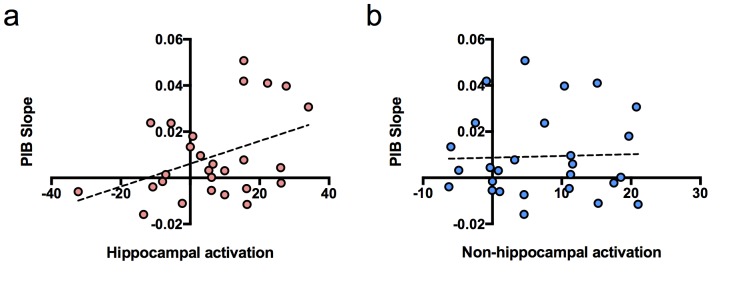
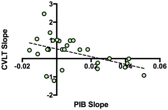
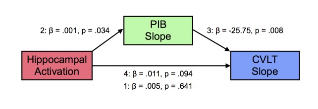
Verification
Claims the paper makes
The claims and statistics of this paper haven’t been checked yet.
Run the check — free, no account needed. It reads the open-access text and checks the sources it cites.
Claims other papers make about this one
Each is checked against this paper’s own text: does it say what the citing paper says it does?
In the citing paper
“and even predicts future cognitive decline in cognitively healthy elderly [178].”
From Apolipoprotein E4, inhibitory network dysfunction, and Alzheimer’s disease (2019) · cites this paper as [178]
Self-check
Percentages, sums and reported differences re-computed from the paper’s own numbers.
The self-check needs the paper’s full text.
Citations
Cites
(2013) · Cited by 44,563
Amyloid oligomers exacerbate tau pathology in a mouse model of tauopathy
Selenica, M.-L. B., Brownlow, M., Jimenez, J. P., Lee, D. C., Pena, G., Dickey, C. A., Gordon, M. N., & Morgan, D. (2013) · Neurodegenerative Diseases · doi:10.1159/000337230 · Cited by 10,359
Alzheimer's Disease Is a Synaptic Failure
Selkoe, D. J. (2002) · Science · doi:10.1126/science.1074069 · Cited by 4,420
Phases of Aβ-deposition in the human brain and its relevance for the development of AD
Thal, D. R., Rüb, U., Orantes, M., & Braak, H. (2002) · Neurology · doi:10.1212/wnl.58.12.1791 · Cited by 3,490
Buckner, R. L., Sepulcre, J., Talukdar, T., Krienen, F. M., Liu, H., Hedden, T., Andrews-Hanna, J. R., Sperling, R. A., & Johnson, K. A. (2009) · The Journal of Neuroscience · doi:10.1523/jneurosci.5062-08.2009 · Cited by 2,862
Staging of alzheimer's disease-related neurofibrillary changes
Braak, H., & Braak, E. (1995) · Neurobiology of Aging · doi:10.1016/0197-4580(95)00021-6 · Cited by 2,357
Palop, J. J., Chin, J., Roberson, E. D., Wang, J., Thwin, M. T., Bien-Ly, N., Yoo, J., Ho, K. O., Yu, G.-Q., Kreitzer, A., et al. (2007) · Neuron · doi:10.1016/j.neuron.2007.07.025 · Cited by 1,653
APP Processing and Synaptic Function
Kamenetz, F., Tomita, T., Hsieh, H., Seabrook, G., Borchelt, D., Iwatsubo, T., Sisodia, S., & Malinow, R. (2003) · Neuron · doi:10.1016/s0896-6273(03)00124-7 · Cited by 1,644
Patterns of Brain Activation in People at Risk for Alzheimer's Disease
Bookheimer, S. Y., Strojwas, M. H., Cohen, M. S., Saunders, A. M., Pericak-Vance, M. A., Mazziotta, J. C., & Small, G. W. (2000) · New England Journal of Medicine · doi:10.1056/nejm200008173430701 · Cited by 1,328
Synaptic Activity Regulates Interstitial Fluid Amyloid-β Levels In Vivo
Cirrito, J. R., Yamada, K. A., Finn, M. B., Sloviter, R. S., Bales, K. R., May, P. C., Schoepp, D. D., Paul, S. M., Mennerick, S., & Holtzman, D. M. (2005) · Neuron · doi:10.1016/j.neuron.2005.10.028 · Cited by 1,265
Cited by
Sorted by citations
Items 101–117 of 117
Skouras, S., Torner, J., Andersson, P., Koush, Y., Falcón, C., Minguillón, C., Fauria, K., Alpiste, F., Blenow, K., Zetterberg, H., et al. (2019) · bioRxiv (Cold Spring Harbor Laboratory) · doi:10.1101/654426 · Cited by 1
Bjorkli, C., Ebbesen, N. C., Julian, J. B., Witter, M. P., Sandvig, A., & Sandvig, I. (2022) · bioRxiv (Cold Spring Harbor Laboratory) · doi:10.1101/2022.07.05.498797 · Cited by 2
Pathological brain states in Alzheimer’s diseasePreprint
Adams, J. N., Kark, S. M., Chappel-Farley, M. G., Escalante, Y. Y., Stith, L., Rapp, P. E., Yassa, M. A., & Initiative, T. A. D. N. (2023) · bioRxiv (Cold Spring Harbor Laboratory) · doi:10.1101/2023.08.30.555617 · Cited by 2
Katiyar, D., & Manish, M. (2025) · Letters in Drug Design & Discovery · doi:10.1016/j.lddd.2025.100126 · Cited by 1
Amyloid-beta relates to default and attention network connectivity during working memory retrieval
He, H., Habeck, C. G., & Stern, Y. (2026) · Brain Communications · doi:10.1093/braincomms/fcag278 · Cited by 1
Wang, T., Lai, X.-X., Xu, L., Shen, Y., Lin, J., Gao, S., & Hu, H. (2020) · Evidence-based Complementary and Alternative Medicine · doi:10.1155/2020/9058135 · Cited by 1
Purohit, P., & Roy, P. K. (2022) · Springer eBooks · doi:10.1007/978-981-19-1352-5_11 · Cited by 1
Chernek, P., Yazdi, B. G., Hojjati, S. H., Ozoria, S., Calimag, J., Wang, X. H., Modarresi, A., Picha, S. G., Chiang, G. C.-Y., & Razlighi, Q. R. (2025) · medRxiv · doi:10.1101/2025.11.02.25339341 · Cited by 1
Neuroimaging Advances in Preclinical Alzheimers disease
Sheng, C., Huang, Y. M., Li, H., Sun, Y., Chen, Q., Ma, C., Liu, F., & Han, Y. (2018) · Neuropsychiatry · doi:10.4172/neuropsychiatry.1000508
Fact, Fiction, or Evolution: Mechanism Hypothesis of Alzheimer’s Disease
Pardo, J. V. (2019) · IntechOpen eBooks · doi:10.5772/intechopen.83824
Cappa, S. F. (2020) · Faculty Opinions – Post-Publication Peer Review of the Biomedical Literature · doi:10.3410/f.735988257.793576095
Branch, A., Glover, L. R., & Gallagher, M. (2023) · Frontiers in Aging Neuroscience · doi:10.3389/fnagi.2023.1238444
Shin, J., Brady, E. S., Chen, C., Lauderdale, K., Agrawal, A., Zhang, Y., Jiang, X., Nambiar, P., Herbert, J., Mallen, D. D., et al. (2025) · bioRxiv (Cold Spring Harbor Laboratory) · doi:10.1101/2025.11.11.687887
D-sérine : un maillon central de la physiopathologie amyloïde ?
Ploux, E. (2022) · theses.fr (ABES) · doi:10.70675/e7b5bba0z9abcz4325zae71z841a60892f8b
Clemens, S. G., Hobbs, D. A., McKay, N. S., Millar, P. R., Morris, J. C., Hassenstab, J. J., Zacks, J. M., Balota, D. A., & Gordon, B. A. (2026) · Brain Communications · doi:10.1093/braincomms/fcag167
Adrita, N., Claassen, I., Huerta, M., Black, S. M., Estévez, Á. G., Roberts, B. R., Beckman, J. S., & Franco, M. C. (2026) · Redox Biology · doi:10.1016/j.redox.2026.104423
Solar, K. G., Kashyap, S., Deom, N., Uludaǧ, K., Zotovic, L., Lanctôt, K. L., Black, S. E., & McAndrews, M. P. (2026) · Brain Imaging and Behavior · doi:10.1007/s11682-026-01208-9
Metadata
- Authors
- Leal, S. L., Landau, S. M., Bell, R. K., & Jagust, W. J.
- Affiliations
- University of California, Berkeley (US)
- Lawrence Berkeley National Laboratory (US)
- Corresponding author
- Leal, Stephanie L
- Jagust, William J
- Venue
- eLife
- Publisher
- eLife Sciences Publications, Ltd
- Published
- 2017-02-08
- Volume
- 6
- ISSN
- 2050-084X (ISSN-L)
- PMC
- PMC5325620
- OpenAlex
- W2586682108
- Cited by
- 117
- References
- 48
- Citations by year
- 2026: 5 · 2025: 10 · 2024: 11 · 2023: 16 · 2022: 11 · 2021: 16 · 2020: 14 · 2019: 20 · 2018: 10 · 2017: 4
- FWCI
- 10.79
- Type
- journal article
- Language
- en
- Open access
- gold
- doi.org
- Licence
- Locations
- eLife (publishedVersion, cc-by)
- PubMed Central (publishedVersion, cc-by)
- PubMed (publishedVersion)
- eScholarship (California Digital Library) (submittedVersion)
- OSTI OAI (U.S. Department of Energy Office of Scientific and Technical Information) (submittedVersion)
- DOAJ (DOAJ: Directory of Open Access Journals) (submittedVersion, cc-by-sa)
- Funders
- National Institute on Aging (AG054116, AG034570)
- Topics
- Health Sciences › Medicine › Psychiatry and Mental health › Dementia and Cognitive Impairment Research
- Life Sciences › Neuroscience › Cellular and Molecular Neuroscience › Alzheimer's disease research and treatments
- Health Sciences › Medicine › Oncology › Cancer-related cognitive impairment studies
- Keywords
- hippocampal activation, amyloid-beta deposition, cognitive decline, Alzheimer's disease, amyloid hypothesis, memory performance, PiB PET, functional MRI, older adults, synaptic activity, longitudinal change
- MeSH terms
- Aged, Aged, 80 and over, Female, Hippocampus, Humans, Longitudinal Studies, Magnetic Resonance Imaging, Male, Amyloid beta-Peptides, Positron-Emission Tomography, Cognitive Dysfunction
About these checks
Registry, retraction, Cites and statistics results are computed, not judged, with one exception: a printed reference that no index lists is read once by a model before it counts as a citation error. Claims are extracted by a model and kept only when their quote appears verbatim in the text; each cited source is then checked, with a model’s help, for whether it says what the claim relies on. Those verdicts can be wrong — read the claim’s quote and the cited source before relying on one.
Statistics and arithmetic are recomputed, not judged.
Confidence bands are shown only for fields calibrated on our annotated test set. None is calibrated yet, so no band is shown.
What counts as a claim ›How we measure confidence ›How we check this ›
Are you an author, or do you see a problem with this page? Request a review
