

The DOI matches its Crossref record; 48 of the 48 references it cites were found in a registry or an index.
Hippocampal activation is associated with longitudinal amyloid accumulation and cognitive decline
Open accessAbstract
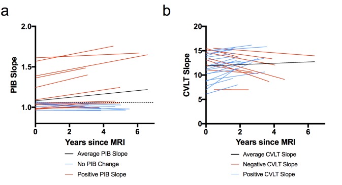
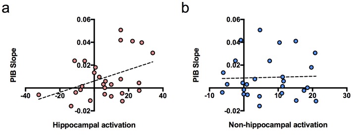
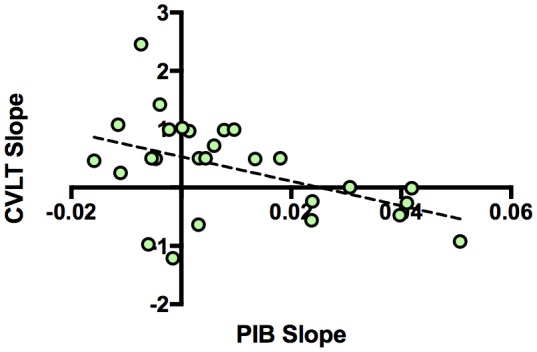
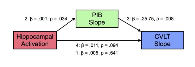
The amyloid hypothesis suggests that beta-amyloid (Aβ) deposition leads to alterations in neural function and ultimately to cognitive decline in Alzheimer’s disease. However, factors that underlie Aβ deposition are incompletely understood. One proposed model suggests that synaptic activity leads to increased Aβ deposition. More specifically, hyperactivity in the hippocampus may be detrimental and could be one factor that drives Aβ deposition. To test this model, we examined the relationship between hippocampal activity during a memory task using fMRI and subsequent longitudinal change in Aβ using PIB-PET imaging in cognitively normal older adults. We found that greater hippocampal activation at baseline was associated with increased Aβ accumulation. Furthermore, increasing Aβ accumulation mediated the influence of hippocampal activation on declining memory performance, demonstrating a crucial role of Aβ in linking hippocampal activation and memory. These findings support a model linking increased hippocampal activation to subsequent Aβ deposition and cognitive decline.
Overview
The claims and statistics of this paper haven’t been checked yet.
Run the check — free, no account needed. It reads the open-access text and checks the sources it cites.
Integrity signals
Registry
Cites
Statistics
Figures
Verification
Claims the paper makes
The claims and statistics of this paper haven’t been checked yet.
Run the check — free, no account needed. It reads the open-access text and checks the sources it cites.
Claims other papers make about this one
Each is checked against this paper’s own text: does it say what the citing paper says it does?
In the citing paper
“and even predicts future cognitive decline in cognitively healthy elderly [178].”
From Apolipoprotein E4, inhibitory network dysfunction, and Alzheimer’s disease (2019) · cites this paper as [178]
Self-check
Percentages, sums and reported differences re-computed from the paper’s own numbers.
The self-check needs the paper’s full text.
Citations
Cites
(2013) · Cited by 44,563
Amyloid oligomers exacerbate tau pathology in a mouse model of tauopathy
Selenica, M.-L. B., Brownlow, M., Jimenez, J. P., Lee, D. C., Pena, G., Dickey, C. A., Gordon, M. N., & Morgan, D. (2013) · Neurodegenerative Diseases · doi:10.1159/000337230 · Cited by 10,359
Alzheimer's Disease Is a Synaptic Failure
Selkoe, D. J. (2002) · Science · doi:10.1126/science.1074069 · Cited by 4,420
Phases of Aβ-deposition in the human brain and its relevance for the development of AD
Thal, D. R., Rüb, U., Orantes, M., & Braak, H. (2002) · Neurology · doi:10.1212/wnl.58.12.1791 · Cited by 3,490
Buckner, R. L., Sepulcre, J., Talukdar, T., Krienen, F. M., Liu, H., Hedden, T., Andrews-Hanna, J. R., Sperling, R. A., & Johnson, K. A. (2009) · The Journal of Neuroscience · doi:10.1523/jneurosci.5062-08.2009 · Cited by 2,862
Staging of alzheimer's disease-related neurofibrillary changes
Braak, H., & Braak, E. (1995) · Neurobiology of Aging · doi:10.1016/0197-4580(95)00021-6 · Cited by 2,357
Palop, J. J., Chin, J., Roberson, E. D., Wang, J., Thwin, M. T., Bien-Ly, N., Yoo, J., Ho, K. O., Yu, G.-Q., Kreitzer, A., et al. (2007) · Neuron · doi:10.1016/j.neuron.2007.07.025 · Cited by 1,653
APP Processing and Synaptic Function
Kamenetz, F., Tomita, T., Hsieh, H., Seabrook, G., Borchelt, D., Iwatsubo, T., Sisodia, S., & Malinow, R. (2003) · Neuron · doi:10.1016/s0896-6273(03)00124-7 · Cited by 1,644
Patterns of Brain Activation in People at Risk for Alzheimer's Disease
Bookheimer, S. Y., Strojwas, M. H., Cohen, M. S., Saunders, A. M., Pericak-Vance, M. A., Mazziotta, J. C., & Small, G. W. (2000) · New England Journal of Medicine · doi:10.1056/nejm200008173430701 · Cited by 1,328
Synaptic Activity Regulates Interstitial Fluid Amyloid-β Levels In Vivo
Cirrito, J. R., Yamada, K. A., Finn, M. B., Sloviter, R. S., Bales, K. R., May, P. C., Schoepp, D. D., Paul, S. M., Mennerick, S., & Holtzman, D. M. (2005) · Neuron · doi:10.1016/j.neuron.2005.10.028 · Cited by 1,265
Cited by
Sorted by citations
Imaging the evolution and pathophysiology of Alzheimer disease
Jagust, W. (2018) · Nature Reviews Neuroscience · doi:10.1038/s41583-018-0067-3 · Cited by 604
Self-citationNeuroimaging biomarkers for Alzheimer’s disease.
Márquez, F., & Yassa, M. A. (2019) · Molecular Neurodegeneration · doi:10.1186/s13024-019-0325-5 · Cited by 289
“Recent evidence also suggests that hippocampal activation is associated with longitudinal amyloid accumulation and cognitive decline [74].”
BackgroundCognition and dementia in older patients with epilepsy
Sen, A., Capelli, V., & Husain, M. (2018) · Brain · doi:10.1093/brain/awy022 · Cited by 280
Apolipoprotein E and Alzheimer’s disease: Findings, hypotheses, and potential mechanisms
Koutsodendris, N., Nelson, M. R., Rao, A., & Huang, Y. (2022) · Annual Review of Pathology Mechanisms of Disease · doi:10.1146/annurev-pathmechdis-030421-112756 · Cited by 231
Imaging biomarkers in neurodegeneration: current and future practices
Young, P. N. E., Estarellas, M., Coomans, E. M., Srikrishna, M., Beaumont, H., Maaß, A., Venkataraman, A. V., Lissaman, R., Jiménez, D., Betts, M. J., et al. (2020) · Alzheimer s Research & Therapy · doi:10.1186/s13195-020-00612-7 · Cited by 244
Self-citationWhat Happens with the Circuit in Alzheimer's Disease in Mice and Humans?
Zott, B., Busche, M. A., Sperling, R. A., & Konnerth, A. (2018) · Annual Review of Neuroscience · doi:10.1146/annurev-neuro-080317-061725 · Cited by 221
Alzheimer’s pathology targets distinct memory networks in the ageing brain
Maaß, A., Berron, D., Harrison, T. M., Adams, J. N., Joie, R. L., Baker, S. L., Mellinger, T. J., Bell, R. K., Swinnerton, K. N., Inglis, B. A., et al. (2019) · Brain · doi:10.1093/brain/awz154 · Cited by 209
Self-citationApolipoprotein E4, inhibitory network dysfunction, and Alzheimer’s disease
Najm, R., Jones, E. A., & Huang, Y. (2019) · Molecular Neurodegeneration · doi:10.1186/s13024-019-0324-6 · Cited by 183
Vogel, J. W., Corriveau‐Lecavalier, N., Franzmeier, N., Pereira, J. B., Brown, J. A., Maaß, A., Botha, H., Seeley, W. W., Bassett, D. S., Jones, D. T., et al. (2023) · Nature reviews. Neuroscience · doi:10.1038/s41583-023-00731-8 · Cited by 180
Harris, S. S., Wolf, F., De Strooper, B., & Busche, M. A. (2020) · Neuron · doi:10.1016/j.neuron.2020.06.005 · Cited by 161
Targeting synapse function and loss for treatment of neurodegenerative diseases
Dejanovic, B., Sheng, M., & Hanson, J. E. (2023) · Nature Reviews Drug Discovery · doi:10.1038/s41573-023-00823-1 · Cited by 177
Koelewijn, L., Lancaster, T. M., Linden, D. E. J., Dima, D. C., Routley, B. C., Magazzini, L., Barawi, K., Brindley, L. M., Adams, R., Tansey, K. E., et al. (2019) · eLife · doi:10.7554/elife.36011 · Cited by 151
Subthreshold Amyloid Predicts Tau Deposition in Aging
Leal, S. L., Lockhart, S. N., Maaß, A., Bell, R. K., & Jagust, W. J. (2018) · Journal of Neuroscience · doi:10.1523/jneurosci.0485-18.2018 · Cited by 137
Self-citationAmyloid-β: a potential link between epilepsy and cognitive decline
Romoli, M., Sen, A., Parnetti, L., Calabresi, P., & Costa, C. (2021) · Nature Reviews Neurology · doi:10.1038/s41582-021-00505-9 · Cited by 133
Gatchel, J. R., Rabin, J. S., Buckley, R. F., Locascio, J. J., Quiroz, Y. T., Yang, H., Vannini, P., Amariglio, R. E., Rentz, D. M., Properzi, M. J., et al. (2019) · JAMA Network Open · doi:10.1001/jamanetworkopen.2019.8964 · Cited by 137
BACE inhibition-dependent repair of Alzheimer’s pathophysiology
Keskin, A. D., Kekuš, M., Adelsberger, H., Neumann, U., Shimshek, D. R., Song, B., Zott, B., Peng, T., Förstl, H., Staufenbiel, M., et al. (2017) · Proceedings of the National Academy of Sciences · doi:10.1073/pnas.1708106114 · Cited by 125
Tau Accumulation in Clinically Normal Older Adults Is Associated with Hippocampal Hyperactivity
Huijbers, W., Schultz, A. P., Papp, K. V., LaPoint, M. R., Hanseeuw, B. J., Chhatwal, J. P., Hedden, T., Johnson, K. A., & Sperling, R. A. (2018) · Journal of Neuroscience · doi:10.1523/jneurosci.1397-18.2018 · Cited by 118
Local Sleep and Alzheimer’s Disease Pathophysiology
Mander, B. A. (2020) · Frontiers in Neuroscience · doi:10.3389/fnins.2020.525970 · Cited by 97
Modulation of Brain Hyperexcitability: Potential New Therapeutic Approaches in Alzheimer’s Disease
Toniolo, S., Sen, A., & Husain, M. (2020) · International Journal of Molecular Sciences · doi:10.3390/ijms21239318 · Cited by 97
Targeting Neural Hyperactivity as a Treatment to Stem Progression of Late-Onset Alzheimer's Disease
Haberman, R. P., Branch, A., & Gallagher, M. (2017) · Neurotherapeutics · doi:10.1007/s13311-017-0541-z · Cited by 98
Hyperactivity Induced by Soluble Amyloid-β Oligomers in the Early Stages of Alzheimer's Disease
Hector, A., & Brouillette, J. (2021) · Frontiers in Molecular Neuroscience · doi:10.3389/fnmol.2020.600084 · Cited by 97
Gallego-Rudolf, J., Wiesman, A. I., Binette, A. P., Villeneuve, S., & Baillet, S. (2024) · Nature Neuroscience · doi:10.1038/s41593-024-01763-8 · Cited by 109
Medial Temporal Lobe Disconnection and Hyperexcitability Across Alzheimer’s Disease Stages
Pasquini, L., Rahmani, F., Balajoo, S. M., Joie, R. L., Zarei, M., Sorg, C., Drzezga, A. E., & Tahmasian, M. (2019) · Journal of Alzheimer s Disease Reports · doi:10.3233/adr-190121 · Cited by 77
Proteostasis failure and cellular senescence in long‐term cultured postmitotic rat neurons
Ishikawa, S., & Ishikawa, F. (2020) · Aging Cell · doi:10.1111/acel.13071 · Cited by 67
Aron, L., Zullo, J., & Yankner, B. A. (2021) · Current Opinion in Neurobiology · doi:10.1016/j.conb.2021.09.009 · Cited by 70
Taksima, T., Chonpathompikunlert, P., Sroyraya, M., Hutamekalin, P., Limpawattana, M., & Klaypradit, W. (2019) · Marine Drugs · doi:10.3390/md17110628 · Cited by 64
Morrone, C. D., Raghuraman, R., Hussaini, S. A., & Yu, W. H. (2023) · Molecular Neurodegeneration · doi:10.1186/s13024-023-00617-4 · Cited by 68
A Neuroimaging Signature of Cognitive Aging from Whole‐Brain Functional Connectivity
Jiang, R., Scheinost, D., Zuo, N., Wu, J., Qi, S., Liang, Q., Zhi, D., Luo, N., Chung, Y., Liu, S., et al. (2022) · Advanced Science · doi:10.1002/advs.202201621 · Cited by 62
Cerebral hyperactivation across the Alzheimer’s disease pathological cascade
Corriveau‐Lecavalier, N., Adams, J. N., Fischer, L., Molloy, E. N., & Maaß, A. (2024) · Brain Communications · doi:10.1093/braincomms/fcae376 · Cited by 71
Brain properties predict proximity to symptom onset in sporadic Alzheimer’s disease
Vogel, J. W., Vachon‐Presseau, É., Binette, A. P., Tam, A., Orban, P., Joie, R. L., Savard, M., Picard, C., Poirier, J., Bellec, P., et al. (2018) · Brain · doi:10.1093/brain/awy093 · Cited by 52
Skouras, S., Falcón, C., Tucholka, A., Rami, L., Sánchez‐Valle, R., Lladó, A., Gispert, J. D., & Molinuevo, J. L. (2019) · NeuroImage Clinical · doi:10.1016/j.nicl.2019.101777 · Cited by 53
Foster, C. M., Kennedy, K. M., Horn, M. M., Hoagey, D. A., & Rodrigue, K. M. (2017) · NeuroImage · doi:10.1016/j.neuroimage.2017.10.068 · Cited by 49
Corriveau‐Lecavalier, N., Mellah, S., Clément, F., & Belleville, S. (2019) · NeuroImage Clinical · doi:10.1016/j.nicl.2019.101958 · Cited by 50
Bollack, A., Pemberton, H., Collij, L. E., Markiewicz, P. J., Cash, D. M., Farrar, G., & Barkhof, F. (2023) · Alzheimer s & Dementia · doi:10.1002/alz.13158 · Cited by 51
Pardillo-Díaz, R., Pérez-García, P., Castro, C., Nunez‐Abades, P., & Carrascal, L. (2022) · Antioxidants · doi:10.3390/antiox11081511 · Cited by 50
Late-in-life neurodegeneration after chronic sleep loss in young adult mice
Owen, J. E., Zhu, Y., Fenik, P., Zhan, G., Bell, P., Liu, X. C., & Veasey, S. C. (2021) · SLEEP · doi:10.1093/sleep/zsab057 · Cited by 51
Cognitive reserve against Alzheimer’s pathology is linked to brain activity during memory formation
Vockert, N., Machts, J., Kleineidam, L., Nemali, A., Incesoy, E. I., Bernal, J., Schütze, H., Yakupov, R., Peters, O., Gref, D., et al. (2024) · Nature Communications · doi:10.1038/s41467-024-53360-9 · Cited by 59
Diersch, N., Valdés-Herrera, J. P., Tempelmann, C., & Wolbers, T. (2021) · Journal of Neuroscience · doi:10.1523/jneurosci.0528-20.2021 · Cited by 46
A quadratic function of activation in individuals at risk of Alzheimer's disease
Corriveau‐Lecavalier, N., Duchesne, S., Gauthier, S. G., Hudon, C., Kergoat, M., Mellah, S., & Belleville, S. (2020) · Alzheimer s & Dementia Diagnosis Assessment & Disease Monitoring · doi:10.1002/dad2.12139 · Cited by 40
Probing neural circuit mechanisms in Alzheimer’s disease using novel technologies
Grieco, S. F., Holmes, T. C., & Xu, X. (2023) · Molecular Psychiatry · doi:10.1038/s41380-023-02018-x · Cited by 37
Multipurpose Virtual Reality Environment for Biomedical and Health Applications
Torner, J., Skouras, S., Molinuevo, J. L., Gispert, J. D., & Alpiste, F. (2019) · IEEE Transactions on Neural Systems and Rehabilitation Engineering · doi:10.1109/tnsre.2019.2926786 · Cited by 32
Li, Q., Marcu, D.-C., Palazzo, O., Turner, F. S., King, D., Spires‐Jones, T. L., Stefan, M. I., & Busch, K. E. (2020) · eLife · doi:10.7554/elife.59711 · Cited by 29
Leal, S. L., Ferguson, L. A., Harrison, T. M., & Jagust, W. J. (2019) · Learning & Memory · doi:10.1101/lm.048967.118 · Cited by 32
Self-citationYoshikawa, M., Soeda, Y., Michikawa, M., Almeida, O. F. X., & Takashima, A. (2018) · Frontiers in Neuroscience · doi:10.3389/fnins.2018.00124 · Cited by 30
Nicolini, P., Lucchi, T. A., Abbate, C., Inglese, S., Tomasini, E., Mari, D., Rossi, P. D., & Vicenzi, M. (2022) · Frontiers in Aging Neuroscience · doi:10.3389/fnagi.2022.886023 · Cited by 31
1 H-MRS metabolites and rate of β-amyloid accumulation on serial PET in clinically normal adults
Nedelská, Z., Przybelski, S. A., Lesnick, T. G., Schwarz, C. G., Lowe, V. J., Machulda, M. M., Kremers, W. K., Mielke, M. M., Roberts, R. O., Boeve, B. F., et al. (2017) · Neurology · doi:10.1212/wnl.0000000000004421 · Cited by 29
Skouras, S., Torner, J., Andersson, P. U., Koush, Y., Falcón, C., Minguillón, C., Fauria, K., Alpiste, F., Blenow, K., Zetterberg, H., et al. (2020) · Brain · doi:10.1093/brain/awaa011 · Cited by 28
What are the threats to successful brain and cognitive aging?
Gallagher, M., Okonkwo, O. C., Resnick, S. M., Jagust, W. J., Benzinger, T. L. S., & Rapp, P. R. (2019) · Neurobiology of Aging · doi:10.1016/j.neurobiolaging.2019.04.016 · Cited by 25
Self-citationGhatak, S., Nakamura, T., & Lipton, S. A. (2023) · Frontiers in Neural Circuits · doi:10.3389/fncir.2023.1099467 · Cited by 26
Branch, A., Monasterio, A., Blair, G. A., Knierim, J. J., Gallagher, M., & Haberman, R. P. (2019) · Neurobiology of Aging · doi:10.1016/j.neurobiolaging.2018.12.015 · Cited by 24
Impairment of entorhinal cortex network activity in Alzheimer’s disease
Karimani, F., Taei, A. A., Abolghasemi-Dehaghani, M.-R., Safari, M.-S., & Dargahi, L. (2024) · Frontiers in Aging Neuroscience · doi:10.3389/fnagi.2024.1402573 · Cited by 27
Chen, X., Farrell, M. E., Rundle, M. M., Chan, M. Y., Moore, W., Wig, G. S., & Park, D. C. (2021) · Neurobiology of Aging · doi:10.1016/j.neurobiolaging.2021.04.020 · Cited by 32
Desmidt, T., Delrieu, J., Lebouvier, T., Robert, G., David, R., Balageas, A., Surget, A., Belzung, C., Arlicot, N., Ribeiro, M. J., et al. (2019) · Neurobiology of Aging · doi:10.1016/j.neurobiolaging.2019.08.008 · Cited by 22
Rasia‐Filho, A. A., Guerra, K. T. K., Vásquez, C. E., Dall’Oglio, A., Reberger, R., Jung, C. R., & Calcagnotto, M. E. (2021) · Frontiers in Synaptic Neuroscience · doi:10.3389/fnsyn.2021.616607 · Cited by 24
Klink, K., Jaun, U., Federspiel, A., Wunderlin, M., Teunissen, C. E., Kiefer, C., Wiest, R., Scharnowski, F., Sladky, R., Haugg, A., et al. (2021) · BMC Psychiatry · doi:10.1186/s12888-021-03091-8 · Cited by 22
Pourzinal, D., Yang, J., Bakker, A., McMahon, K. L., Byrne, G. J., Pontone, G. M., Mari, Z. K., & Dissanayaka, N. N. (2021) · Journal of Neuroscience Research · doi:10.1002/jnr.24863 · Cited by 19
Corriveau‐Lecavalier, N., Rajah, M. N., Mellah, S., & Belleville, S. (2021) · NeuroImage Clinical · doi:10.1016/j.nicl.2021.102643 · Cited by 24
Sleep: The Tip of the Iceberg in the Bidirectional Link Between Alzheimer's Disease and Epilepsy
Szabo, A. B., Cretin, B., Gérard, F., Curot, J., Barbeau, E. J., Pariente, J., Dahan, L., & Valton, L. (2022) · Frontiers in Neurology · doi:10.3389/fneur.2022.836292 · Cited by 21
Parent, C., Rousseau, L., Predovan, D., Duchesne, S., & Hudon, C. (2023) · Aging Brain · doi:10.1016/j.nbas.2023.100074 · Cited by 21
Samudra, N. P., Ranasinghe, K. G., Kirsch, H. E., Rankin, K. P., & Miller, B. L. (2023) · Journal of Alzheimer s Disease · doi:10.3233/jad-220983 · Cited by 18
Emerging links between cell competition and Alzheimer's disease
Coelho, D. S., & Moreno, E. (2019) · Journal of Cell Science · doi:10.1242/jcs.231258 · Cited by 19
Meyer, P., McSweeney, M., Gonneaud, J., & Villeneuve, S. (2019) · Progress in molecular biology and translational science · doi:10.1016/bs.pmbts.2019.05.001 · Cited by 20
Hu, S., & Li, C.-S. R. (2020) · Brain Sciences · doi:10.3390/brainsci10121013 · Cited by 16
Kennedy, K. M., Foster, C. M., & Rodrigue, K. M. (2018) · NeuroImage · doi:10.1016/j.neuroimage.2018.08.017 · Cited by 14
Aging Atlas Reveals Cell-Type-Specific Regulation of Pro-longevity StrategiesPreprint
Gao, S. M., Qi, Y., Zhang, Q., Mohammed, A. S., Lee, Y.-T., Guan, Y., Li, H., Fu, Y., & Wang, M. C. (2023) · bioRxiv (Cold Spring Harbor Laboratory) · doi:10.1101/2023.02.28.530490 · Cited by 14
Hou, M., de Chastelaine, M., Jayakumar, M., Donley, B. E., & Rugg, M. D. (2020) · Neuropsychologia · doi:10.1016/j.neuropsychologia.2020.107537 · Cited by 17
Eisenstein, T., Giladi, N., Hendler, T., Havakuk, O., & Lerner, Y. (2021) · Frontiers in Aging Neuroscience · doi:10.3389/fnagi.2021.720990 · Cited by 11
Bagheri, S., Saboury, A. A., Haertlé, T., Rongioletti, M. C. A., & Saso, L. (2022) · Inorganics · doi:10.3390/inorganics10010006 · Cited by 14
Li, J., Liu, Y., Yin, C., Zeng, Y., & Mei, Y. (2024) · Ageing Research Reviews · doi:10.1016/j.arr.2024.102468 · Cited by 13
Chen, B. (2020) · Aging Clinical and Experimental Research · doi:10.1007/s40520-020-01539-7 · Cited by 11
Fischer, L., Parker, D. M., Maboudian, S. A., Fonseca, C. S., Tato-Fernández, C., Annen, L., Arunachalam, P., Bacci, J. R., Barboure, M. C., Capelli, S., et al. (2025) · Alzheimer s Research & Therapy · doi:10.1186/s13195-025-01920-6 · Cited by 13
Self-citationMolecular and neuroimaging biomarkers in the diagnosis of Alzheimer's disease
Sethi, P., Singh, D. K. K., Amist, A. D., Singh, M. P., Sinha, J. K., & Ghosh, S. (2024) · Elsevier eBooks · doi:10.1016/b978-0-443-15540-6.00006-9 · Cited by 10
Agosta, F., Canu, E., & Filippi, M. (2020) · Brain · doi:10.1093/brain/awaa038 · Cited by 10
Tyraminergic corollary discharge filters reafferent perception in a chemosensory neuron
Riedl, J., Fieseler, C., & Zimmer, M. (2022) · Current Biology · doi:10.1016/j.cub.2022.05.051 · Cited by 12
Fischer, L., Molloy, E. N., Binette, A. P., Vockert, N., Marquardt, J., Pilar, A. P., Kreissl, M. C., Remz, J., Tremblay‐Mercier, J., Poirier, J., et al. (2025) · Journal of Neuroscience · doi:10.1523/jneurosci.1408-24.2024 · Cited by 13
Michaelian, J. C., Duffy, S. L., Mowszowski, L., Guastella, A. J., McCade, D., McKinnon, A. C., & Naismith, S. L. (2021) · Journal of Alzheimer s Disease · doi:10.3233/jad-201284 · Cited by 12
Argiris, G., Stern, Y., & Habeck, C. G. (2021) · eNeuro · doi:10.1523/eneuro.0273-21.2021 · Cited by 10
Voltage-Gated Na+ Channels in Alzheimer’s Disease: Physiological Roles and Therapeutic Potential
Baumgartner, T. J., Haghighijoo, Z., Goode, N. A., Dvorak, N. M., Arman, P., & Laezza, F. (2023) · Life · doi:10.3390/life13081655 · Cited by 10
Alzheimer Disease Biomarkers and Subjective Cognitive Decline Among Hispanic and/or Latino Adults
Márquez, F., Tarraf, W., González, K. A., Valencia, D. F., Stickel, A. M., Anita, N. Z., Sotres‐Alvarez, D., Levin, B. E., Yassa, M. A., Zhou, H., et al. (2025) · JAMA Network Open · doi:10.1001/jamanetworkopen.2025.31038 · Cited by 8
Targeting hippocampal amyloidogenesis with SV2A protein modulator levetiracetam
Kasatkina, L. A., Gumenyuk, V. P., Lisakovska, O. O., & Trikash, I. O. (2022) · Biochemical Pharmacology · doi:10.1016/j.bcp.2022.114927 · Cited by 7
Anwar, U., Arslan, T., Hussain, A., & Lomax, P. (2022) · IEEE Access · doi:10.1109/access.2022.3195875 · Cited by 7
Investigating Compensatory Brain Activity in Older Adults with Subjective Cognitive Decline
Krebs, C. R., Brill, E., Minkova, L., Federspiel, A., Kellner‐Weldon, F., Wyss, P., Teunissen, C. E., van Harten, A. C., Seydell‐Greenwald, A., Klink, K., et al. (2023) · Journal of Alzheimer s Disease · doi:10.3233/jad-221001 · Cited by 8
Alterations of Audiovisual Integration in Alzheimer’s Disease
Liu, Y., Wang, Z., Wei, T., Zhou, S., Yin, Y., Mi, Y., Liu, X., & Tang, Y. (2023) · Neuroscience Bulletin · doi:10.1007/s12264-023-01125-7 · Cited by 6
Liao, K., Martin, L. E., Fakorede, S., Brooks, W. M., Burns, J. M., & Devos, H. (2024) · Clinical Neurophysiology · doi:10.1016/j.clinph.2024.11.013 · Cited by 6
Optimizing Longitudinal Amyloid-β PET Measurement: The Challenges of Intensity Normalization
Landau, S. M. (2018) · Journal of Nuclear Medicine · doi:10.2967/jnumed.118.212662 · Cited by 6
Self-citationCorona-Long, C. A., Tran, T., Chang, E. H. T., Speck, C. L., Gallagher, M., & Bakker, A. (2020) · Alzheimer s & Dementia Diagnosis Assessment & Disease Monitoring · doi:10.1002/dad2.12043 · Cited by 6
Vahid, A., Zamani, J., & Hosseini, S. M. H. (2025) · npj Dementia · doi:10.1038/s44400-025-00041-z · Cited by 5
Default mode network failure across the Alzheimer’s disease spectrum
Corriveau‐Lecavalier, N., Dicks, E., Martin, P. R., Wiste, H. J., Gunter, J. L., Kamykowski, M. G., Senjem, M. L., Schwarz, C. G., Botha, H., Graff‐Radford, J., et al. (2026) · Brain · doi:10.1093/brain/awag146 · Cited by 6
Sciaccaluga, M., Ruffolo, G., Palma, E., & Costa, C. (2023) · Current Neuropharmacology · doi:10.2174/1570159x21666230504160948 · Cited by 7
Cognitive Impairment Induced by Gestational Diabetes: The Role of Oxidative Stress
Campo-Rota, I. M. D., Delgado-Casillas, O. M., & Ibarra, A. (2024) · Archives of Medical Research · doi:10.1016/j.arcmed.2024.103016 · Cited by 5
Recent developments in translational imaging of in vivo gene therapy outcomes
Day, I. L., Tamboline, M., Lipshutz, G. S., & Xu, S. (2024) · Molecular Therapy · doi:10.1016/j.ymthe.2024.12.049 · Cited by 4
The Role of Epileptic Activity in Alzheimer’s Disease
Chen, T., Lai, M., Hong, W., & Huang, C. (2024) · American Journal of Alzheimer s Disease & Other Dementias® · doi:10.1177/15333175241303569 · Cited by 4
Shu, H., Chen, G., Ward, B. D., Chen, G., Wang, Z., Liu, D., Su, F., Gu, L., Xu, Z., Li, S., et al. (2021) · European Archives of Psychiatry and Clinical Neuroscience · doi:10.1007/s00406-021-01310-7 · Cited by 4
Skouras, S., Torner, J., Anderson, P., Koush, Y., Falcón, C., Minguillón, C., Fauria, K., Alpiste, F., Gispert, J. D., Molinuevo, J. L., et al. (2019) · bioRxiv (Cold Spring Harbor Laboratory) · doi:10.1101/643577 · Cited by 4
APOE4 Increases Susceptibility to Amyloid, Accelerating Episodic Memory DeclinePreprint
Vanderlip, C. R., Stark, C. E. L., & Initiative, T. A. D. N. (2024) · bioRxiv (Cold Spring Harbor Laboratory) · doi:10.1101/2024.12.23.630203 · Cited by 4
He, H., Razlighi, Q. R., Gazes, Y., Habeck, C. G., Stern, Y., Initiative, A. D. N., Weiner, M. W., Aisen, P. S. S., Petersen, R. C., Jack, C. R., et al. (2025) · Communications Medicine · doi:10.1038/s43856-025-01170-5 · Cited by 5
Self-citationVanderlip, C. R., & Stark, C. E. L. (2025) · Neurology · doi:10.1212/wnl.0000000000213853 · Cited by 2
Barber, D., Hasselmo, M. E., & Rice, H. C. (2025) · Alzheimer s & Dementia · doi:10.1002/alz.70748 · Cited by 1
Costigan, A. G., Umla‐Runge, K., Evans, J., Raybould, R., Graham, K. S., & Lawrence, A. D. (2021) · Neuroimage Reports · doi:10.1016/j.ynirp.2021.100059 · Cited by 2
Functional MRI Studies of Memory in Aging, Mild Cognitive Impairment, and Alzheimer’s Disease
Zhu, J. K., Risacher, S. L., Wishart, H. A., & Saykin, A. J. (2023) · Springer eBooks · doi:10.1007/978-3-031-10909-6_30 · Cited by 2
Metadata
- Authors
- Leal, S. L., Landau, S. M., Bell, R. K., & Jagust, W. J.
- Affiliations
- University of California, Berkeley (US)
- Lawrence Berkeley National Laboratory (US)
- Corresponding author
- Leal, Stephanie L
- Jagust, William J
- Venue
- eLife
- Publisher
- eLife Sciences Publications, Ltd
- Published
- 2017-02-08
- Volume
- 6
- ISSN
- 2050-084X (ISSN-L)
- PMC
- PMC5325620
- OpenAlex
- W2586682108
- Cited by
- 117
- References
- 48
- Citations by year
- 2026: 5 · 2025: 10 · 2024: 11 · 2023: 16 · 2022: 11 · 2021: 16 · 2020: 14 · 2019: 20 · 2018: 10 · 2017: 4
- FWCI
- 10.79
- Type
- journal article
- Language
- en
- Open access
- gold
- doi.org
- Licence
- Locations
- eLife (publishedVersion, cc-by)
- PubMed Central (publishedVersion, cc-by)
- PubMed (publishedVersion)
- eScholarship (California Digital Library) (submittedVersion)
- OSTI OAI (U.S. Department of Energy Office of Scientific and Technical Information) (submittedVersion)
- DOAJ (DOAJ: Directory of Open Access Journals) (submittedVersion, cc-by-sa)
- Funders
- National Institute on Aging (AG054116, AG034570)
- Topics
- Health Sciences › Medicine › Psychiatry and Mental health › Dementia and Cognitive Impairment Research
- Life Sciences › Neuroscience › Cellular and Molecular Neuroscience › Alzheimer's disease research and treatments
- Health Sciences › Medicine › Oncology › Cancer-related cognitive impairment studies
- Keywords
- hippocampal activation, amyloid-beta deposition, cognitive decline, Alzheimer's disease, amyloid hypothesis, memory performance, PiB PET, functional MRI, older adults, synaptic activity, longitudinal change
- MeSH terms
- Aged, Aged, 80 and over, Female, Hippocampus, Humans, Longitudinal Studies, Magnetic Resonance Imaging, Male, Amyloid beta-Peptides, Positron-Emission Tomography, Cognitive Dysfunction
About these checks
Registry, retraction, Cites and statistics results are computed, not judged, with one exception: a printed reference that no index lists is read once by a model before it counts as a citation error. Claims are extracted by a model and kept only when their quote appears verbatim in the text; each cited source is then checked, with a model’s help, for whether it says what the claim relies on. Those verdicts can be wrong — read the claim’s quote and the cited source before relying on one.
Statistics and arithmetic are recomputed, not judged.
Confidence bands are shown only for fields calibrated on our annotated test set. None is calibrated yet, so no band is shown.
What counts as a claim ›How we measure confidence ›How we check this ›
Are you an author, or do you see a problem with this page? Request a review
