

The DOI matches its Crossref record; 9 of the 9 references it cites were found in a registry or an index.
SynEM, automated synapse detection for connectomics
Published version of a preprint
Abstract
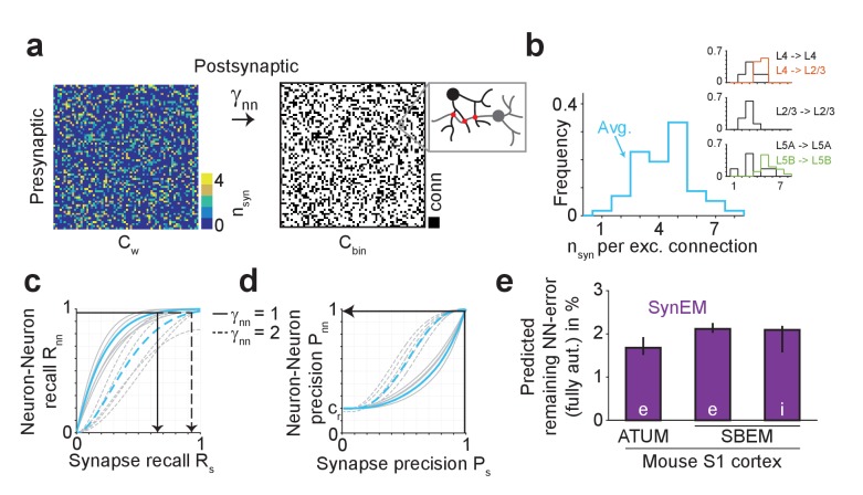
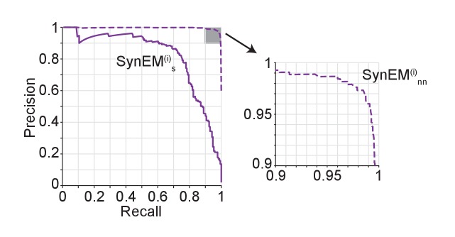
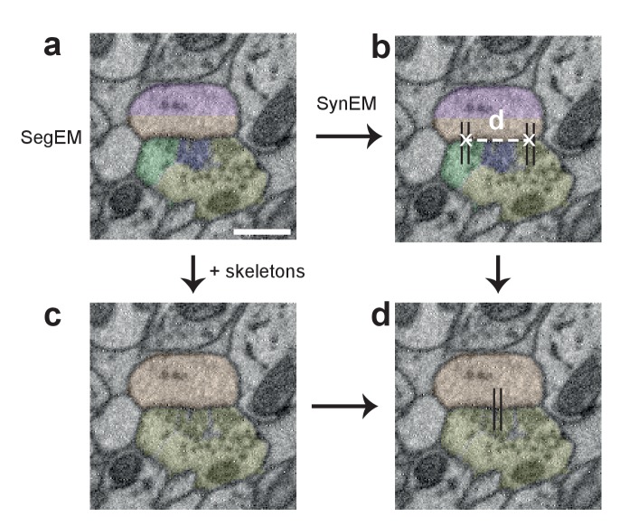
Nerve tissue contains a high density of chemical synapses, about 1 per µm3 in the mammalian cerebral cortex. Thus, even for small blocks of nerve tissue, dense connectomic mapping requires the identification of millions to billions of synapses. While the focus of connectomic data analysis has been on neurite reconstruction, synapse detection becomes limiting when datasets grow in size and dense mapping is required. Here, we report SynEM, a method for automated detection of synapses from conventionally en-bloc stained 3D electron microscopy image stacks. The approach is based on a segmentation of the image data and focuses on classifying borders between neuronal processes as synaptic or non-synaptic. SynEM yields 97% precision and recall in binary cortical connectomes with no user interaction. It scales to large volumes of cortical neuropil, plausibly even whole-brain datasets. SynEM removes the burden of manual synapse annotation for large densely mapped connectomes.
Overview
The claims and statistics of this paper haven’t been checked yet.
Run the check — free, no account needed. It reads the open-access text and checks the sources it cites.
Integrity signals
Registry
Cites
Statistics
Figures
Verification
Claims the paper makes
The claims and statistics of this paper haven’t been checked yet.
Run the check — free, no account needed. It reads the open-access text and checks the sources it cites.
Claims other papers make about this one
Each is checked against this paper’s own text: does it say what the citing paper says it does?
The source backs part of the claim, not all of it — or backs it for a different population, condition or comparison.
In the citing paper
“Several methods have been proposed for synapse detection in large EM datasets 30–35 but only a few solved the problem of partner assignments in polyadic synapses in the fly 30,34,36,37.”
From FlyWire: online community for whole-brain connectomics (2022) · cites this paper as [33]
Experimental — accuracy not yet measured
Self-check
Percentages, sums and reported differences re-computed from the paper’s own numbers.
The self-check needs the paper’s full text.
Citations
Cites
Interneurons of the neocortical inhibitory system
Markram, H., Toledo-Rodriguez, M., Wang, Y., Gupta, A., Silberberg, G., & Wu, C. (2004) · Nature Reviews Neuroscience · doi:10.1038/nrn1519 · Cited by 2,954
Serial Block-Face Scanning Electron Microscopy to Reconstruct Three-Dimensional Tissue Nanostructure
Denk, W., & Horstmann, H. (2004) · PLoS Biology · doi:10.1371/journal.pbio.0020329 · Cited by 1,644
Cortex: Statistics and Geometry of Neuronal Connectivity
Braitenberg, V., & Schüz, A. (1998) · doi:10.1007/978-3-662-03733-1 · Cited by 1,178 · cited 2×
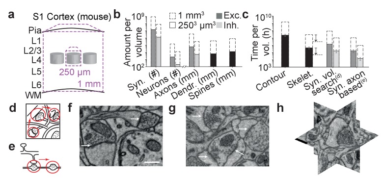
“(b) Number of synapses and neurons, total axonal, dendritic and spine path length for the example datasets in (a) (White and Peters, 1993; Braitenberg and Schüz, 1998; Merchán-Pérez et al., 2014).”
BackgroundMethodConnectomic reconstruction of the inner plexiform layer in the mouse retina
Helmstaedter, M., Briggman, K. L., Turaga, S. C., Jain, V., Seung, H. S., & Denk, W. (2013) · Nature · doi:10.1038/nature12346 · Cited by 997
Self-citationColonnier, M. (1968) · Brain Research · doi:10.1016/0006-8993(68)90234-5 · Cited by 946
Wiring specificity in the direction-selectivity circuit of the retina
Briggman, K. L., Helmstaedter, M., & Denk, W. (2011) · Nature · doi:10.1038/nature09818 · Cited by 883
Self-citationUltrastructural evidence for synaptic scaling across the wake/sleep cycle
de Vivo, L., Bellesi, M., Marshall, W., Bushong, E. A., Ellisman, M. H., Tononi, G., & Cirelli, C. (2017) · Science · doi:10.1126/science.aah5982 · Cited by 574
Feldmeyer, D., Egger, V., Lübke, J., & Sakmann, B. (1999) · The Journal of Physiology · doi:10.1111/j.1469-7793.1999.00169.x · Cited by 441
Automated synaptic connectivity inference for volume electron microscopy
Dorkenwald, S., Schubert, P. J., Killinger, M. F., Urban, G., Mikula, S., Svara, F., & Kornfeld, J. (2017) · Nature Methods · doi:10.1038/nmeth.4206 · Cited by 132
Research works printed but not linked: 40
- Bartol, 2015, Nanoconnectomic upper bound on the variability of synaptic plasticity, eLife, 4, e10778, 10.7554/eLife.10778 · DOI
- Becker, 2012, Medical Image Computing and Computer-Assisted Intervention – MICCAI 2012: 15th International Conference, Nice, France, October 1-5, 2012, Proceedings, Part I, 585, 10.1007/978-3-642-33415-3_72 · DOI
- Becker, 2013, Learning context cues for synapse segmentation, IEEE Transactions on Medical Imaging, 32, 1864, 10.1109/TMI.2013.2267747 · DOI
- Berning, 2015, SegEM: efficient image analysis for High-Resolution Connectomics, Neuron, 87, 1193, 10.1016/j.neuron.2015.09.003 · DOI
- Boergens, 2017, webKnossos: efficient online 3D data annotation for connectomics, Nature Methods, 14, 691, 10.1038/nmeth.4331 · DOI
- Eberle, 2015, High-resolution, high-throughput imaging with a multibeam scanning electron microscope, Journal of Microscopy, 259, 114, 10.1111/jmi.12224 · DOI
- Feldmeyer, 2006, Efficacy and connectivity of intracolumnar pairs of layer 2/3 pyramidal cells in the barrel cortex of juvenile rats, The Journal of Physiology, 575, 583, 10.1113/jphysiol.2006.105106 · DOI · Paper page
- Feldmeyer, 2002, Synaptic connections between layer 4 spiny neurone- layer 2/3 pyramidal cell pairs in juvenile rat barrel cortex: physiology and anatomy of interlaminar signalling within a cortical column, The Journal of Physiology, 538, 803, 10.1113/jphysiol.2001.012959 · DOI · Paper page
- Freund, 1997, A Decision-Theoretic generalization of On-Line Learning and an application to boosting, Journal of Computer and System Sciences, 55, 119, 10.1006/jcss.1997.1504 · DOI · Paper page
- Frick, 2008, Monosynaptic connections between pairs of L5A pyramidal neurons in columns of juvenile rat somatosensory cortex, Cerebral Cortex, 18, 397, 10.1093/cercor/bhm074 · DOI
- Friedman, 2000, Additive logistic regression: a statistical view of boosting, Annals of Statistics, 28, 337, 10.1214/aos/1016218223 · DOI · Paper page
- Gibson, 1999, Two networks of electrically coupled inhibitory neurons in neocortex, Nature, 402, 75, 10.1038/47035 · DOI · Paper page
- Gray, 1959, Axo-somatic and axo-dendritic synapses of the cerebral cortex: an electron microscope study, Journal of Anatomy, 93, 420
- Gupta, 2000, Organizing principles for a diversity of GABAergic interneurons and synapses in the Neocortex, Science, 287, 273, 10.1126/science.287.5451.273 · DOI · Paper page
- Hayworth, 2006, Automating the collection of Ultrathin serial sections for large volume TEM reconstructions, Microscopy and Microanalysis, 12, 86, 10.1017/S1431927606066268 · DOI
- Helmstaedter, 2011, High-accuracy neurite reconstruction for high-throughput neuroanatomy, Nature Neuroscience, 14, 1081, 10.1038/nn.2868 · DOI
- Helmstaedter, 2013, Cellular-resolution connectomics: challenges of dense neural circuit reconstruction, Nature Methods, 10, 501, 10.1038/nmeth.2476 · DOI
- Hoffmann, 2015, Synaptic Conductance estimates of the connection between Local Inhibitor Interneurons and Pyramidal Neurons in Layer 2/3 of a Cortical column, Cerebral Cortex, 25, 4415, 10.1093/cercor/bhv039 · DOI
- Hua, 2015, Large-volume en-bloc staining for electron microscopy-based connectomics, Nature Communications, 6, 7923, 10.1038/ncomms8923 · DOI
- Huang GB, Scheffer LK, Plaza SM. 2016. Fully-automatic synapse prediction and validation on a large data set. arXiv. https://arxiv.org/abs/1604.03075. · arXiv
- Jagadeesh, 2014, Synapse classification and localization in Electron Micrographs, Pattern Recognition Letters, 43, 17, 10.1016/j.patrec.2013.06.001 · DOI
- Kasthuri, 2015, Saturated reconstruction of a volume of Neocortex, Cell, 162, 648, 10.1016/j.cell.2015.06.054 · DOI · Paper page
- Knott, 2008, Serial section scanning electron microscopy of adult brain tissue using focused ion beam milling, Journal of Neuroscience, 28, 2959, 10.1523/JNEUROSCI.3189-07.2008 · DOI · Paper page
- Koelbl, 2015, A barrel-related interneuron in layer 4 of rat somatosensory cortex with a high intrabarrel connectivity, Cerebral Cortex, 25, 713, 10.1093/cercor/bht263 · DOI · Paper page
- Kreshuk, 2015, Medical Image Computing and Computer-Assisted Intervention - MICCAI 2015: 18th International Conference, Munich, Germany, October 5-9, 2015, Proceedings, Part I, 661, 10.1007/978-3-319-24553-9_81 · DOI · Paper page
- Kreshuk, 2014, Automated detection of synapses in serial section transmission electron microscopy image stacks, PLoS One, 9, e87351, 10.1371/journal.pone.0087351 · DOI
- Kreshuk, 2011, Automated detection and segmentation of synaptic contacts in nearly isotropic serial electron microscopy images, PLoS One, 6, e24899, 10.1371/journal.pone.0024899 · DOI
- Markram, 1997, Physiology and anatomy of synaptic connections between thick tufted pyramidal neurones in the developing rat neocortex, The Journal of Physiology, 500, 409, 10.1113/jphysiol.1997.sp022031 · DOI · Paper page
- Márquez Neila, 2016, A fast method for the segmentation of synaptic Junctions and Mitochondria in serial electron microscopic images of the brain, Neuroinformatics, 14, 235-50, 10.1007/s12021-015-9288-z · DOI
- Merchán-Pérez, 2014, Three-dimensional spatial distribution of synapses in the neocortex: a dual-beam electron microscopy study, Cerebral Cortex, 24, 1579, 10.1093/cercor/bht018 · DOI
- Mikula, 2012, Staining and embedding the whole mouse brain for electron microscopy, Nature Methods, 9, 1198, 10.1038/nmeth.2213 · DOI
- Mikula, 2015, High-resolution whole-brain staining for electron microscopic circuit reconstruction, Nature Methods, 12, 541, 10.1038/nmeth.3361 · DOI
- Mishchenko, 2010, Ultrastructural analysis of hippocampal neuropil from the connectomics perspective, Neuron, 67, 1009, 10.1016/j.neuron.2010.08.014 · DOI
- Pallotto, 2015, Extracellular space preservation aids the connectomic analysis of neural circuits, eLife, 4, e08206, 10.7554/eLife.08206 · DOI
- Perez, 2014, A workflow for the automatic segmentation of organelles in electron microscopy image stacks, Frontiers in Neuroanatomy, 8, 126, 10.3389/fnana.2014.00126 · DOI
- Plaza SM, Parag T, Huang GB, Olbris DJ, Saunders MA, Rivlin PK. 2014. Annotating synapses in large EM datasets. arXiv. https://arxiv.org/abs/1409.1801. · arXiv
- Roncal WG, Pekala M, Kaynig-Fittkau V, Kleissas DM, Vogelstein JT, Pfister H, Hager GD. 2015. VESICLE: Volumetric Evaluation of Synaptic Interfaces using Computer Vision at Large Scale. BMVA Press. Proceedings of the British Machine Vision Conference.
- Sommer C, Straehle C, Kothe U, Hamprecht FA. 2011. Ilastik: interactive Learning and Segmentation Toolkit. 2011 8th Ieee International Symposium on Biomedical Imaging: From Nano to Macro.
- Staffler B, Berning M, Boergens KM, Gour A, van der Smagt P, Helmstaedter M. 2017. SynEM: automated synapse detection for connectomics. bioRxiv. http://www.biorxiv.org/content/early/2017/01/22/099994.
- White, 1993, Cortical modules in the posteromedial barrel subfield (Sml) of the mouse, The Journal of Comparative Neurology, 334, 86, 10.1002/cne.903340107 · DOI
Cited by
Sorted by citations
Dense connectomic reconstruction in layer 4 of the somatosensory cortex
Motta, A., Berning, M., Boergens, K. M., Staffler, B., Beining, M., Loomba, S., Hennig, P., Wissler, H., & Helmstaedter, M. (2019) · Science · doi:10.1126/science.aay3134 · Cited by 333
Self-citationFlyWire: online community for whole-brain connectomics
Dorkenwald, S., McKellar, C. E., Macrina, T., Kemnitz, N., Lee, K., Lu, R., Wu, J., Popovych, S., Mitchell, E., Nehoran, B., et al. (2022) · Nature Methods · doi:10.1038/s41592-021-01330-0 · Cited by 289
Eckstein, N., Bates, A. S., Champion, A., Du, M., Yin, Y., Schlegel, P., Lu, A. K.-Y., Rymer, T., Finley-May, S., Paterson, T., et al. (2024) · Cell · doi:10.1016/j.cell.2024.03.016 · Cited by 277
“…et al., 2018; Lee et al., 2019; Sheridan et al., 2022; Schmidt et al., 2022; Dorkenwald et al., 2022; Svara et al., 2022) or to detect synapses (Buhmann et al., 2018; Kreshuk et al., 2015; Staffler et al., 2017; Buhmann et al., 2019; Ryan et al., 2016; Dorkenwald et al., 2017; Svara et al., 2022).”
BackgroundMethodInfluentialEckstein, N., Bates, A. S., Champion, A., Du, M., Yin, Y., Schlegel, P., Lu, A. K.-Y., Rymer, T., Finley-May, S., Paterson, T., et al. (2020) · doi:10.1101/2020.06.12.148775 · Cited by 115
Heinrich, L., Funke, J., Pape, C., Nunez-Iglesias, J., & Saalfeld, S. (2018) · Lecture Notes in Computer Science · doi:10.1007/978-3-030-00934-2_36 · Cited by 117
Metadata
- Authors
- Staffler, B., Berning, M., Boergens, K. M., Gour, A., Smagt, P. V. D., & Helmstaedter, M.
- Affiliations
- Department of Connectomics, Max Planck Institute for Brain Research, Frankfurt, Germany
- Biomimetic Robotics and Machine Learning, Munich, Germany
- Venue
- eLife
- Publisher
- eLife Sciences Publications, Ltd
- Published
- 2017-07-14
- Volume
- 6
- ISSN
- 2050-084X
- PMC
- PMC5658066
- Cited by
- 62
- References
- 9
- Language
- en
- Licence
- Funders
- Max-Planck Society (Open access funding)
About these checks
Registry, retraction, Cites and statistics results are computed, not judged, with one exception: a printed reference that no index lists is read once by a model before it counts as a citation error. Claims are extracted by a model and kept only when their quote appears verbatim in the text; each cited source is then checked, with a model’s help, for whether it says what the claim relies on. Those verdicts can be wrong — read the claim’s quote and the cited source before relying on one.
Statistics and arithmetic are recomputed, not judged.
Confidence bands are shown only for fields calibrated on our annotated test set. None is calibrated yet, so no band is shown.
What counts as a claim ›How we measure confidence ›How we check this ›
Are you an author, or do you see a problem with this page? Request a review
