Cited by 146Neurogenesis and neuroplasticity mechanismsMultiple Sclerosis Research StudiesNeuroinflammation and Neurodegeneration Mechanisms

The DOI matches its Crossref record; 76 of the 76 references it cites were found in a registry or an index.

Microglial transglutaminase-2 drives myelination and myelin repair via GPR56/ADGRG1 in oligodendrocyte precursor cells

Open access

Abstract

In the central nervous system (CNS), myelin formation and repair are regulated by oligodendrocyte (OL) lineage cells, which sense and integrate signals from their environment, including from other glial cells and the extracellular matrix (ECM). The signaling pathways that coordinate this complex communication, however, remain poorly understood. The adhesion G protein-coupled receptor ADGRG1 (also known as GPR56) is an evolutionarily conserved regulator of OL development in humans, mice, and zebrafish, although its activating ligand for OL lineage cells is unknown. Here, we report that microglia-derived transglutaminase-2 (TG2) signals to ADGRG1 on OL precursor cells (OPCs) in the presence of the ECM protein laminin and that TG2/laminin-dependent activation of ADGRG1 promotes OPC proliferation. Signaling by TG2/laminin to ADGRG1 on OPCs additionally improves remyelination in two murine models of demyelination. These findings identify a novel glia-to-glia signaling pathway that promotes myelin formation and repair, and suggest new strategies to enhance remyelination.

Overview

The claims and statistics of this paper haven’t been checked yet.

Run the check — free, no account needed. It reads the open-access text and checks the sources it cites.

A few free checks a day · papers already verified open instantly · results are public

Integrity signals

Registry

Crossref · 10.7554/elife.33385

Cites

76 of 76 references verified0 citation errors · 0 not checkable

Statistics

Figures

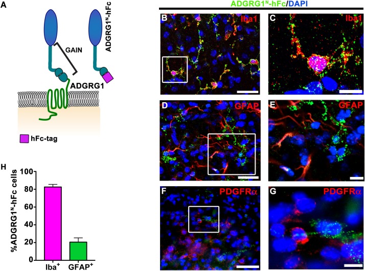
Figure 1. Microglia express the putative ligand of ADGRG1. (A) Schema of ADGRG1 receptor. GAIN domain and ADGRG1N-hFc are shown. (B, D, and F) Double labeling of ADGRG1N-hFc (green) and Iba1 (B, red), GFAP (D, red), PDGFRα (F, red) in P5 wt corpus callosum. DAPI, blue. Scale bar, 25 µm. (C, E, and G) Higher magnification of the boxed region in (B, D and F). Scale bar, 10 µm. Staining was repeated N = 3–4 animals. (H) Quantification of double positive ADGRG1N-hFc+ and Iba+ cells and ADGRG1N-hFc+ and GFAP+ cells. N = 3–4 per staining.
Figure 1. Microglia express the putative ligand of ADGRG1. (A) Schema of ADGRG1 receptor. GAIN domain and ADGRG1N-hFc are shown. (B, D, and F) Double labeling of ADGRG1N-hFc (green) and Iba1 (B, red), GFAP (D, red), PDGFRα (F, red) in P5 wt corpus callosum. DAPI, blue. Scale bar, 25 µm. (C, E, and G) Higher magnification of the boxed region in (B, D and F). Scale bar, 10 µm. Staining was repeated N = 3–4 animals. (H) Quantification of double positive ADGRG1N-hFc+ and Iba+ cells and ADGRG1N-hFc+ and GFAP+ cells. N = 3–4 per staining.CC BY · PubMed Central
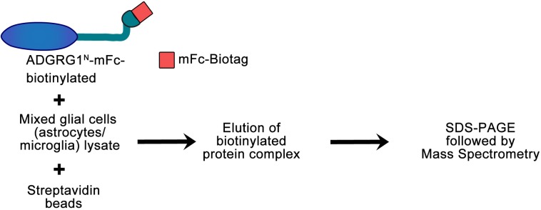
Figure 1—figure supplement 1. Flow chart of the in vitro biotinylation/proteomic approach. Biotinylated ADGRG1N-mFc was used to purify ADGRG1 binding partner(s) from mixed glial cell (enriched in microglia and astrocytes) lysate. Protein complexes were subjected to SDS PAGE and tryptic peptide mass spectrometry protein identification after one-step purification with immobilized streptavidin. The whole lane was excised and subjected to tryptic peptide mass spectrometry (MS) protein identification analysis.
Figure 1—figure supplement 1. Flow chart of the in vitro biotinylation/proteomic approach. Biotinylated ADGRG1N-mFc was used to purify ADGRG1 binding partner(s) from mixed glial cell (enriched in microglia and astrocytes) lysate. Protein complexes were subjected to SDS PAGE and tryptic peptide mass spectrometry protein identification after one-step purification with immobilized streptavidin. The whole lane was excised and subjected to tryptic peptide mass spectrometry (MS) protein identification analysis.CC BY · PubMed Central
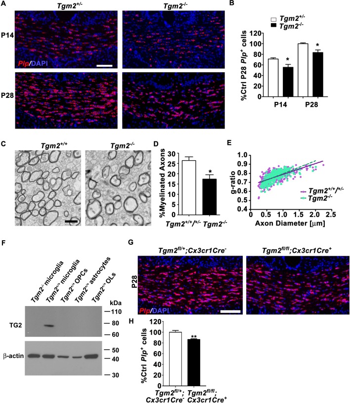
Figure 2. Loss of Tgm2 leads to fewer mature OLs and hypomyelination in the CC. (A) Representative in situ hybridization (ISH) images of Plp (red) in the CC of P14 (top panel) and P28 (bottom panel) Tgm2+/- and Tgm2-/- mice. DAPI, blue. Scale bar, 100 µm. (B) Quantification of Plp+ oligodendrocytes from the CC of Tgm2+/- and Tgm2-/- P14 and P28 mice. * P=0.0484; N = 5 per genotype (P14); *p=0.0378; N = 4 per genotype (P28), unpaired t-test. (C) Representative TEM images from P28 CC of Tgm2+/+ and Tgm2-/- mice. Scale bar, 1 µm. (D) Percentage of myelinated axons in the CC of control (Tgm2+/+ and Tgm2+/-) and Tgm2-/- mice. *p=0.0299; N = 3 per genotype; unpaired t-test. (E) Scatter plot displaying g-ratio values in the CC of control and Tgm2-/- mice. (F) TG2 western blot on acutely isolated microglia, OPCs, astrocytes, and mature oligodendrocytes from the P7 neonatal brain. β-actin was used as loading control. (G) Representative ISH images of Plp (red) in the CC of P28 Tgm2fl/+;Cx3cr1Cre- and Tgm2fl/fl;Cx3cr1Cre + mice. DAPI, blue. Scale bar, 100 µm. (H) Quantification of Plp+ oligodendrocytes in the CC of P28 Tgm2fl/+;Cx3cr1Cre- and Tgm2fl/fl;Cx3cr1Cre + mice. **p=0.00850; N = 4 per genotype, unpaired t-test. Error bars are means ± s.e.m (B, D, H).
Figure 2. Loss of Tgm2 leads to fewer mature OLs and hypomyelination in the CC. (A) Representative in situ hybridization (ISH) images of Plp (red) in the CC of P14 (top panel) and P28 (bottom panel) Tgm2+/- and Tgm2-/- mice. DAPI, blue. Scale bar, 100 µm. (B) Quantification of Plp+ oligodendrocytes from the CC of Tgm2+/- and Tgm2-/- P14 and P28 mice. * P=0.0484; N = 5 per genotype (P14); *p=0.0378; N = 4 per genotype (P28), unpaired t-test. (C) Representative TEM images from P28 CC of Tgm2+/+ and Tgm2-/- mice. Scale bar, 1 µm. (D) Percentage of myelinated axons in the CC of control (Tgm2+/+ and Tgm2+/-) and Tgm2-/- mice. *p=0.0299; N = 3 per genotype; unpaired t-test. (E) Scatter plot displaying g-ratio values in the CC of control and Tgm2-/- mice. (F) TG2 western blot on acutely isolated microglia, OPCs, astrocytes, and mature oligodendrocytes from the P7 neonatal brain. β-actin was used as loading control. (G) Representative ISH images of Plp (red) in the CC of P28 Tgm2fl/+;Cx3cr1Cre- and Tgm2fl/fl;Cx3cr1Cre + mice. DAPI, blue. Scale bar, 100 µm. (H) Quantification of Plp+ oligodendrocytes in the CC of P28 Tgm2fl/+;Cx3cr1Cre- and Tgm2fl/fl;Cx3cr1Cre + mice. **p=0.00850; N = 4 per genotype, unpaired t-test. Error bars are means ± s.e.m (B, D, H).CC BY · PubMed Central
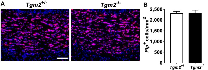
Figure 2—figure supplement 1. Oligodendrocyte number recovers by 5 month in Tgm2 knockout mice. (A) Representative images of ISH of Plp in the CC of 4–5 month old Tgm2+/- and Tgm2-/- mice. Scale bar, 100 µm. DAPI, blue. (B) Quantification of Plp+ oligodendrocytes in the CC reveals similar numbers of OLs in the CC of Tgm2+/- and Tgm2-/- mice. * P=0.807, unpaired t-test, N = 3 per genotype. Error bars are means ± s.e.m.
Figure 2—figure supplement 1. Oligodendrocyte number recovers by 5 month in Tgm2 knockout mice. (A) Representative images of ISH of Plp in the CC of 4–5 month old Tgm2+/- and Tgm2-/- mice. Scale bar, 100 µm. DAPI, blue. (B) Quantification of Plp+ oligodendrocytes in the CC reveals similar numbers of OLs in the CC of Tgm2+/- and Tgm2-/- mice. * P=0.807, unpaired t-test, N = 3 per genotype. Error bars are means ± s.e.m.CC BY · PubMed Central
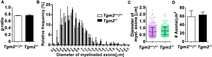
Figure 2—figure supplement 2. Loss of Tgm2 does not affect g-ratio, myelinated axon distribution, axon diameter or axon number. (A) Average g-ratio of myelin sheath thickness was comparable in control and Tgm2-/- mice. p=0.728, unpaired t-test, N = 3 per genotype. (B) The distribution of myelinated axons with respect to the axon diameter in the CC of control and Tgm2-/- mice. p=0.8291, two-way ANOVA, p=0.2524, Gaussian non-linear curve fit, N = 3 per genotype. (C) Axon diameter in the CC is comparable in control and Tgm2-/- mice. Mean ± s.d.; p=0.8771; unpaired t-test; N = 3 per genotype. (D) Number of total axons (myelinated and unmyelinated) is comparable in the CC of in control and Tgm2-/- mice at P28. p=0.7947, unpaired t-test, N = 3 per genotype. Error bars are means ± s.e.m (A–D).
Figure 2—figure supplement 2. Loss of Tgm2 does not affect g-ratio, myelinated axon distribution, axon diameter or axon number. (A) Average g-ratio of myelin sheath thickness was comparable in control and Tgm2-/- mice. p=0.728, unpaired t-test, N = 3 per genotype. (B) The distribution of myelinated axons with respect to the axon diameter in the CC of control and Tgm2-/- mice. p=0.8291, two-way ANOVA, p=0.2524, Gaussian non-linear curve fit, N = 3 per genotype. (C) Axon diameter in the CC is comparable in control and Tgm2-/- mice. Mean ± s.d.; p=0.8771; unpaired t-test; N = 3 per genotype. (D) Number of total axons (myelinated and unmyelinated) is comparable in the CC of in control and Tgm2-/- mice at P28. p=0.7947, unpaired t-test, N = 3 per genotype. Error bars are means ± s.e.m (A–D).CC BY · PubMed Central
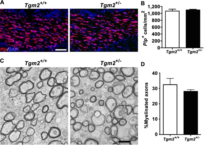
Figure 2—figure supplement 3. Deleting one allele of Tgm2 has no effect on the number of Plp+ + and myelinated axons. (A) Representative images of ISH of Plp on P28 in the CC in Tgm2+/+ and Tgm2+/- 100 µm. Scale bar, 100 µm. DAPI, blue. (B) Quantification of Plp+ oligodendrocytes in the CC reveals similar numbers of OLs in the CC of Tgm2+/+ and Tgm2+/- mice. *p=0.6005, unpaired t-test, N = 3 per genotype. (C) Representative TEM images from P28 CC of Tgm2+/+ and Tgm2+/- mice. Scale bar, 1 µm. (D) Percentage of myelinated axons was quantified in the CC in Tgm2+/+ and Tgm2+/- mice. p=0.5129, unpaired t-test, N = 3 per genotype. Error bars are means ± s.e.m (B, D).
Figure 2—figure supplement 3. Deleting one allele of Tgm2 has no effect on the number of Plp+ + and myelinated axons. (A) Representative images of ISH of Plp on P28 in the CC in Tgm2+/+ and Tgm2+/- 100 µm. Scale bar, 100 µm. DAPI, blue. (B) Quantification of Plp+ oligodendrocytes in the CC reveals similar numbers of OLs in the CC of Tgm2+/+ and Tgm2+/- mice. *p=0.6005, unpaired t-test, N = 3 per genotype. (C) Representative TEM images from P28 CC of Tgm2+/+ and Tgm2+/- mice. Scale bar, 1 µm. (D) Percentage of myelinated axons was quantified in the CC in Tgm2+/+ and Tgm2+/- mice. p=0.5129, unpaired t-test, N = 3 per genotype. Error bars are means ± s.e.m (B, D).CC BY · PubMed Central
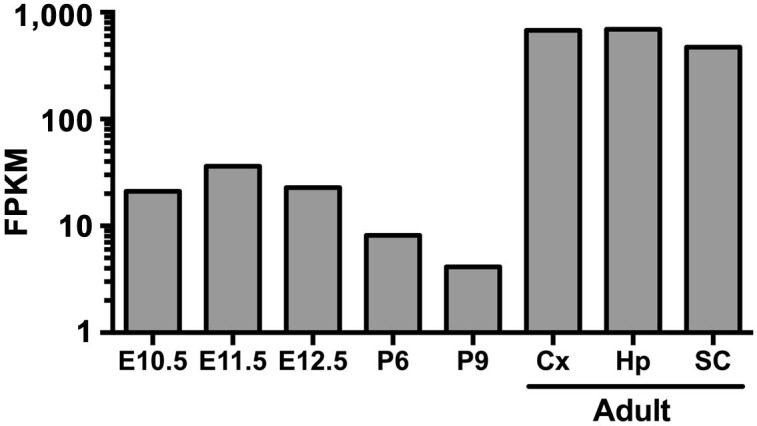
Figure 2—figure supplement 4. Developmental expression of Tgm2. Developmental expression of Tgm2 in murine microglia purified from whole brains (E10.5 through P) or brain regions (adult) at the indicated embryonic and postnatal ages. RNA-sequencing data was extracted from public database of (Matcovitch-Natan et al., 2016). Cx: cortex, Hp: hippocampus, SC: spinal cord.
Figure 2—figure supplement 4. Developmental expression of Tgm2. Developmental expression of Tgm2 in murine microglia purified from whole brains (E10.5 through P) or brain regions (adult) at the indicated embryonic and postnatal ages. RNA-sequencing data was extracted from public database of (Matcovitch-Natan et al., 2016). Cx: cortex, Hp: hippocampus, SC: spinal cord.CC BY · PubMed Central
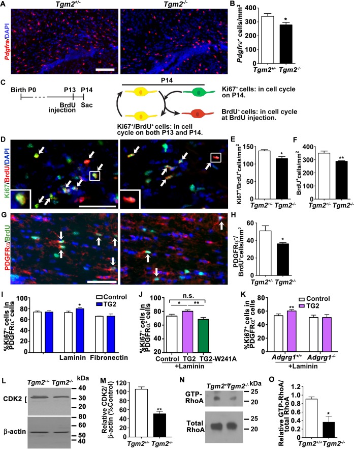
Figure 3. TG2 regulates OPC proliferation through ADGRG1. (A) Representative ISH images of Pdgfra (red) in the CC of P14 Tgm2+/- and Tgm2-/- mice. DAPI, blue. Scale bar, 100 µm. (B) Quantification of Pdgfra+ OPCs in the CC of control and Tgm2-/- mice. *p=0.0320, unpaired t-test, N = 6 per genotype. (C) Cartoon showing the cell cycle exit assay. (D) Representative images of double IHC of BrdU (red) and Ki67 (green) in the CC of P14 Tgm2+/- and Tgm2-/- mice that were pulsed with BrdU 24 hr earlier (arrows mark double positive Ki67+/BrdU+ cells). DAPI, blue. Scale bar, 50 µm. (E) The density of Ki67+/BrdU+ cells in the CC is lower in Tgm2-/- mice. *p=0.0207, unpaired t-test, N = 5 per genotype. (F) The density of BrdU+ cells in the CC is reduced in Tgm2-/- mice. **p=0.0099, unpaired t-test, N = 5 per genotype. (G) Representative images of double IHC of PDGFRα (red) and BrdU (green) in the CC of P14 Tgm2+/- and Tgm2-/- mice (arrows mark double positive PDGFRα+/BrdU+cells). DAPI, blue. Scale bar, 50 µm. (H) The number of BrdU+/PDGFRα+ cells in the CC is reduced in Tgm2-/- mice. *p=0.0465, unpaired t-test, N = 4 per genotype. Error bars are means ± s.e.m. (I) The effect of recombinant TG2 (rTG2) on OPC proliferation in basal condition, laminin-111 or fibronectin. rTG2 stimulates OPC proliferation only in the presence of laminin-111. *p=0.039, paired t-test, N = 3–6 per group. (J) The effect of wild type TG2 and its enzymatic dead mutant TG2-W241A proteins on OPC proliferation. Only wild type TG2 stimulates OPC proliferation. **p=0.0091; One-way ANOVA followed by Tukey post-hoc test, F(2,14) = 6.7, *p=0.05 (control vs. rTG2); n.s. p=0.70 (control vs. TG2-W241A); **p=0.0088 (TG2 vs. TG2-W241A); (K) Adgrg1-/- OPCs fail to respond to rTG2-enhanced proliferation. *p=0.0063, paired t-test, N = 5 per genotype. (L) Western blot analyses of CDK2 in acutely isolated OPCs from P7 Tgm2+/- and Tgm2-/- brains. The bracket indicates CDK2 protein isoform bands. β-actin was used as loading control. (M) CDK2 protein levels are reduced in the Tgm2-/- mice. ±=0.002, unpaired t-test, N = 3 per genotype. (N) Western blot of active RhoA (top panel) and total RhoA (bottom panel) in the CC of Tgm2+/+ and Tgm2-/- mice. (O) The relative level of active RhoA to total RhoA was diminished in the CC of Tgm2-/- mice compared to Tgm2+/+ control mice. *p=0.0207, unpaired t-test, N = 3 per genotype. Error bars are means ± s.e.m (B, E, F, H, I, J, K, M, O).
Figure 3. TG2 regulates OPC proliferation through ADGRG1. (A) Representative ISH images of Pdgfra (red) in the CC of P14 Tgm2+/- and Tgm2-/- mice. DAPI, blue. Scale bar, 100 µm. (B) Quantification of Pdgfra+ OPCs in the CC of control and Tgm2-/- mice. *p=0.0320, unpaired t-test, N = 6 per genotype. (C) Cartoon showing the cell cycle exit assay. (D) Representative images of double IHC of BrdU (red) and Ki67 (green) in the CC of P14 Tgm2+/- and Tgm2-/- mice that were pulsed with BrdU 24 hr earlier (arrows mark double positive Ki67+/BrdU+ cells). DAPI, blue. Scale bar, 50 µm. (E) The density of Ki67+/BrdU+ cells in the CC is lower in Tgm2-/- mice. *p=0.0207, unpaired t-test, N = 5 per genotype. (F) The density of BrdU+ cells in the CC is reduced in Tgm2-/- mice. **p=0.0099, unpaired t-test, N = 5 per genotype. (G) Representative images of double IHC of PDGFRα (red) and BrdU (green) in the CC of P14 Tgm2+/- and Tgm2-/- mice (arrows mark double positive PDGFRα+/BrdU+cells). DAPI, blue. Scale bar, 50 µm. (H) The number of BrdU+/PDGFRα+ cells in the CC is reduced in Tgm2-/- mice. *p=0.0465, unpaired t-test, N = 4 per genotype. Error bars are means ± s.e.m. (I) The effect of recombinant TG2 (rTG2) on OPC proliferation in basal condition, laminin-111 or fibronectin. rTG2 stimulates OPC proliferation only in the presence of laminin-111. *p=0.039, paired t-test, N = 3–6 per group. (J) The effect of wild type TG2 and its enzymatic dead mutant TG2-W241A proteins on OPC proliferation. Only wild type TG2 stimulates OPC proliferation. **p=0.0091; One-way ANOVA followed by Tukey post-hoc test, F(2,14) = 6.7, *p=0.05 (control vs. rTG2); n.s. p=0.70 (control vs. TG2-W241A); **p=0.0088 (TG2 vs. TG2-W241A); (K) Adgrg1-/- OPCs fail to respond to rTG2-enhanced proliferation. *p=0.0063, paired t-test, N = 5 per genotype. (L) Western blot analyses of CDK2 in acutely isolated OPCs from P7 Tgm2+/- and Tgm2-/- brains. The bracket indicates CDK2 protein isoform bands. β-actin was used as loading control. (M) CDK2 protein levels are reduced in the Tgm2-/- mice. ±=0.002, unpaired t-test, N = 3 per genotype. (N) Western blot of active RhoA (top panel) and total RhoA (bottom panel) in the CC of Tgm2+/+ and Tgm2-/- mice. (O) The relative level of active RhoA to total RhoA was diminished in the CC of Tgm2-/- mice compared to Tgm2+/+ control mice. *p=0.0207, unpaired t-test, N = 3 per genotype. Error bars are means ± s.e.m (B, E, F, H, I, J, K, M, O).CC BY · PubMed Central
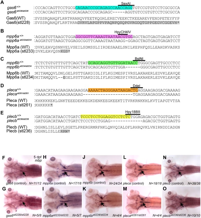
Figure 3—figure supplement 1. Mutations in gas6, mpp6, and plec do not affect CNS Mbp expression. (A–E) CRISPR-Cas9 technology was used to generate mutations in zebrafish gas6 (A), mpp6a (B), mpp6b (C), pleca (D), and plecb (E). (A) A CRISPR/Cas9 system guide RNA (gRNA) designed to disrupt exon 2 (targeted sequence highlighted blue and SexAI restriction enzyme site used for genotyping denoted) of gas6 generated gas6stl228, a 4 bp deletion predicted to result in a frameshift and premature STOP. Alignments of wt and mutant nucleotide sequences (top), and predicted wt (706 aa) and mutant (78 aa) protein sequences (bottom) shown. (B) A gRNA designed to disrupt the 3rd coding exon 5 (targeted sequence highlighted purple, and HpyCH4IV restriction enzyme site used for genotyping denoted) of mpp6a generated mpp6astl233, a 1 bp deletion predicted to result in a frameshift and premature STOP. Alignments of wt and mutant nucleotide sequences (top), and predicted wt (550 aa) and mutant (69 aa) protein sequences (bottom) shown. (C) A gRNA designed to disrupt exon 2 (targeted sequence highlighted green and BstNI restriction enzyme site used for genotyping denoted) of mpp6b generated mpp6bstl234, a 6 bp deletion predicted to result in a two amino acid deletion and a single amino acid change within the receptor targeting domain. Alignments of wt and mutant nucleotide sequences (top), and predicted wt (539 aa) and mutant (537 aa) protein sequences (bottom) shown. (D) A gRNA designed to disrupt exon 3 (targeted sequence highlighted orange, and DdeI restriction enzyme site used for genotyping denoted) of pleca generated plecastl261, a 20 bp deletion predicted to result in a frameshift and premature STOP. Alignments of wt and mutant nucleotide sequences (top), and predicted wt (4752 aa) and mutant (103 aa) protein sequences (bottom) shown. (E) A gRNA designed to disrupt the 3rd coding exon (targeted sequence highlighted yellow, and Hpy188III restriction enzyme site used for genotyping denoted) of plecb generated plecbstl236, a 13 bp deletion predicted to result in a frameshift and premature STOP. Alignments of wt and mutant nucleotide sequences (top), and predicted wt (4530 aa) and mutant (111 aa) protein sequences (bottom) shown. (F– O) Whole mount ISH showing mbp expression (CNS denoted by black arrows) at 5 days post-fertilization (dpf) in (F) gas6 control larva (wt and gas6stl228/+, N = 11/12), (G) gasstl228/stl228 mutant larva (N = 5/8), (H) mpp6a control larva (wt and mpp6astl233/+, N = 17/18), (I) mpp6astl233/stl233 mutant larva (N = 5/7), (J) mpp6b control larva (wt and mpp6bstl234/+, N = 24/24), (K) mpp6bstl234/stl234 (N = 4/4), (L) pleca control larva (wt and plecastl261/+, N = 18/18), (M) plecastl261/stl261mutant larva (N = 4/4), (N) plecb control larva (wt and plecbstl236/+, N = 36/38), and (O) plecbstl236/st’236 mutant larva (N = 10/10). Dorsal views are shown for all, anterior to the left. No gross changes in mbp expression were observed in putative ligand mutants relative to sibling controls.
Figure 3—figure supplement 1. Mutations in gas6, mpp6, and plec do not affect CNS Mbp expression. (A–E) CRISPR-Cas9 technology was used to generate mutations in zebrafish gas6 (A), mpp6a (B), mpp6b (C), pleca (D), and plecb (E). (A) A CRISPR/Cas9 system guide RNA (gRNA) designed to disrupt exon 2 (targeted sequence highlighted blue and SexAI restriction enzyme site used for genotyping denoted) of gas6 generated gas6stl228, a 4 bp deletion predicted to result in a frameshift and premature STOP. Alignments of wt and mutant nucleotide sequences (top), and predicted wt (706 aa) and mutant (78 aa) protein sequences (bottom) shown. (B) A gRNA designed to disrupt the 3rd coding exon 5 (targeted sequence highlighted purple, and HpyCH4IV restriction enzyme site used for genotyping denoted) of mpp6a generated mpp6astl233, a 1 bp deletion predicted to result in a frameshift and premature STOP. Alignments of wt and mutant nucleotide sequences (top), and predicted wt (550 aa) and mutant (69 aa) protein sequences (bottom) shown. (C) A gRNA designed to disrupt exon 2 (targeted sequence highlighted green and BstNI restriction enzyme site used for genotyping denoted) of mpp6b generated mpp6bstl234, a 6 bp deletion predicted to result in a two amino acid deletion and a single amino acid change within the receptor targeting domain. Alignments of wt and mutant nucleotide sequences (top), and predicted wt (539 aa) and mutant (537 aa) protein sequences (bottom) shown. (D) A gRNA designed to disrupt exon 3 (targeted sequence highlighted orange, and DdeI restriction enzyme site used for genotyping denoted) of pleca generated plecastl261, a 20 bp deletion predicted to result in a frameshift and premature STOP. Alignments of wt and mutant nucleotide sequences (top), and predicted wt (4752 aa) and mutant (103 aa) protein sequences (bottom) shown. (E) A gRNA designed to disrupt the 3rd coding exon (targeted sequence highlighted yellow, and Hpy188III restriction enzyme site used for genotyping denoted) of plecb generated plecbstl236, a 13 bp deletion predicted to result in a frameshift and premature STOP. Alignments of wt and mutant nucleotide sequences (top), and predicted wt (4530 aa) and mutant (111 aa) protein sequences (bottom) shown. (F– O) Whole mount ISH showing mbp expression (CNS denoted by black arrows) at 5 days post-fertilization (dpf) in (F) gas6 control larva (wt and gas6stl228/+, N = 11/12), (G) gasstl228/stl228 mutant larva (N = 5/8), (H) mpp6a control larva (wt and mpp6astl233/+, N = 17/18), (I) mpp6astl233/stl233 mutant larva (N = 5/7), (J) mpp6b control larva (wt and mpp6bstl234/+, N = 24/24), (K) mpp6bstl234/stl234 (N = 4/4), (L) pleca control larva (wt and plecastl261/+, N = 18/18), (M) plecastl261/stl261mutant larva (N = 4/4), (N) plecb control larva (wt and plecbstl236/+, N = 36/38), and (O) plecbstl236/st’236 mutant larva (N = 10/10). Dorsal views are shown for all, anterior to the left. No gross changes in mbp expression were observed in putative ligand mutants relative to sibling controls.CC BY · PubMed Central
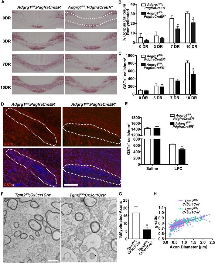
Figure 4. ADGRG1 is required for remyelination in vivo. (A) Representative images of Black-Gold myelin staining of the corpus callosum of Adgrg1fl/fl;PdgfraCreER- and Adgrg1fl/fl;PdgfraCreER+ + after cuprizone feeding for 6 weeks followed by recovery for 3 d (3 DR), 7 d (7 DR) and 10 d (10 DR) (dotted line outlines quantified region). Scale bar, 250 µm. (B) Percentage of remyelinated corpus callosum displayed significant decrease in myelination at 7 DR and 10 DR between Adgrg1fl/fl;PdgfraCreER- and Adgrg1fl/fl;PdgfraCreER+ +. *p=0.0285 (7 DR), N = 4 (cre-); N = 4 (cre+); *p=0.0416 (10 DR), N = 3 (cre-); N = 4 (cre+), unpaired t-test. (C) Number of GSTπ+ OLs are significantly decreased at 10 DR between Adgrg1fl/fl;PdgfraCreER- and Adgrg1fl/fl;PdgfraCreER+ +. *p=0.0457 (10 DR), N = 3 (cre-); N = 3 (cre+), unpaired t-test. (D) Representative images of GSTπ+ OLs in the corpus callosum of Adgrg1fl/fl;PdgfraCreER- and Adgrg1fl/fl;PdgfraCreER+ + 14 days post-lesion (dotted line outlines quantified region). Scale bar, 200 µm. (E) Number of GSTπ+ OLs are significantly decreased 14 days post-lesion between Adgrg1fl/fl;PdgfraCreER- and Adgrg1fl/fl;PdgfraCreER+ +. *p=0.0133, N = 3 (cre-); N = 3 (cre+), unpaired t-test. (F) Representative TEM images from the CC of Tgm2fl/fl;Cx3cr1Cre- and Tgm2fl/fl;Cx3cr1Cre + mice. Scale bar, 1 µm. (D) Percentage of myelinated axons in the CC of Tgm2fl/fl;Cx3cr1Cre- and Tgm2fl/fl;Cx3cr1Cre + mice. *p=0.0493; N = 4 per genotype; unpaired t-test. (E) Scatter plot displaying g-ratio values in the CC of Tgm2fl/fl;Cx3cr1Cre- and Tgm2fl/fl;Cx3cr1Cre + mice.
Figure 4. ADGRG1 is required for remyelination in vivo. (A) Representative images of Black-Gold myelin staining of the corpus callosum of Adgrg1fl/fl;PdgfraCreER- and Adgrg1fl/fl;PdgfraCreER+ + after cuprizone feeding for 6 weeks followed by recovery for 3 d (3 DR), 7 d (7 DR) and 10 d (10 DR) (dotted line outlines quantified region). Scale bar, 250 µm. (B) Percentage of remyelinated corpus callosum displayed significant decrease in myelination at 7 DR and 10 DR between Adgrg1fl/fl;PdgfraCreER- and Adgrg1fl/fl;PdgfraCreER+ +. *p=0.0285 (7 DR), N = 4 (cre-); N = 4 (cre+); *p=0.0416 (10 DR), N = 3 (cre-); N = 4 (cre+), unpaired t-test. (C) Number of GSTπ+ OLs are significantly decreased at 10 DR between Adgrg1fl/fl;PdgfraCreER- and Adgrg1fl/fl;PdgfraCreER+ +. *p=0.0457 (10 DR), N = 3 (cre-); N = 3 (cre+), unpaired t-test. (D) Representative images of GSTπ+ OLs in the corpus callosum of Adgrg1fl/fl;PdgfraCreER- and Adgrg1fl/fl;PdgfraCreER+ + 14 days post-lesion (dotted line outlines quantified region). Scale bar, 200 µm. (E) Number of GSTπ+ OLs are significantly decreased 14 days post-lesion between Adgrg1fl/fl;PdgfraCreER- and Adgrg1fl/fl;PdgfraCreER+ +. *p=0.0133, N = 3 (cre-); N = 3 (cre+), unpaired t-test. (F) Representative TEM images from the CC of Tgm2fl/fl;Cx3cr1Cre- and Tgm2fl/fl;Cx3cr1Cre + mice. Scale bar, 1 µm. (D) Percentage of myelinated axons in the CC of Tgm2fl/fl;Cx3cr1Cre- and Tgm2fl/fl;Cx3cr1Cre + mice. *p=0.0493; N = 4 per genotype; unpaired t-test. (E) Scatter plot displaying g-ratio values in the CC of Tgm2fl/fl;Cx3cr1Cre- and Tgm2fl/fl;Cx3cr1Cre + mice.CC BY · PubMed Central
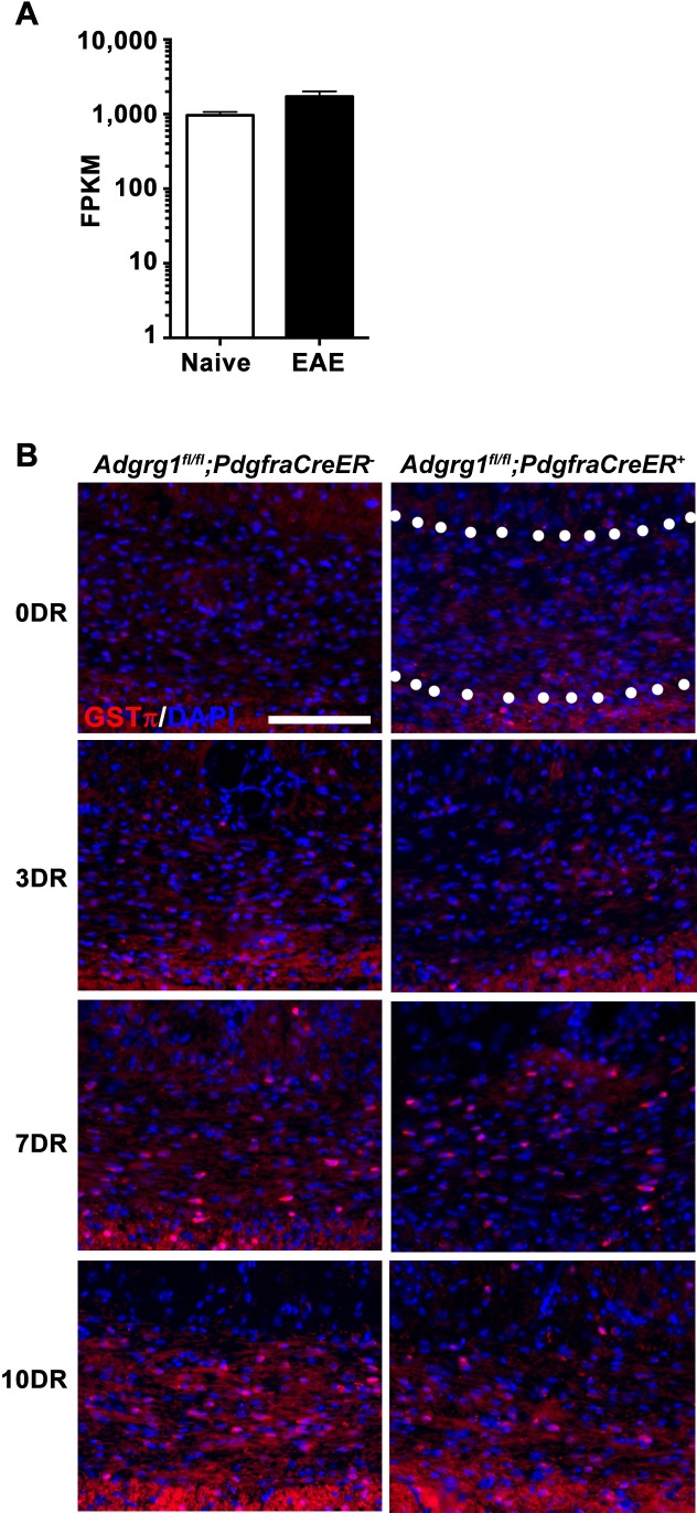
Figure 4—figure supplement 1. Loss of Adgrg1 in the OL lineage leads to reduced numbers of myelinating OLs. (A) Expression of Tgm2 in murine microglia purified from naive or EAE brains. RNA-sequencing data was extracted from public database of (Wlodarczyk et al., 2017). (B) Representative images of GSTπ+ OLs of the corpus callosum of Adgrg1fl/fl;PdgfraCreER- and Adgrg1fl/fl;PdgfraCreER + mice after cuprizone feeding for 6 weeks followed by recovery for 3 d (3 DR), 7 d (7 DR) and 10 d (10 DR) (dotted line outlines quantified region). Scale bar, 100 µm.
Figure 4—figure supplement 1. Loss of Adgrg1 in the OL lineage leads to reduced numbers of myelinating OLs. (A) Expression of Tgm2 in murine microglia purified from naive or EAE brains. RNA-sequencing data was extracted from public database of (Wlodarczyk et al., 2017). (B) Representative images of GSTπ+ OLs of the corpus callosum of Adgrg1fl/fl;PdgfraCreER- and Adgrg1fl/fl;PdgfraCreER + mice after cuprizone feeding for 6 weeks followed by recovery for 3 d (3 DR), 7 d (7 DR) and 10 d (10 DR) (dotted line outlines quantified region). Scale bar, 100 µm.CC BY · PubMed Central
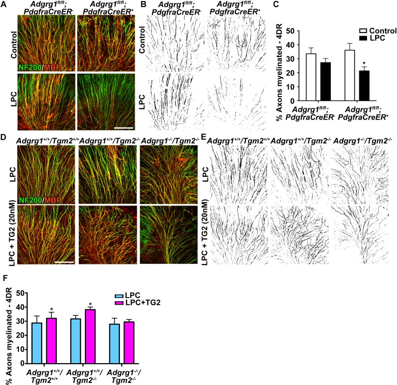
Figure 5. ADGRG1 and its ligand TG2 are required for remyelination. (A) Representative images of cerebellar slice cultures from P10 Adgrg1fl/fl;PdgfraCreER- and Adgrg1fl/fl;PdgfraCreER+ + cerebella that were demyelinated with LPC for 24 hr followed by 4 days of recovery (4DR). Slices were labeled with NF200 (green) and MBP (red); myelinated fibers appear yellow in merged images. Scale bar, 100 µm. (B) Composite images created in Image J show myelinated axons (black) for quantification. (C) Percentage of myelinated axons after remyelination. Remyelination is reduced in cerebellar slices that lack OPC-derived ADGRG1. *p=0.0135, paired t-test, N = 5 per genotype. (D) Representative images of cerebellar slice cultures from P10 Adgrg1+/+;Tgm2+/+, Adgrg1+/+;Tgm2-/- and Adgrg1-/-;Tgm2-/- mouse cerebella that were demyelinated with LPC for 24 hr followed by 4 days of remyelination in the presence or absence of rTG2 (20 nM). Slices were immunostained with NF200 (green) and MBP (red); myelinated fibers appear yellow in merged images. Scale bar, 100 µm. (E) Composite images created in Image J show myelinated axons (black) for quantification. (F) Percentage of myelinated axons. Recombinant TG2 promotes remyelination in Adgrg1+/+;Tgm2+/+ and Adgrg1+/+;Tgm2-/-, but not Adgrg1-/-;Tgm2-/- cerebellar slices. *p=0.0475 (Adgrg1+/+;Tgm2+/+), *p=0.0197 (Adgrg1+/+;Tgm2-/-), paired t-test, N = 5–6 per genotype. Error bars are mean ± s.e.m (C, F)..
Figure 5. ADGRG1 and its ligand TG2 are required for remyelination. (A) Representative images of cerebellar slice cultures from P10 Adgrg1fl/fl;PdgfraCreER- and Adgrg1fl/fl;PdgfraCreER+ + cerebella that were demyelinated with LPC for 24 hr followed by 4 days of recovery (4DR). Slices were labeled with NF200 (green) and MBP (red); myelinated fibers appear yellow in merged images. Scale bar, 100 µm. (B) Composite images created in Image J show myelinated axons (black) for quantification. (C) Percentage of myelinated axons after remyelination. Remyelination is reduced in cerebellar slices that lack OPC-derived ADGRG1. *p=0.0135, paired t-test, N = 5 per genotype. (D) Representative images of cerebellar slice cultures from P10 Adgrg1+/+;Tgm2+/+, Adgrg1+/+;Tgm2-/- and Adgrg1-/-;Tgm2-/- mouse cerebella that were demyelinated with LPC for 24 hr followed by 4 days of remyelination in the presence or absence of rTG2 (20 nM). Slices were immunostained with NF200 (green) and MBP (red); myelinated fibers appear yellow in merged images. Scale bar, 100 µm. (E) Composite images created in Image J show myelinated axons (black) for quantification. (F) Percentage of myelinated axons. Recombinant TG2 promotes remyelination in Adgrg1+/+;Tgm2+/+ and Adgrg1+/+;Tgm2-/-, but not Adgrg1-/-;Tgm2-/- cerebellar slices. *p=0.0475 (Adgrg1+/+;Tgm2+/+), *p=0.0197 (Adgrg1+/+;Tgm2-/-), paired t-test, N = 5–6 per genotype. Error bars are mean ± s.e.m (C, F)..CC BY · PubMed Central
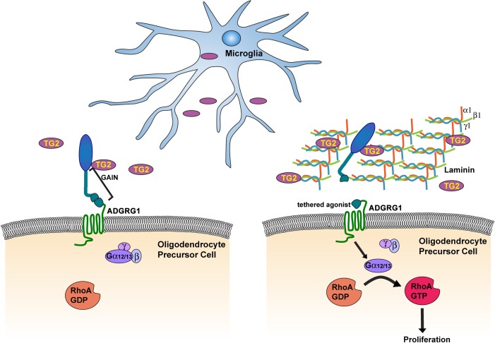
Figure 6. Microglia promote OPC proliferation via ADGRG1 signaling. TG2, secreted by microglia, binds ADGRG1 but fails to activate the receptor in the absence of ECM protein laminin-111. The binding of TG2 and laminin-111 to ADGRG1 leads to the dissociation of the ADGRG1 NTF from its CTF, allowing the tethered agonist to initiate G-protein signaling, culminating in activated RhoA, which promotes OPC proliferation. GAIN, GPCR-Autoproteolysis-INducing.
Figure 6. Microglia promote OPC proliferation via ADGRG1 signaling. TG2, secreted by microglia, binds ADGRG1 but fails to activate the receptor in the absence of ECM protein laminin-111. The binding of TG2 and laminin-111 to ADGRG1 leads to the dissociation of the ADGRG1 NTF from its CTF, allowing the tethered agonist to initiate G-protein signaling, culminating in activated RhoA, which promotes OPC proliferation. GAIN, GPCR-Autoproteolysis-INducing.CC BY · PubMed Central

Verification

Claims the paper makes

The claims and statistics of this paper haven’t been checked yet.

Run the check — free, no account needed. It reads the open-access text and checks the sources it cites.

A few free checks a day · papers already verified open instantly · results are public

Claims other papers make about this one

Each is checked against this paper’s own text: does it say what the citing paper says it does?

Self-check

Percentages, sums and reported differences re-computed from the paper’s own numbers.

The self-check needs the paper’s full text.

Citations

Cites

1 retracted

Show all 76 references

Cited by

Sorted by citations

See all 146

Metadata

Authors
Giera, S., Luo, R., Ying, Y., Ackerman, S. D., Jeong, S.-J., Stoveken, H. M., Folts, C. J., Welsh, C. A., Tall, G. G., Stevens, B., Monk, K. R., & Piao, X.
Affiliations
  • Boston Children's Hospital (US)
  • University of Oregon (US)
  • University of Michigan (US)
  • Oregon Health & Science University (US)
Corresponding author
  • Monk, Kelly R
  • Piao, Xianhua
Venue
eLife
Published
2018-05-29
Volume
7
ISSN
2050-084X (ISSN-L)
OpenAlex
W2805257681
Cited by
145
References
76
Citations by year
2026: 18 · 2025: 18 · 2024: 19 · 2023: 10 · 2022: 20 · 2021: 20 · 2020: 19 · 2019: 17 · 2018: 2
FWCI
5.33
Type
journal article
Language
en
Open access
Locations
  • eLife (publishedVersion, cc-by)
  • Europe PMC (PubMed Central) (publishedVersion, cc-by)
  • PubMed Central (publishedVersion, cc-by)
  • PubMed (publishedVersion)
  • Digital Commons@Becker (Washington University School of Medicine) (submittedVersion)
  • Digital Access to Scholarship at Harvard (DASH) (Harvard University) (submittedVersion, cc-by)
  • DOAJ (DOAJ: Directory of Open Access Journals) (submittedVersion, cc-by-sa)
Funders
  • National Institute of Neurological Disorders and Stroke (NS085201, NS085201 (R56), NS087801, NS079445)
  • National Multiple Sclerosis Society (FG 2063-A1/2, Harry Weaver Neuroscience Fellowship, RG-1501-02577)
  • National Research Foundation of Korea (2015M3C7A1029037)
  • Ministry of Science, ICT and Future Planning (KBRI Basic Research program 18-BR-02-02)
Topics
  • Life Sciences › Neuroscience › Developmental Neuroscience › Neurogenesis and neuroplasticity mechanisms
  • Health Sciences › Medicine › Clinical Neurology › Multiple Sclerosis Research Studies
  • Life Sciences › Neuroscience › Neurology › Neuroinflammation and Neurodegeneration Mechanisms
Keywords
tissue transglutaminase, ADGRG1, GPR56, oligodendrocyte precursor cells, myelination, remyelination, demyelination, laminin, extracellular matrix, microglia, OPC proliferation
MeSH terms
Oligodendrocyte Precursor Cells, CX3C Chemokine Receptor 1, Remyelination, Protein Glutamine gamma Glutamyltransferase 2, Animals, Cell Differentiation, Cerebellum, Demyelinating Diseases, Female, Humans, Laminin, Male, Oligodendroglia, Transglutaminases, Signal Transduction, Prosencephalon, Microglia, Mice, Knockout, Gene Expression Regulation, Developmental, Cell Lineage, GTP-Binding Proteins, Receptor, Platelet-Derived Growth Factor alpha, Receptors, G-Protein-Coupled, Mice, Neurogenesis

About these checks

Registry, retraction, Cites and statistics results are computed, not judged, with one exception: a printed reference that no index lists is read once by a model before it counts as a citation error. Claims are extracted by a model and kept only when their quote appears verbatim in the text; each cited source is then checked, with a model’s help, for whether it says what the claim relies on. Those verdicts can be wrong — read the claim’s quote and the cited source before relying on one.

Statistics and arithmetic are recomputed, not judged.

Confidence bands are shown only for fields calibrated on our annotated test set. None is calibrated yet, so no band is shown.

What counts as a claim ›How we measure confidence ›How we check this ›

Are you an author, or do you see a problem with this page? Request a review