Cited by 10Cellular transport and secretionFungal and yeast genetics researchAutophagy in Disease and Therapy

The DOI matches its Crossref record; 1 of the 1 references it cites were found in a registry or an index.

The Uso1 globular head interacts with SNAREs to maintain viability even in the absence of the coiled-coil domain

Open access

Published version of a preprint

Abstract

Uso1/p115 and RAB1 tether ER-derived vesicles to the Golgi. Uso1/p115 contains a globular-head-domain (GHD), a coiled-coil (CC) mediating dimerization/tethering, and a C-terminal region (CTR) interacting with golgins. Uso1/p115 is recruited to vesicles by RAB1. Genetic studies placed Uso1 paradoxically acting upstream of, or in conjunction with RAB1 (Sapperstein et al., 1996). We selected two missense mutations in uso1 resulting in E6K and G540S in the GHD that rescued lethality of rab1 -deficient Aspergillus nidulans . The mutations are phenotypically additive, their combination suppressing the complete absence of RAB1, which emphasizes the key physiological role of the GHD. In living hyphae Uso1 recurs on puncta (60 s half-life) colocalizing partially with the Golgi markers RAB1, Sed5, and GeaA/Gea1/Gea2, and totally with the retrograde cargo receptor Rer1, consistent with Uso1 dwelling in a very early Golgi compartment from which ER residents reaching the Golgi recycle back to the ER. Localization of Uso1, but not of Uso1 E6K/G540S , to puncta is abolished by compromising RAB1 function, indicating that E6K/G540S creates interactions bypassing RAB1. That Uso1 delocalization correlates with a decrease in the number of Gea1 cisternae supports that Uso1-and-Rer1-containing puncta are where the protein exerts its physiological role. In S-tag-coprecipitation experiments, Uso1 is an associate of the Sed5/Bos1/Bet1/Sec22 SNARE complex zippering vesicles with the Golgi, with Uso1 E6K/G540S showing a stronger association. Using purified proteins, we show that Bos1 and Bet1 bind the Uso1 GHD directly. However, Bet1 is a strong E6K/G540S-independent binder, whereas Bos1 is weaker but becomes as strong as Bet1 when the GHD carries E6K/G540S. G540S alone markedly increases GHD binding to Bos1, whereas E6K causes a weaker effect, correlating with their phenotypic contributions. AlphaFold2 predicts that G540S increases the binding of the GHD to the Bos1 Habc domain. In contrast, E6K lies in an N-terminal, potentially alpha-helical, region that sensitive genetic tests indicate as required for full Uso1 function. Remarkably, this region is at the end of the GHD basket opposite to the end predicted to interact with Bos1. We show that, unlike dimeric full-length and CTR∆ Uso1 proteins, the GHD lacking the CC/CTR dimerization domain, whether originating from bacteria or Aspergillus extracts and irrespective of whether it carries or not E6K/G540S, would appear to be monomeric. With the finding that overexpression of E6K/G540S and wild-type GHD complement uso1∆ , our data indicate that the GHD monomer is capable of providing, at least partially, the essential Uso1 functions, and that long-range tethering activity is dispensable. Rather, these findings strongly suggest that the essential role of Uso1 involves the regulation of SNAREs.

Overview

The claims and statistics of this paper haven’t been checked yet.

Run the check — free, no account needed. It reads the open-access text and checks the sources it cites.

A few free checks a day · papers already verified open instantly · results are public

Integrity signals

Registry

Crossref · 10.7554/elife.85079

Cites

1 of 1 references verified0 citation errors · 0 not checkable

Statistics

Figures
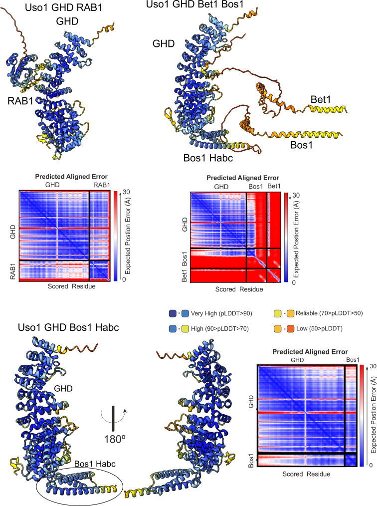
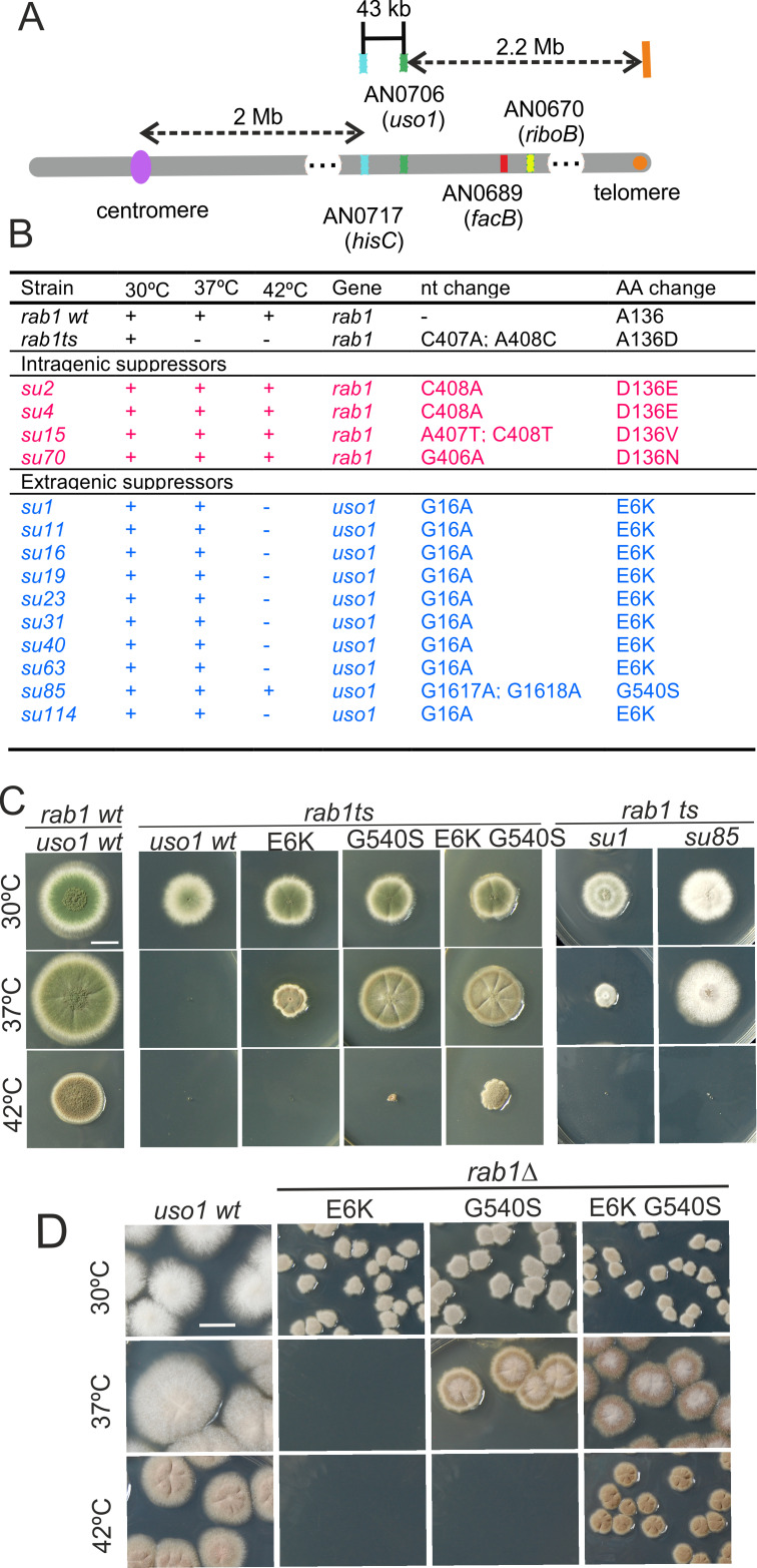
Figure 1. Characterization of mutations bypassing the essential role of RAB1. (A). Genetic map in the region surrounding uso1 with genetic markers used as landmarks for mapping. (B). Molecular identification of the nucleotide changes in suArab1ts strains. (C) and (D): growth tests showing rab1ts- and rab1∆-rescuing phenotypes, respectively, of individual mutations, and synthetic positive interaction between E6K and G540S. Strains produce either green or white conidiospores (conidiospore colors are used as genetic markers). In (C), strains were point-inoculated. In (D) conidiospores were spread on agar plates to give individual colonies.
Figure 1. Characterization of mutations bypassing the essential role of RAB1. (A). Genetic map in the region surrounding uso1 with genetic markers used as landmarks for mapping. (B). Molecular identification of the nucleotide changes in suArab1ts strains. (C) and (D): growth tests showing rab1ts- and rab1∆-rescuing phenotypes, respectively, of individual mutations, and synthetic positive interaction between E6K and G540S. Strains produce either green or white conidiospores (conidiospore colors are used as genetic markers). In (C), strains were point-inoculated. In (D) conidiospores were spread on agar plates to give individual colonies.CC BY · PubMed Central
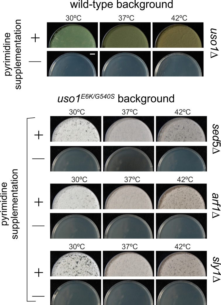
Figure 1—figure supplement 1. E6K/G540S do not rescue lethality resulting from arf1∆, sly1∆, or sed5∆. Top, uso1 is an essential gene. Singly-nucleated conidiospores derived from a heterokaryotic strain in which one class of nuclei carries a deficient pyrG uracil biosynthetic gene whereas the second class contains a uso1∆ allele tagged with functional pyrG were unable to grow on medium lacking pyrimidines at any of the tested temperatures. Bottom: Similar experiments show that unlike rab1∆ strains, strains carrying lethal arf1∆, sly1∆, and sed5∆ alleles cannot be rescued by uso1E6K/G540S. Top panel, strains with green conidiospores; bottom, strains with white conidiospores.
Figure 1—figure supplement 1. E6K/G540S do not rescue lethality resulting from arf1∆, sly1∆, or sed5∆. Top, uso1 is an essential gene. Singly-nucleated conidiospores derived from a heterokaryotic strain in which one class of nuclei carries a deficient pyrG uracil biosynthetic gene whereas the second class contains a uso1∆ allele tagged with functional pyrG were unable to grow on medium lacking pyrimidines at any of the tested temperatures. Bottom: Similar experiments show that unlike rab1∆ strains, strains carrying lethal arf1∆, sly1∆, and sed5∆ alleles cannot be rescued by uso1E6K/G540S. Top panel, strains with green conidiospores; bottom, strains with white conidiospores.CC BY · PubMed Central
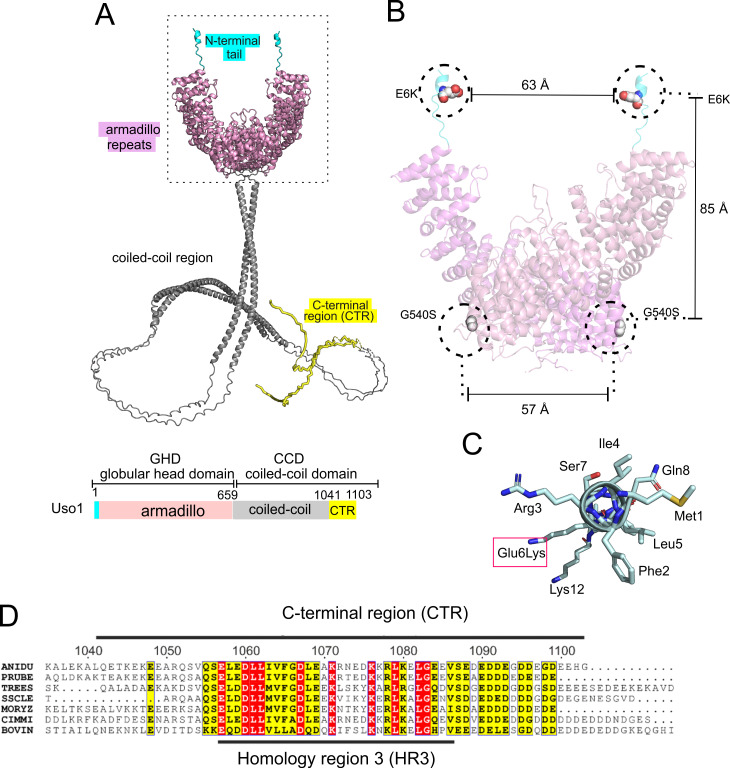
Figure 2. Localization of the amino acid substitutions within the Uso1 AlphaFold2 structure. (A). AlphaFold2 cartoon representations of A. nidulans Uso1 dimer. Note that the depiction of the coiled-coil domain (CCD) is highly speculative. It is included with the sole purpose of visually appreciating the relative sizes of the globular-head-domain (GHD) and the CCD domains. Confidence estimations for Uso1 models are detailed in Figure 2—figure supplement 1. Colors are as in the scheme below: Cyan, N-terminal tail; pink, globular head domain; gray, coiled-coil; yellow, C-terminal region (CTR). (B). Position of the Gly6Lys and Gly540Ser substitutions. Only the GHD of dimeric full-length Uso1 is shown. Distances between mutated residues are displayed in angstroms. (C). The N-terminal amphipathic α-helix affected by the Glu6Lys substitution. (D). Amino acid alignment of fungal sequences with mammalian p115 showing strong conservation within the CTR: ANIDU, Aspergillus nidulans; PRUBE, Penicillium rubens; TREES, Thrichoderma ressei; SSCLE, Sclerotinia scleriotorum; MORYZ, Magnaporthe oryzae; CIMM, Coccidioides immitis; BOVIN, Bos taurus.
Figure 2. Localization of the amino acid substitutions within the Uso1 AlphaFold2 structure. (A). AlphaFold2 cartoon representations of A. nidulans Uso1 dimer. Note that the depiction of the coiled-coil domain (CCD) is highly speculative. It is included with the sole purpose of visually appreciating the relative sizes of the globular-head-domain (GHD) and the CCD domains. Confidence estimations for Uso1 models are detailed in Figure 2—figure supplement 1. Colors are as in the scheme below: Cyan, N-terminal tail; pink, globular head domain; gray, coiled-coil; yellow, C-terminal region (CTR). (B). Position of the Gly6Lys and Gly540Ser substitutions. Only the GHD of dimeric full-length Uso1 is shown. Distances between mutated residues are displayed in angstroms. (C). The N-terminal amphipathic α-helix affected by the Glu6Lys substitution. (D). Amino acid alignment of fungal sequences with mammalian p115 showing strong conservation within the CTR: ANIDU, Aspergillus nidulans; PRUBE, Penicillium rubens; TREES, Thrichoderma ressei; SSCLE, Sclerotinia scleriotorum; MORYZ, Magnaporthe oryzae; CIMM, Coccidioides immitis; BOVIN, Bos taurus.CC BY · PubMed Central
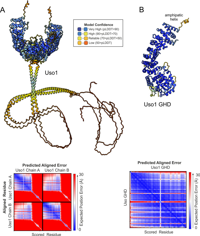
Figure 2—figure supplement 1. AlphaFold2 predictions of Uso1. Ribbon representation of AlphaFold 2-predicted structures of the full-length Uso1 dimer (A) and the Uso1 globular-head-domain(GHD), represented as a monomer (B), color-coded by pLDDT values. Graphs at the bottom are the corresponding plots of predicted aligned error of the residues (PAE).
Figure 2—figure supplement 1. AlphaFold2 predictions of Uso1. Ribbon representation of AlphaFold 2-predicted structures of the full-length Uso1 dimer (A) and the Uso1 globular-head-domain(GHD), represented as a monomer (B), color-coded by pLDDT values. Graphs at the bottom are the corresponding plots of predicted aligned error of the residues (PAE).CC BY · PubMed Central
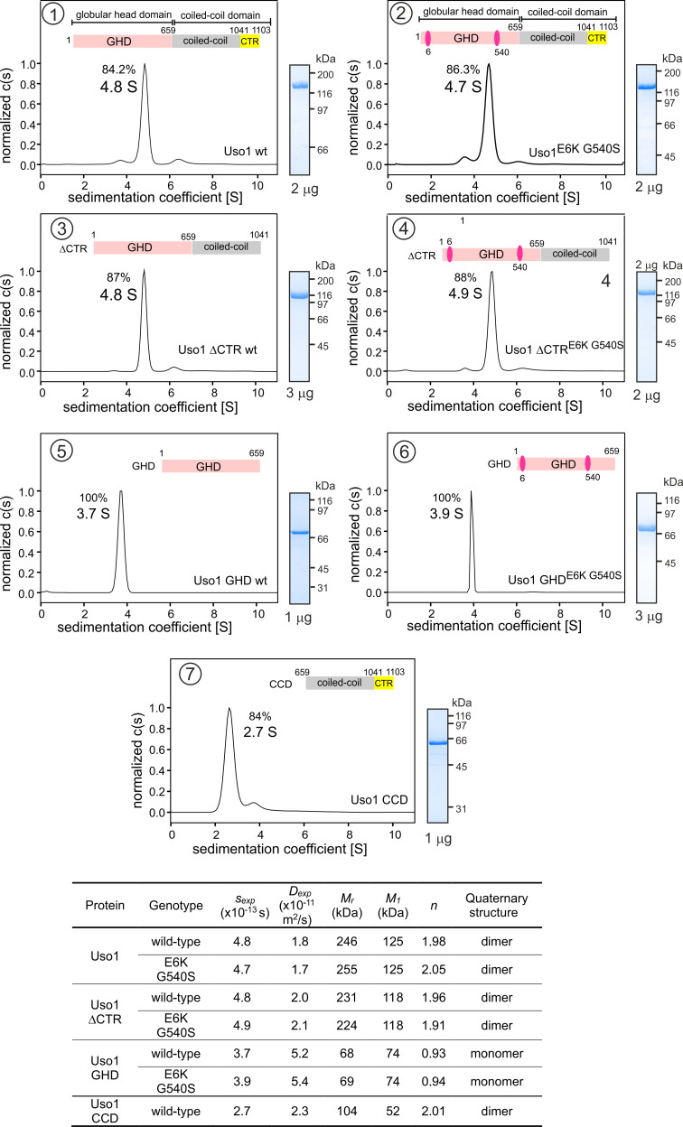
Figure 3. Determining molecular masses and oligomerization status of the different Uso1 constructs by velocity sedimentation analysis. The different panels display the sedimentation profiles of the protein being analyzed, with % of the main species, scheme of the corresponding constructs (color matching those in Figure 2), and pictures of Coomassie stained gels showing the purity of the protein preparations. The bottom table depicts the biophysical parameters of the constructs used to obtain relative molecular masses. sexp is the experimentally determined Svedberg coefficient; Dexp, translational diffusion coefficient of the main species; Mr, molecular mass deduced from Svedberg equation; M1 predicted molecular mass of the monomer; n = (Mr /M1).
Figure 3. Determining molecular masses and oligomerization status of the different Uso1 constructs by velocity sedimentation analysis. The different panels display the sedimentation profiles of the protein being analyzed, with % of the main species, scheme of the corresponding constructs (color matching those in Figure 2), and pictures of Coomassie stained gels showing the purity of the protein preparations. The bottom table depicts the biophysical parameters of the constructs used to obtain relative molecular masses. sexp is the experimentally determined Svedberg coefficient; Dexp, translational diffusion coefficient of the main species; Mr, molecular mass deduced from Svedberg equation; M1 predicted molecular mass of the monomer; n = (Mr /M1).CC BY · PubMed Central
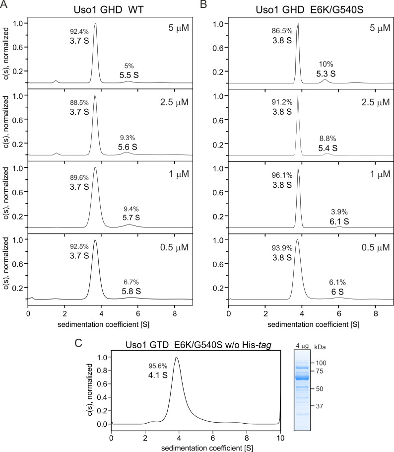
Figure 3—figure supplement 1. Globular-head-domain (GHD) is a monomer across a range of concentrations. (A) and (B). Sedimentation velocity experiments with wild-type and E6K/G540S mutant GHD, respectively, show that they behave as monomers at concentrations up to 5 μM. (C). Sedimentation velocity profile of E6K/G540S mutant GHD lacking the His-tag, showing that the presence of the latter does not interfere with oligomerization, and a picture of a Coomassie-stained gel showing the purity of the protein preparation on the right.
Figure 3—figure supplement 1. Globular-head-domain (GHD) is a monomer across a range of concentrations. (A) and (B). Sedimentation velocity experiments with wild-type and E6K/G540S mutant GHD, respectively, show that they behave as monomers at concentrations up to 5 μM. (C). Sedimentation velocity profile of E6K/G540S mutant GHD lacking the His-tag, showing that the presence of the latter does not interfere with oligomerization, and a picture of a Coomassie-stained gel showing the purity of the protein preparation on the right.CC BY · PubMed Central
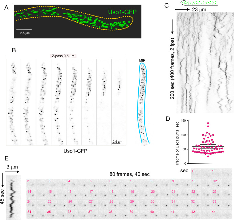
Figure 4. Subcellular localization of Uso1. (A). Uso1-GFP localizing to punctate cytoplasmic structures, 3D shaded by software. (B). Sections of a deconvolved Z-stack and its corresponding maximal intensity projection (MIP). Uso1-GFP in inverted grayscale for clarity. (C). Kymograph showing the transient recruitment of Uso1 to punctate cytoplasmic structures. (D). Average time of residence of Uso1 in these structures. Error bars, 95% CI. (E). Example of one such structure visualized with a kymograph and with the corresponding movie frames (Video 4).
Figure 4. Subcellular localization of Uso1. (A). Uso1-GFP localizing to punctate cytoplasmic structures, 3D shaded by software. (B). Sections of a deconvolved Z-stack and its corresponding maximal intensity projection (MIP). Uso1-GFP in inverted grayscale for clarity. (C). Kymograph showing the transient recruitment of Uso1 to punctate cytoplasmic structures. (D). Average time of residence of Uso1 in these structures. Error bars, 95% CI. (E). Example of one such structure visualized with a kymograph and with the corresponding movie frames (Video 4).CC BY · PubMed Central
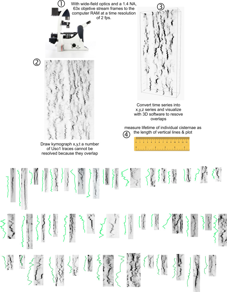
Figure 4—figure supplement 1. Methodology for tracking the half-life of Uso1-GFP on punctate structures. 3D movies were acquired by streaming pictures to the computer RAM at 2 fps. Appropriate reduction of excitation light intensity permitted the acquisition of 400 frames without apparent phototoxicity. The behavior of punctate structures over time was represented in kymographs, in which vertical lines represent the time of residence of Uso1 on membranes. (3). As vertical lines frequently overlapped, jeopardizing the quality of this analysis, we imported the time series into a 3D viewer as if they were (x, y, z) series. Rotation across the different axes facilitated unambiguous tracking of the trajectories across time. (4). The length of the trajectories was measured and converted to time units. Bottom graphs display examples of time trajectories.
Figure 4—figure supplement 1. Methodology for tracking the half-life of Uso1-GFP on punctate structures. 3D movies were acquired by streaming pictures to the computer RAM at 2 fps. Appropriate reduction of excitation light intensity permitted the acquisition of 400 frames without apparent phototoxicity. The behavior of punctate structures over time was represented in kymographs, in which vertical lines represent the time of residence of Uso1 on membranes. (3). As vertical lines frequently overlapped, jeopardizing the quality of this analysis, we imported the time series into a 3D viewer as if they were (x, y, z) series. Rotation across the different axes facilitated unambiguous tracking of the trajectories across time. (4). The length of the trajectories was measured and converted to time units. Bottom graphs display examples of time trajectories.CC BY · PubMed Central
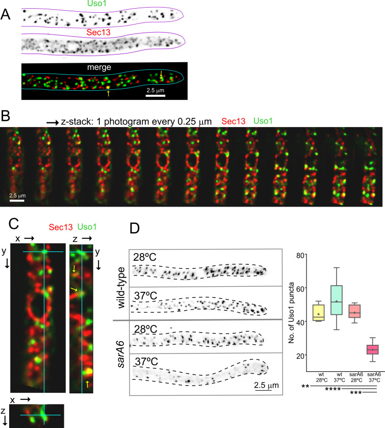
Figure 5. Uso1 puncta do not colocalize with ER exit sites (ERESs). (A). Low extent co-localization of Sec13 ERES and Uso1 structures. Z-stacks for the two channels were acquired simultaneously, deconvolved, and represented as maximal intensity projections (MIPs). Two rare examples of colocalization are arrowed. (B). Photograms of a dual channel Z-stack with a Sec13-labeled nuclear envelope focused in the middle plane, illustrating that while some puncta show colocalization, the red Sec13 signal, and the green Uso1 signal do not usually overlap. (C). A MIP of the same z-stack showing orthogonal views with some overlapping puncta (arrows). (D). A ts mutation in the sarA gene encoding the SarASar1 GTPase governing ER exit markedly reduces the number of Uso1-GFP puncta upon shifting cells to restrictive conditions. Box-and-whisker plots: Statistical comparison was made using one-way ANOVA with Dunn’s test for multiple comparisons. Whiskers are in Tukey’s style: Only significant differences were indicated, using asterisks.
Figure 5. Uso1 puncta do not colocalize with ER exit sites (ERESs). (A). Low extent co-localization of Sec13 ERES and Uso1 structures. Z-stacks for the two channels were acquired simultaneously, deconvolved, and represented as maximal intensity projections (MIPs). Two rare examples of colocalization are arrowed. (B). Photograms of a dual channel Z-stack with a Sec13-labeled nuclear envelope focused in the middle plane, illustrating that while some puncta show colocalization, the red Sec13 signal, and the green Uso1 signal do not usually overlap. (C). A MIP of the same z-stack showing orthogonal views with some overlapping puncta (arrows). (D). A ts mutation in the sarA gene encoding the SarASar1 GTPase governing ER exit markedly reduces the number of Uso1-GFP puncta upon shifting cells to restrictive conditions. Box-and-whisker plots: Statistical comparison was made using one-way ANOVA with Dunn’s test for multiple comparisons. Whiskers are in Tukey’s style: Only significant differences were indicated, using asterisks.CC BY · PubMed Central
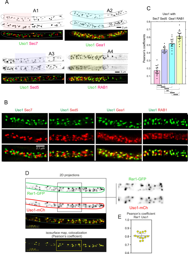
Figure 6. Uso1 localizes to Rer1-containing Golgi cisternae. (A). Tip cells showing Uso1 colocalization with the indicated subcellular markers. Images are maximal intensity projections (MIPs) of deconvolved Z-stacks. (B). Magnified images of the color-coded shaded regions of the cells shown in A. (C). Pearson’s coefficients of the different combinations. (D) Gray scale: MIPs of deconvolved z-stacks of the individual Rer1-GFP and Uso1-mCh channels, with their merge at the bottom. The boxed section of the channels is magnified on the right. The bottom shows a yellow isosurface map corresponding to the regions with high Pearson’s coefficient (constructed with Huygens software). (E) Pearson’s coefficient in n=12 cells. Error bars indicate 95% CI.
Figure 6. Uso1 localizes to Rer1-containing Golgi cisternae. (A). Tip cells showing Uso1 colocalization with the indicated subcellular markers. Images are maximal intensity projections (MIPs) of deconvolved Z-stacks. (B). Magnified images of the color-coded shaded regions of the cells shown in A. (C). Pearson’s coefficients of the different combinations. (D) Gray scale: MIPs of deconvolved z-stacks of the individual Rer1-GFP and Uso1-mCh channels, with their merge at the bottom. The boxed section of the channels is magnified on the right. The bottom shows a yellow isosurface map corresponding to the regions with high Pearson’s coefficient (constructed with Huygens software). (E) Pearson’s coefficient in n=12 cells. Error bars indicate 95% CI.CC BY · PubMed Central
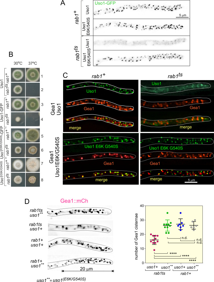
Figure 7. Uso1 localization to punctate structures is dependent on RAB1. (A). Complete de-localization of Uso1-GFP to the cytosol by rab1ts and relocalization by E6K/G540S. (B). Uso1-GFP and rab1ts show a synthetic negative interaction that is rescued by the E6K/G540S double substitution. Strains in lanes 7 and 8 carry the wA2 mutation resulting in white conidiospores. (C) In rab1ts cells, punctate Uso1-GFP structures largely disappear, correlating with Golgi disorganization (GeaA/Gea1-mCh, see text). The presence of E6K/G540S relocalizes Uso1-GFP to puncta and restores Golgi organization. (D) Representative images (maximal intensity projections(MIPs) of deconvolved Z-stacks) of hyphae used to quantify the number of cisternae within the apex-proximal 20 µm. Datasets were compared by one-way ANOVA with Tukey’s multiple comparison test. Error bars represent S.D.
Figure 7. Uso1 localization to punctate structures is dependent on RAB1. (A). Complete de-localization of Uso1-GFP to the cytosol by rab1ts and relocalization by E6K/G540S. (B). Uso1-GFP and rab1ts show a synthetic negative interaction that is rescued by the E6K/G540S double substitution. Strains in lanes 7 and 8 carry the wA2 mutation resulting in white conidiospores. (C) In rab1ts cells, punctate Uso1-GFP structures largely disappear, correlating with Golgi disorganization (GeaA/Gea1-mCh, see text). The presence of E6K/G540S relocalizes Uso1-GFP to puncta and restores Golgi organization. (D) Representative images (maximal intensity projections(MIPs) of deconvolved Z-stacks) of hyphae used to quantify the number of cisternae within the apex-proximal 20 µm. Datasets were compared by one-way ANOVA with Tukey’s multiple comparison test. Error bars represent S.D.CC BY · PubMed Central
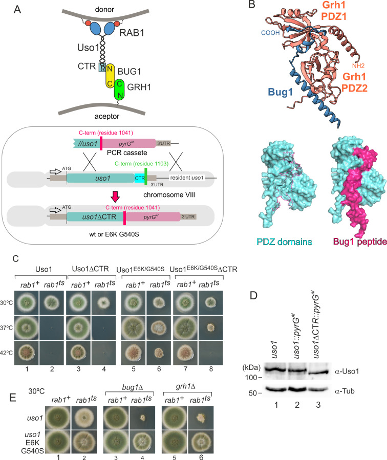
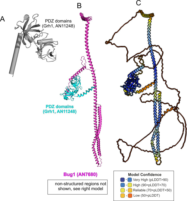
Figure 8. Genetic evidence shows that the C-terminal region (CTR) region of Uso1 contributes to its recruitment to membranes. (A). Top, scheme of the predicted interactions. Bottom, engineering a gene-replaced allele lacking the CTR domain by homologous recombination. (B). The Bug1 C-terminal residues fit into the groove formed between the two Grh1 PDZ domains and into the pocket of the N-terminal PDZ domain (PDZ1). (C). A gene-replaced uso1∆CTR allele encoding a protein truncated for the CTR domain shows a synthetic negative interaction with rab1ts. (D). Western blot analysis. Removal of the CTR does not result in Uso1 instability. (E). bug1∆ and grh1∆ show a synthetic negative interaction with rab1ts that is rescued by the double E6K/G540S substitution in Uso1.
Figure 8. Genetic evidence shows that the C-terminal region (CTR) region of Uso1 contributes to its recruitment to membranes. (A). Top, scheme of the predicted interactions. Bottom, engineering a gene-replaced allele lacking the CTR domain by homologous recombination. (B). The Bug1 C-terminal residues fit into the groove formed between the two Grh1 PDZ domains and into the pocket of the N-terminal PDZ domain (PDZ1). (C). A gene-replaced uso1∆CTR allele encoding a protein truncated for the CTR domain shows a synthetic negative interaction with rab1ts. (D). Western blot analysis. Removal of the CTR does not result in Uso1 instability. (E). bug1∆ and grh1∆ show a synthetic negative interaction with rab1ts that is rescued by the double E6K/G540S substitution in Uso1.CC BY · PubMed Central
Figure 8—figure supplement 1. AlphaFold2 modeling of Grh1-Bug1. (A). Cartoon, with α-helices shown as cylinders, of the nearly N-terminal PDZ domains of Grh1. (B). AlphaFold 2 prediction of a 1:1 Grh1-Bug1 complex, trimmed of disordered regions. (C). complete AlphaFold2 model of Grh1-Bug1 with color-coded model confidence values.
Figure 8—figure supplement 1. AlphaFold2 modeling of Grh1-Bug1. (A). Cartoon, with α-helices shown as cylinders, of the nearly N-terminal PDZ domains of Grh1. (B). AlphaFold 2 prediction of a 1:1 Grh1-Bug1 complex, trimmed of disordered regions. (C). complete AlphaFold2 model of Grh1-Bug1 with color-coded model confidence values.CC BY · PubMed Central
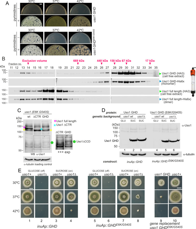
Figure 9. The globular-head-domain (GHD) of Uso1 is sufficient to support cell viability. (A). Gene-replaced uso1GHD allele carrying the double E6K/G540S substitution is sufficient to rescue viability at 30 °C, but not at higher temperatures. (B). The Uso1 GHD is a monomer in cell extracts. Fractions collected from Superose columns loaded with the indicated protein extracts and reference His-tagged proteins were collected and analyzed by western blotting with α-HA and α-His antibodies. (C) Truncating Uso1 after the GHD results in markedly reduced protein levels, as determined by α-Uso1 GHD western blotting. The band (yellow asterisks) moving slower than Uso1 (magenta asterisk) and at nearly the same position as Uso1 ∆CTR (blue asterisk) represents cross-reacting contaminants unrelated to Uso1. The right panel shows a longer exposure for the indicated region, to reveal the faint GHD band (green dot). (D). Overexpression of wild-type and E6K/G540S mutant GHD, under the control of the inuA promoter, which is turned off on glucose and induced on sucrose. Western blots reacted with α-Uso1 GHD antiserum. (E). Overexpressed GHD, be it E6K/G540S or wild-type, as the only source of Uso1 supports viability.
Figure 9. The globular-head-domain (GHD) of Uso1 is sufficient to support cell viability. (A). Gene-replaced uso1GHD allele carrying the double E6K/G540S substitution is sufficient to rescue viability at 30 °C, but not at higher temperatures. (B). The Uso1 GHD is a monomer in cell extracts. Fractions collected from Superose columns loaded with the indicated protein extracts and reference His-tagged proteins were collected and analyzed by western blotting with α-HA and α-His antibodies. (C) Truncating Uso1 after the GHD results in markedly reduced protein levels, as determined by α-Uso1 GHD western blotting. The band (yellow asterisks) moving slower than Uso1 (magenta asterisk) and at nearly the same position as Uso1 ∆CTR (blue asterisk) represents cross-reacting contaminants unrelated to Uso1. The right panel shows a longer exposure for the indicated region, to reveal the faint GHD band (green dot). (D). Overexpression of wild-type and E6K/G540S mutant GHD, under the control of the inuA promoter, which is turned off on glucose and induced on sucrose. Western blots reacted with α-Uso1 GHD antiserum. (E). Overexpressed GHD, be it E6K/G540S or wild-type, as the only source of Uso1 supports viability.CC BY · PubMed Central
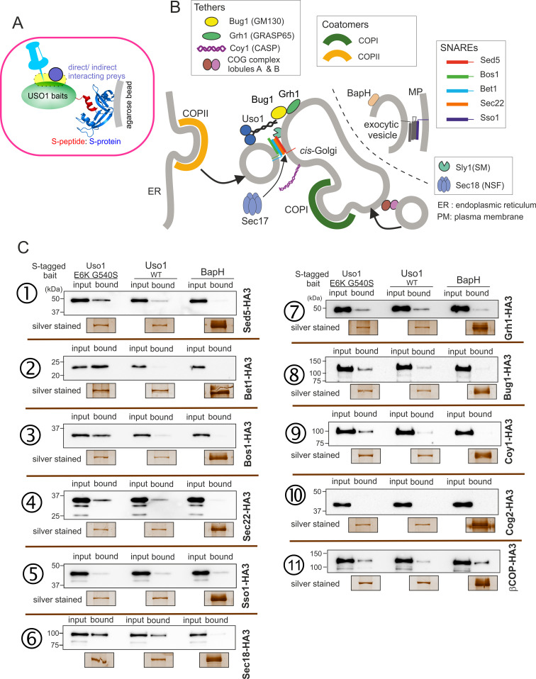
Figure 10. Screening the preferential association of proteins acting in the ER/Golgi interface with E6K/G540S Uso1. (A) S-tagged baits (Uso1, wt, and E6K/G540S, and the unrelated protein BapH), expressed after gene replacement, were captured with their associated polypeptides on S-protein agarose beads. Candidate associates, also expressed after gene replacement, were tagged with HA3. (B) Schematic depiction of the proteins listed in these experiments showing their sites of action. (C). Anti-HA3 western blot analysis of the indicated S-bait and HA3-prey combinations. Equal loading of Uso1 proteins was confirmed by silver staining of precipitates. Note that BapH, chosen as a negative control, is expressed at much higher levels than Uso1 proteins. Each panel is a representative experiment of three experimental replicates.
Figure 10. Screening the preferential association of proteins acting in the ER/Golgi interface with E6K/G540S Uso1. (A) S-tagged baits (Uso1, wt, and E6K/G540S, and the unrelated protein BapH), expressed after gene replacement, were captured with their associated polypeptides on S-protein agarose beads. Candidate associates, also expressed after gene replacement, were tagged with HA3. (B) Schematic depiction of the proteins listed in these experiments showing their sites of action. (C). Anti-HA3 western blot analysis of the indicated S-bait and HA3-prey combinations. Equal loading of Uso1 proteins was confirmed by silver staining of precipitates. Note that BapH, chosen as a negative control, is expressed at much higher levels than Uso1 proteins. Each panel is a representative experiment of three experimental replicates.CC BY · PubMed Central
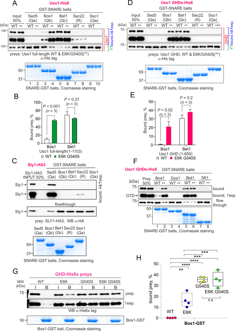
Figure 11. The Uso1 globular-head-domain (GHD) interacts directly with Bos1 and Bet1 SNAREs acting in the ER/Golgi interface. (A). Purified fusion proteins in which the complete cytosolic domains of the indicated SNAREs have been fused to GST were used in pulldown experiments with His-tagged, purified wild-type and E6K/G540S Uso1. The plasma membrane Qa syntaxin Sso1 was used as a negative control. Pulled-down material was analyzed by anti-His western blotting. In this and other panels ↑exp. and ↓exp. indicate high and low exposure to the chemiluminescent western blots, respectively. (B) Quantitation of the above experiment; significance was determined by unpaired t-student tests. Error bars represent SEM. (C) As in (A), but using in vitro synthesized, HA3-tagged Sly1 as prey. Samples were analyzed by anti-HA western blotting. (D) As in (A), but using wild-type and mutant GHD as preys, rather than full-length Uso1. (E). Quantitation of the experiment in (D). (F). GST pull-down experiment comparing the ability of the GHD to interact with the early Golgi Qb and Qc SNAREs (Bos1 and Bet1), with that of their medial Golgi counterparts (Qb Gos1 and Qc Sft1). (G) Bos1-GST pull-down experiments comparing single E6K and G540S mutants with the double mutant. (I and B) represent input and bound fractions. (H) Percentage of the input prey that is retained by the Bos1-GST beads. Data were analyzed by one-way ANOVA with Tukey’s multiple comparisons. Whiskers indicate minimal and maximal values.
Figure 11. The Uso1 globular-head-domain (GHD) interacts directly with Bos1 and Bet1 SNAREs acting in the ER/Golgi interface. (A). Purified fusion proteins in which the complete cytosolic domains of the indicated SNAREs have been fused to GST were used in pulldown experiments with His-tagged, purified wild-type and E6K/G540S Uso1. The plasma membrane Qa syntaxin Sso1 was used as a negative control. Pulled-down material was analyzed by anti-His western blotting. In this and other panels ↑exp. and ↓exp. indicate high and low exposure to the chemiluminescent western blots, respectively. (B) Quantitation of the above experiment; significance was determined by unpaired t-student tests. Error bars represent SEM. (C) As in (A), but using in vitro synthesized, HA3-tagged Sly1 as prey. Samples were analyzed by anti-HA western blotting. (D) As in (A), but using wild-type and mutant GHD as preys, rather than full-length Uso1. (E). Quantitation of the experiment in (D). (F). GST pull-down experiment comparing the ability of the GHD to interact with the early Golgi Qb and Qc SNAREs (Bos1 and Bet1), with that of their medial Golgi counterparts (Qb Gos1 and Qc Sft1). (G) Bos1-GST pull-down experiments comparing single E6K and G540S mutants with the double mutant. (I and B) represent input and bound fractions. (H) Percentage of the input prey that is retained by the Bos1-GST beads. Data were analyzed by one-way ANOVA with Tukey’s multiple comparisons. Whiskers indicate minimal and maximal values.CC BY · PubMed Central
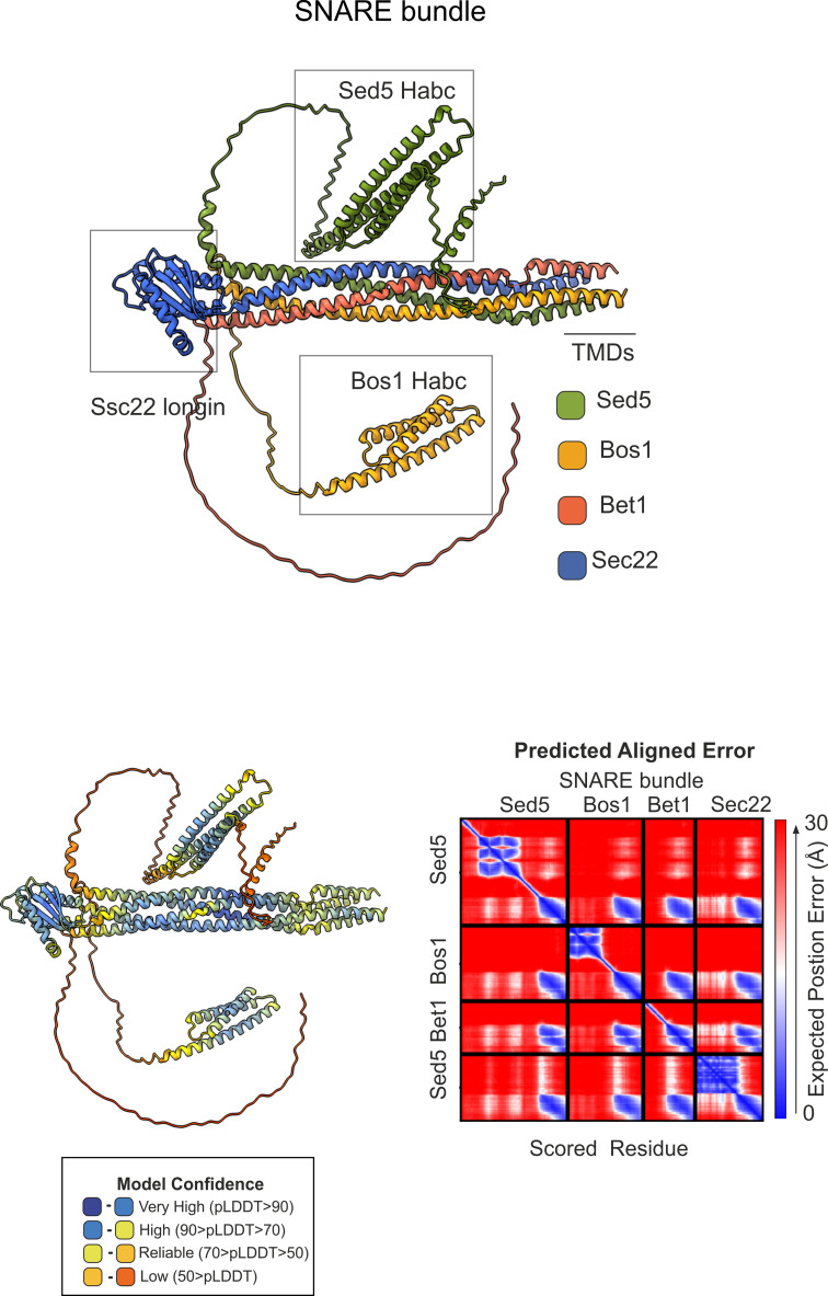
Figure 11—figure supplement 1. AlphaFold2 prediction of the ER/Golgi SNARE bundle. (A) Sec5/Bos1/Bet1/Sec22 predicted SNARE bundle. (B) Quality control (pLDDT, color-coded, and predicted aligned error (PAE)) of the model.
Figure 11—figure supplement 1. AlphaFold2 prediction of the ER/Golgi SNARE bundle. (A) Sec5/Bos1/Bet1/Sec22 predicted SNARE bundle. (B) Quality control (pLDDT, color-coded, and predicted aligned error (PAE)) of the model.CC BY · PubMed Central
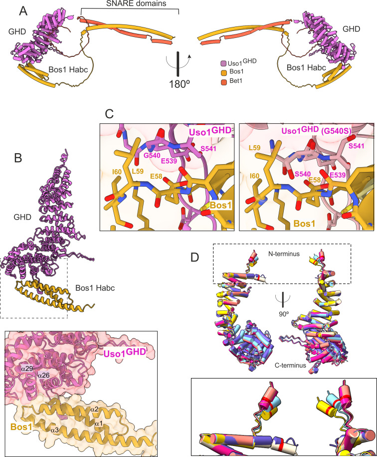
Figure 12. AlphaFold2 models provide insight into the mechanisms of suppression by E6K and G540S. (A). Model of full-length Uso1 bound to the ER/Golgi SNAREs Bos1 and Bet1. (B). Top, ribbon representation of the Bos1 N-terminal Habc domain and Uso1 globular-head-domain (GHD). Bottom, Inset combining surface and ribbon depiction. (C). Increased binding of Bos1 to G540S Uso1 appears to involve the insertion of Ser540 into a pocket located in the Habc domain of the Qb SNARE. Partial view of the Bos1-Uso1 GHD surface of interaction in the wild-type (left) and mutant (right) models. G540 and S540 are annotated. (D). The N-terminal amphipathic α-helix of Uso1 comprising the E6K substitution lies within a flexible stretch of the protein that might facilitate its insertion into membranes. Alignment of six independent predictions, each depicted in a different color, with Glu6 highlighted as a red collar surrounding the cylinders of the N-terminal α-helix. The Uso1 GHD was modeled alone, in a complex with SNARE proteins or with Ypt1. The N-terminal α-helix (boxed) adopts different positions, suggesting high flexibility.
Figure 12. AlphaFold2 models provide insight into the mechanisms of suppression by E6K and G540S. (A). Model of full-length Uso1 bound to the ER/Golgi SNAREs Bos1 and Bet1. (B). Top, ribbon representation of the Bos1 N-terminal Habc domain and Uso1 globular-head-domain (GHD). Bottom, Inset combining surface and ribbon depiction. (C). Increased binding of Bos1 to G540S Uso1 appears to involve the insertion of Ser540 into a pocket located in the Habc domain of the Qb SNARE. Partial view of the Bos1-Uso1 GHD surface of interaction in the wild-type (left) and mutant (right) models. G540 and S540 are annotated. (D). The N-terminal amphipathic α-helix of Uso1 comprising the E6K substitution lies within a flexible stretch of the protein that might facilitate its insertion into membranes. Alignment of six independent predictions, each depicted in a different color, with Glu6 highlighted as a red collar surrounding the cylinders of the N-terminal α-helix. The Uso1 GHD was modeled alone, in a complex with SNARE proteins or with Ypt1. The N-terminal α-helix (boxed) adopts different positions, suggesting high flexibility.CC BY · PubMed Central
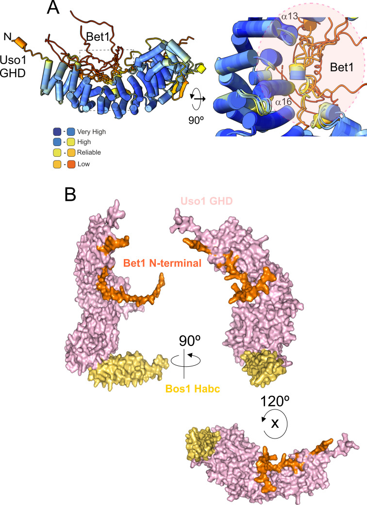
Figure 12—figure supplement 1. AlphaFold2 prediction of the Bet1-GHD interaction. (A) The putative binding surface of Bet1 and Uso1 as determined by AlphaFold2. Top images, a cartoon of Bet1-GHD interactions, colored by pLDDT score. Alignment of four independent predictions involving the Bet1 N-terminal region and Uso1 globular-head-domain (GHD). A single model for Uso1 GHD is shown on the top representation for simplicity. In spite of the disordered nature of the N-terminal Bet1 region, the Bet1-Uso1 binding interface is consistent among models. (B) Bottom, surface representation of the N-terminal Bet1 region (orange) in complex with the GHD. Also indicated is the Habc domain of Bos1 (yellow) bound to the GHD.
Figure 12—figure supplement 1. AlphaFold2 prediction of the Bet1-GHD interaction. (A) The putative binding surface of Bet1 and Uso1 as determined by AlphaFold2. Top images, a cartoon of Bet1-GHD interactions, colored by pLDDT score. Alignment of four independent predictions involving the Bet1 N-terminal region and Uso1 globular-head-domain (GHD). A single model for Uso1 GHD is shown on the top representation for simplicity. In spite of the disordered nature of the N-terminal Bet1 region, the Bet1-Uso1 binding interface is consistent among models. (B) Bottom, surface representation of the N-terminal Bet1 region (orange) in complex with the GHD. Also indicated is the Habc domain of Bos1 (yellow) bound to the GHD.CC BY · PubMed Central
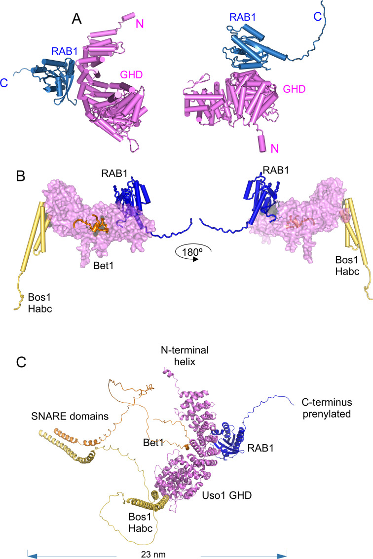
Figure 12—figure supplement 2. AlphaFold 2 predictions of the RAB1 binding site on the Bet1/Bos1/Uso1 globular-head-domain (GHD) complex. (A) cartoon representations of the AlphaFold 2-predicted GHD-RAB1 complex, depicted as pipes and planks. (B): Orthogonal views of the Uso1 GHD-RAB1-Bet1-Bos1Habc structural model. The Uso1 GHD is shown as a surface to emphasize the distant predicted binding sites of the Bos1 Habc domain, RAB1, and the Bet1 N-terminal region. (C): Ribbon representation of the model shown in C but including the full-length SNARE subunits, i.e., the GHD domain of Uso1, RAB1, and the SNARES Bet1 and Bos1. Proteins are in the correct orientation to connect membranes separated by 23 nm, counting from the SNARE TMDs to the prenylated RAB1 residues.
Figure 12—figure supplement 2. AlphaFold 2 predictions of the RAB1 binding site on the Bet1/Bos1/Uso1 globular-head-domain (GHD) complex. (A) cartoon representations of the AlphaFold 2-predicted GHD-RAB1 complex, depicted as pipes and planks. (B): Orthogonal views of the Uso1 GHD-RAB1-Bet1-Bos1Habc structural model. The Uso1 GHD is shown as a surface to emphasize the distant predicted binding sites of the Bos1 Habc domain, RAB1, and the Bet1 N-terminal region. (C): Ribbon representation of the model shown in C but including the full-length SNARE subunits, i.e., the GHD domain of Uso1, RAB1, and the SNARES Bet1 and Bos1. Proteins are in the correct orientation to connect membranes separated by 23 nm, counting from the SNARE TMDs to the prenylated RAB1 residues.CC BY · PubMed Central
Figure 12—figure supplement 3. Quality control assessment of AlphaFold2 predictions for the indicated complexes.
Figure 12—figure supplement 3. Quality control assessment of AlphaFold2 predictions for the indicated complexes.CC BY · PubMed Central
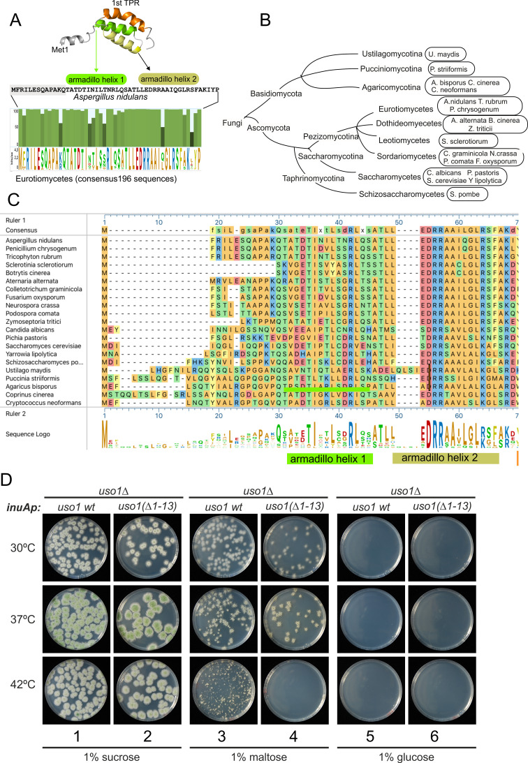
Figure 13. The N-terminal region of Aspergillus Uso1 containing Glu6 is required for full function. (A) Sequence comparison of N-terminal regions up to the second helix of the first armadillo repeat including 196 Uso1 proteins of Eurotiomycetes available in https://fungidb.org/fungidb/app (B) Phylogenetic tree of the Fungi with the indication of species that were used for the alignment in panel (C). (C) Multiple sequence alignment of indicated Uso1 sequences, constructed with ClustalW and Mega Align Pro of the DNAstar package. (D) The 13 N-terminal amino acids of Uso1 are required for full function (see text and Figure 13—figure supplement 2).
Figure 13. The N-terminal region of Aspergillus Uso1 containing Glu6 is required for full function. (A) Sequence comparison of N-terminal regions up to the second helix of the first armadillo repeat including 196 Uso1 proteins of Eurotiomycetes available in https://fungidb.org/fungidb/app (B) Phylogenetic tree of the Fungi with the indication of species that were used for the alignment in panel (C). (C) Multiple sequence alignment of indicated Uso1 sequences, constructed with ClustalW and Mega Align Pro of the DNAstar package. (D) The 13 N-terminal amino acids of Uso1 are required for full function (see text and Figure 13—figure supplement 2).CC BY · PubMed Central
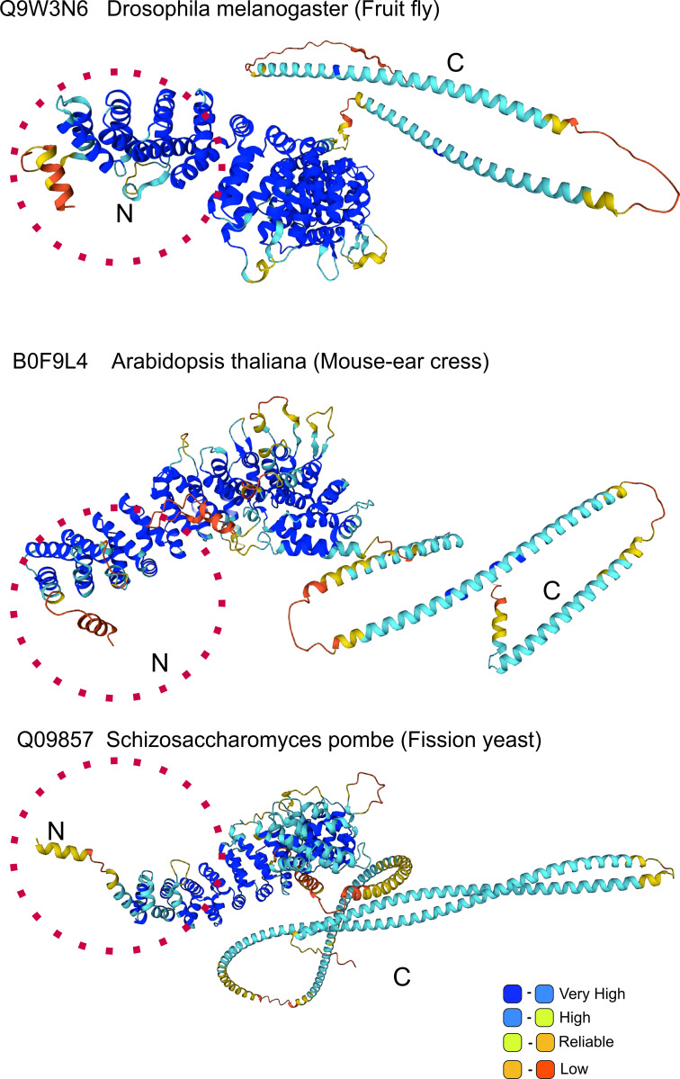
Figure 13—figure supplement 1. AlphaFold prediction of N-terminal helix: model organisms. Alpha-Fold Uso1 structures of the indicated model organisms contain an N-terminal extension which includes a short α-helix akin to that detected in Aspergillus.
Figure 13—figure supplement 1. AlphaFold prediction of N-terminal helix: model organisms. Alpha-Fold Uso1 structures of the indicated model organisms contain an N-terminal extension which includes a short α-helix akin to that detected in Aspergillus.CC BY · PubMed Central
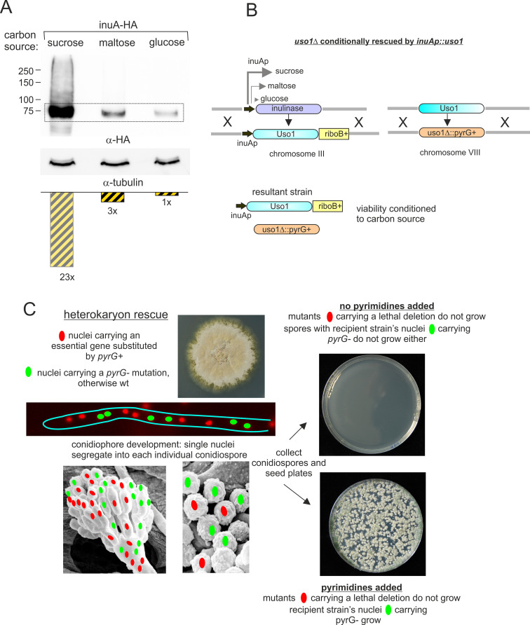
Figure 13—figure supplement 2. Explanation of key genetic manipulations used for this work. (A) Relative levels of inulinase (InuA) on different C sources, as determined by western blotting. (B) Genetically engineering of a strain conditionally expressing Uso1. The inulinase coding region was substituted by the Uso1 coding region and kept in a sucrose medium to delete the endogenous uso1 gene. The resulting strain is unviable on glucose, grows like wild-type on sucrose, and less vigorously on maltose (see Figure 13D). (C) Heterokaryon rescue technique. Nuclei carrying a recessive lethal allele can be maintained in heterokaryosis. Its phenotype can be assessed after the development of asexual spores (conidiospores) into which single nuclei segregate.
Figure 13—figure supplement 2. Explanation of key genetic manipulations used for this work. (A) Relative levels of inulinase (InuA) on different C sources, as determined by western blotting. (B) Genetically engineering of a strain conditionally expressing Uso1. The inulinase coding region was substituted by the Uso1 coding region and kept in a sucrose medium to delete the endogenous uso1 gene. The resulting strain is unviable on glucose, grows like wild-type on sucrose, and less vigorously on maltose (see Figure 13D). (C) Heterokaryon rescue technique. Nuclei carrying a recessive lethal allele can be maintained in heterokaryosis. Its phenotype can be assessed after the development of asexual spores (conidiospores) into which single nuclei segregate.CC BY · PubMed Central
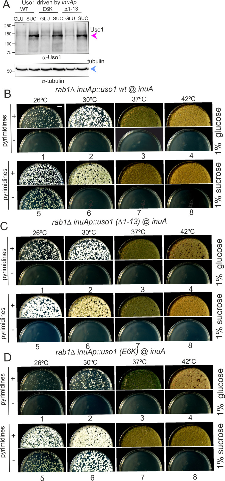
Figure 14. Over-expression of Uso1, but not of Uso1 (∆1–13) rescues the lethality of rab1∆, and rescue is stimulated by E6K. (A) Western blot showing that all three alleles used in growth tests shown below result in similar levels of protein when driven by the inuAp on sucrose (suc). The protein detected on glucose (glu) corresponds to the endogenous gene. (B) Overexpression of Uso1 wild-type permits growth of rab1∆ at 28 °C (C) Overexpression of Uso1 (∆1–13) does not rescue rab1∆ at any temperature,. (D) Overexpression of Uso1 E6K permits growth at 26°C and 30°C.
Figure 14. Over-expression of Uso1, but not of Uso1 (∆1–13) rescues the lethality of rab1∆, and rescue is stimulated by E6K. (A) Western blot showing that all three alleles used in growth tests shown below result in similar levels of protein when driven by the inuAp on sucrose (suc). The protein detected on glucose (glu) corresponds to the endogenous gene. (B) Overexpression of Uso1 wild-type permits growth of rab1∆ at 28 °C (C) Overexpression of Uso1 (∆1–13) does not rescue rab1∆ at any temperature,. (D) Overexpression of Uso1 E6K permits growth at 26°C and 30°C.CC BY · PubMed Central

Verification

Claims the paper makes

The claims and statistics of this paper haven’t been checked yet.

Run the check — free, no account needed. It reads the open-access text and checks the sources it cites.

A few free checks a day · papers already verified open instantly · results are public

Self-check

Percentages, sums and reported differences re-computed from the paper’s own numbers.

The self-check needs the paper’s full text.

Citations

Cites

Show all 1 references

Research works printed but not linked: 93

  1. Allan, 2000, Rab1 recruitment of P115 into a cis-SNARE complex: programming budding COPII vesicles for fusion, Science, 289, 444, 10.1126/science.289.5478.444 · DOI · Paper page
  2. An, 2009, Structural and functional analysis of the Globular head domain of P115 provides insight into membrane tethering, Journal of Molecular Biology, 391, 26, 10.1016/j.jmb.2009.04.062 · DOI
  3. Arst, 2014, GBF/Gea mutant with a single substitution sustains fungal growth in the absence of BIG/Sec7, FEBS Letters, 588, 4799, 10.1016/j.febslet.2014.11.014 · DOI
  4. Baker, 2015, A direct role for the Sec1/Munc18-family protein Vps33 as a template for SNARE assembly, Science, 349, 1111, 10.1126/science.aac7906 · DOI
  5. Baker, 2016, Chaperoning SNARE assembly and disassembly, Nature Reviews. Molecular Cell Biology, 17, 465, 10.1038/nrm.2016.65 · DOI
  6. Banfield, 1995, A SNARE-like protein required for traffic through the Golgi complex, Nature, 375, 10.1038/375806a0 · DOI
  7. Barlowe, 2013, Secretory protein Biogenesis and traffic in the early secretory pathway, Genetics, 193, 383, 10.1534/genetics.112.142810 · DOI
  8. Beard, 2005, A cryptic Rab1-binding site in the P115 tethering protein, The Journal of Biological Chemistry, 280, 25840, 10.1074/jbc.M503925200 · DOI
  9. Behnia, 2007, The yeast orthologue of Grasp65 forms a complex with a coiled-coil protein that contributes to ER to Golgi traffic, The Journal of Cell Biology, 176, 255, 10.1083/jcb.200607151 · DOI
  10. Bentley, 2006, SNARE status regulates tether recruitment and function in homotypic COPII vesicle fusion, The Journal of Biological Chemistry, 281, 38825, 10.1074/jbc.M606044200 · DOI
  11. Bracher, 2002, Structural basis for the Golgi membrane recruitment of Sly1P by Sed5P, The EMBO Journal, 21, 6114, 10.1093/emboj/cdf608 · DOI
  12. Bravo-Plaza, 2019, Identification of the Guanine nucleotide exchange factor for Sar1 in the filamentous fungal model Aspergillus nidulans, Biochimica et Biophysica Acta. Molecular Cell Research, 1866, 10.1016/j.bbamcr.2019.118551 · DOI
  13. Cai, 2007, TRAPPI tethers COPII vesicles by binding the coat subunit Sec23, Nature, 445, 941, 10.1038/nature05527 · DOI
  14. Cao, 1998, Initial docking of ER-derived Vesicles requires Uso1P and Ypt1P but is independent of SNARE proteins, The EMBO Journal, 17, 2156, 10.1093/emboj/17.8.2156 · DOI
  15. Cove, 1966, The induction and repression of nitrate reductase in the fungus Aspergillus nidulans, Biochimica et Biophysica Acta, 113, 51, 10.1016/s0926-6593(66)80120-0 · DOI
  16. Cruz-Garcia, 2014, Remodeling of secretory compartments creates CUPS during nutrient starvation, The Journal of Cell Biology, 207, 695, 10.1083/jcb.201407119 · DOI
  17. Dascher, 1991, Identification and structure of four yeast genes (SLY) that are able to suppress the functional loss of Ypt1, a member of the RAS superfamily, Mol Cell Biol, 11, 872
  18. Day, 2013, A three-stage model of Golgi structure and function, Histochemistry and Cell Biology, 140, 239, 10.1007/s00418-013-1128-3 · DOI
  19. Day, 2018, Budding yeast has a minimal endomembrane system, Developmental Cell, 44, 56, 10.1016/j.devcel.2017.12.014 · DOI
  20. Demircioglu, 2014, The SM protein Sly1 accelerates assembly of the ER-Golgi SNARE complex, PNAS, 111, 13828, 10.1073/pnas.1408254111 · DOI
  21. Galindo, 2016, Structural insights into Arl1-mediated targeting of the ARF-GEF Big1 to the trans-Golgi, Cell Reports, 16, 839, 10.1016/j.celrep.2016.06.022 · DOI
  22. Galindo, 2021, Cryo-EM structure of metazoan TRAPPIII, the multi-subunit complex that activates the GTPase Rab1, The EMBO Journal, 40, 10.15252/embj.2020107608 · DOI
  23. Garcia-Mata, 2003, The membrane-Tethering protein P115 interacts with Gbf1, an ARF Guanine-nucleotide-exchange factor, EMBO Reports, 4, 10.1038/sj.embor.embor762 · DOI
  24. Gustafson, 2017, Regulation of ARF activation occurs via distinct mechanisms at early and late Golgi compartments, Mol Biol Cell, 28, 10.1091/mbc.E17-06-0370 · DOI
  25. Halaby, 2018, The HUS box is required for allosteric regulation of the Sec7 ARF-GEF, The Journal of Biological Chemistry, 293, 6682, 10.1074/jbc.RA117.001318 · DOI
  26. Heo, 2020, Crystal structures of Uso1 membrane tether reveal an alternative conformation in the Globular head domain, Scientific Reports, 10, 10.1038/s41598-020-66480-1 · DOI
  27. Hernández-González, 2015, Conditional inactivation of Aspergillus nidulans sarA(SAR1) uncovers the Morphogenetic potential of regulating endoplasmic reticulum (ER) exit, Molecular Microbiology, 95, 491, 10.1111/mmi.12880 · DOI
  28. Hernández-González, 2018, Genetic dissection of the secretory route followed by a fungal extracellular Glycosyl Hydrolase, Molecular Microbiology, 109, 781, 10.1111/mmi.14073 · DOI
  29. Hernández-González, 2019, COPI Localizes to the early Golgi in Aspergillus nidulans, Fungal Genetics and Biology, 123, 78, 10.1016/j.fgb.2018.12.003 · DOI
  30. Hu, 2015, Structural basis for the interaction between the Golgi reassembly-stacking protein Grasp65 and the Golgi matrix protein Gm130, The Journal of Biological Chemistry, 290, 26373, 10.1074/jbc.M115.657940 · DOI
  31. Jedd, 1995, The Ypt1 Gtpase is essential for the first two steps of the yeast secretory pathway, The Journal of Cell Biology, 131, 583, 10.1083/jcb.131.3.583 · DOI
  32. Joiner, 2021, Structural basis of TRAPPIII-mediated Rab1 activation, The EMBO Journal, 40, 10.15252/embj.2020107607 · DOI
  33. Kurokawa, 2014, Contact of cis-Golgi with ER exit sites executes cargo capture and delivery from the ER, Nature Communications, 5, 10.1038/ncomms4653 · DOI
  34. Lord, 2011, Sequential interactions with Sec23 control the direction of vesicle traffic, Nature, 473, 181, 10.1038/nature09969 · DOI
  35. Losev, 2006, Golgi maturation visualized in living yeast, Nature, 441, 1002, 10.1038/nature04717 · DOI
  36. Lupashin, 1997, T-SNARE activation through transient interaction with a rab-like guanosine triphosphatase, Science, 276, 1255, 10.1126/science.276.5316.1255 · DOI
  37. Malsam, 2011, Organization of SNAREs within the Golgi stack, Cold Spring Harbor Perspectives in Biology, 3, 10.1101/cshperspect.a005249 · DOI
  38. Matsuura-Tokita, 2006, Live imaging of yeast Golgi cisternal maturation, Nature, 441, 1007, 10.1038/nature04737 · DOI
  39. McDonold, 2014, Four Gtpases Differentially regulate the Sec7 ARF-GEF to direct traffic at the trans-Golgi network, Developmental Cell, 30, 759, 10.1016/j.devcel.2014.07.016 · DOI
  40. McNew, 2000, Compartmental specificity of cellular membrane fusion encoded in SNARE proteins, Nature, 407, 153, 10.1038/35025000 · DOI
  41. Mirdita, 2022, Colabfold: making protein folding accessible to all, Nature Methods, 19, 679, 10.1038/s41592-022-01488-1 · DOI · Paper page
  42. Muccini, 2022, Structural basis for activation of Arf1 at the Golgi complex, Cell Reports, 40, 10.1016/j.celrep.2022.111282 · DOI
  43. Nakajima, 1991, A cytoskeleton-related Gene, uso1, is required for intracellular protein transport in Saccharomyces cerevisiae, The Journal of Cell Biology, 113, 245, 10.1083/jcb.113.2.245 · DOI
  44. Nayak, 2006, A versatile and efficient gene targeting system for Aspergillus nidulans, Genetics, 172, 1557, 10.1534/genetics.105.052563 · DOI
  45. Osmani, 1988, Spindle formation and chromatin condensation in cells blocked in interphase by mutation of a negative cell cycle control Gene, Cell, 52, 241, 10.1016/0092-8674(88)90513-2 · DOI
  46. Pantazopoulou, 2011, Characterization of Aspergillus nidulans RabcRab6, Traffic, 12, 386, 10.1111/j.1600-0854.2011.01164.x · DOI
  47. Pantazopoulou, 2016, The Golgi apparatus: insights from filamentous fungi, Mycologia, 108, 603, 10.3852/15-309 · DOI
  48. Pantazopoulou, 2019, A kinetic view of membrane traffic pathways can transcend the classical view of Golgi compartments, Frontiers in Cell and Developmental Biology, 7, 10.3389/fcell.2019.00153 · DOI
  49. Park, 2005, Mutations in a highly conserved region of the Arf1P activator Gea2 block anterograde Golgi transport but not COPI recruitment to membranes, Molecular Biology of the Cell, 16, 3786, 10.1091/mbc.e05-04-0289 · DOI
  50. Parlati, 2002, Distinct SNARE complexes mediating membrane fusion in Golgi transport based on combinatorial specificity, PNAS, 99, 5424, 10.1073/pnas.082100899 · DOI
  51. Pelham, 1999, Snares and the secretory pathway-lessons from yeast, Experimental Cell Research, 247, 1, 10.1006/excr.1998.4356 · DOI
  52. Pelham, 2001, Snares and the specificity of membrane fusion, Trends in Cell Biology, 11, 99, 10.1016/s0962-8924(01)01929-8 · DOI
  53. Peñalva, 2005, Tracing the endocytic pathway of Aspergillus nidulans with FM4-64, Fungal Genetics and Biology, 42, 963, 10.1016/j.fgb.2005.09.004 · DOI
  54. Peñalva, 2020, Tracking Exocytosis of a GPI-anchored protein in Aspergillus nidulans, Traffic, 21, 675, 10.1111/tra.12761 · DOI
  55. Peng, 2002, Sly1 protein bound to Golgi Syntaxin Sed5P allows assembly and contributes to specificity of SNARE fusion complexes, The Journal of Cell Biology, 157, 645, 10.1083/jcb.200202006 · DOI
  56. Pinar, 2013, Acute inactivation of the Aspergillus nidulans Golgi membrane fusion machinery: correlation of apical extension arrest and tip swelling with cisternal disorganization, Molecular Microbiology, 89, 228, 10.1111/mmi.12280 · DOI
  57. Pinar, 2015, TRAPPII regulates exocytic Golgi exit by mediating nucleotide exchange on the Ypt31 orthologue Rabe/Rab11, PNAS, 112, 4346, 10.1073/pnas.1419168112 · DOI
  58. Pinar, 2017, Aspergillus nidulans Baph is a Rab11 Effector that connects membranes in the Spitzenkörper with basal autophagy, Molecular Microbiology, 106, 452, 10.1111/mmi.13777 · DOI
  59. Pinar, 2019, Characterization of Aspergillus nidulans Trapps Uncovers unprecedented similarities with Metazoans and reveals the modular assembly of TRAPPII, PLOS Genetics, 15, 10.1371/journal.pgen.1008557 · DOI
  60. Pinar, 2020, En bloc TGN recruitment of Aspergillus TRAPPII reveals TRAPP maturation as unlikely to drive Rab1-to-Rab11 transition, Journal of Cell Science, 133, 10.1242/jcs.241141 · DOI
  61. Pinar, 2021, The fungal RABOME: RAB Gtpases acting in the Endocytic and Exocytic pathways of Aspergillus nidulans (with excursions to other Filamentous Fungi), Molecular Microbiology, 116, 53, 10.1111/mmi.14716 · DOI
  62. Pinar, 2022, The type V myosin-containing complex HUM is a Rab11 Effector Powering movement of Secretory Vesicles, IScience, 25, 10.1016/j.isci.2022.104514 · DOI
  63. Ren, 2009, A structure-based mechanism for Vesicle capture by the Multisubunit Tethering complex Dsl1, Cell, 139, 1119, 10.1016/j.cell.2009.11.002 · DOI
  64. Rexach, 1994, Characteristics of Endoplasmic Reticulum-derived transport Vesicles, The Journal of Cell Biology, 126, 1133, 10.1083/jcb.126.5.1133 · DOI · Paper page
  65. Richardson, 2012, The Sec7 ARF-GEF is recruited to the Trans-Golgi network by positive feedback, Dev Cell, 22, 799, 10.1016/j.devcel.2012.02.006 · DOI
  66. Richardson, 2016, The Sec7 N-terminal regulatory domains facilitate membrane-proximal activation of the Arf1 Gtpase, eLife, 5, 10.7554/eLife.18204 · DOI
  67. Riedel, 2018, The two TRAPP complexes of Metazoans have distinct roles and act on different Rab Gtpases, The Journal of Cell Biology, 217, 601, 10.1083/jcb.201705068 · DOI
  68. Rizo, 2012, The membrane fusion enigma: Snares, Sec1/Munc18 proteins, and their accomplices--guilty as charged, Annual Review of Cell and Developmental Biology, 28, 279, 10.1146/annurev-cellbio-101011-155818 · DOI
  69. Sánchez-León, 2015, The Rab Gtpase YPT-1 associates with Golgi cisternae and Spitzenkörper microvesicles in Neurospora Crassa, Molecular Microbiology, 95, 472, 10.1111/mmi.12878 · DOI
  70. Sapperstein, 1995, P115 is a general vesicular transport factor related to the yeast Endoplasmic Reticulum to Golgi transport factor Uso1P, PNAS, 92, 522, 10.1073/pnas.92.2.522 · DOI
  71. Sapperstein, 1996, Assembly of the ER to Golgi SNARE complex requires Uso1P, The Journal of Cell Biology, 132, 755, 10.1083/jcb.132.5.755 · DOI
  72. Sato, 2003, Rer1P, a retrieval receptor for ER membrane proteins, recognizes Transmembrane domains in multiple modes, Molecular Biology of the Cell, 14, 3605, 10.1091/mbc.e02-12-0777 · DOI
  73. Schuck, 2000, Size-distribution analysis of macromolecules by sedimentation velocity ultracentrifugation and Lamm equation modeling, Biophysical Journal, 78, 1606, 10.1016/S0006-3495(00)76713-0 · DOI
  74. Seog, 1994, Uso1 protein contains a coiled-coil rod region essential for protein transport from the ER to the Golgi apparatus in Saccharomyces cerevisiae, Journal of Biochemistry, 116, 1341, 10.1093/oxfordjournals.jbchem.a124685 · DOI
  75. Shorter, 2002, Sequential Tethering of Golgins and Catalysis of Snarepin assembly by the Vesicle-Tethering protein P115, The Journal of Cell Biology, 157, 45, 10.1083/jcb.200112127 · DOI
  76. Søgaard, 1994, A Rab protein is required for the assembly of SNARE complexes in the docking of transport Vesicles, Cell, 78, 937, 10.1016/0092-8674(94)90270-4 · DOI
  77. Striegl, 2009, Unusual armadillo fold in the human general vesicular transport factor P115, PLOS ONE, 4, 10.1371/journal.pone.0004656 · DOI
  78. Szewczyk, 2006, Fusion PCR and Gene targeting in Aspergillus nidulans, Nature Protocols, 1, 3111, 10.1038/nprot.2006.405 · DOI
  79. Thomas, 2018, The TRAPPIII complex activates the Gtpase Ypt1 (Rab1) in the Secretory pathway, The Journal of Cell Biology, 217, 283, 10.1083/jcb.201705214 · DOI
  80. Thomas, 2019, A Steric gating mechanism dictates the substrate specificity of a Rab-GEF, Developmental Cell, 48, 100, 10.1016/j.devcel.2018.11.013 · DOI
  81. Tilburn, 1983, Transformation by integration in Aspergillus nidulans, Gene, 26, 205, 10.1016/0378-1119(83)90191-9 · DOI
  82. Todd, 2007, Genetic manipulation of Aspergillus nidulans: Meiotic progeny for genetic analysis and strain construction, Nature Protocols, 2, 811, 10.1038/nprot.2007.112 · DOI
  83. Travis, 2020, Structural basis for the binding of Snares to the Multisubunit Tethering complex Dsl1, J Biol Chem, 295, 10.1074/jbc.RA120.013654 · DOI
  84. Tsui, 2001, Selective formation of Sed5P-containing SNARE complexes is mediated by Combinatorial binding interactions, Molecular Biology of the Cell, 12, 521, 10.1091/mbc.12.3.521 · DOI
  85. Wang, 2014, P115-SNARE interactions: A dynamic cycle of P115 binding monomeric SNARE motifs and releasing assembled bundles, Traffic, 16, 148, 10.1111/tra.12242 · DOI
  86. Weigel, 2021, ER-to-Golgi protein delivery through an interwoven, tubular network extending from ER, Cell, 184, 2412, 10.1016/j.cell.2021.03.035 · DOI
  87. Wooding, 1998, The Dynamics of Golgi protein traffic visualized in living yeast cells, Molecular Biology of the Cell, 9, 2667, 10.1091/mbc.9.9.2667 · DOI
  88. Wright, 2014, Regulating the large Sec7 ARF Guanine nucleotide exchange factors: the when, where and how of activation, Cell Mol Life Sci, 71, 10.1007/s00018-014-1602-7 · DOI
  89. Yamakawa, 1996, Uso1 protein is a Dimer with two Globular heads and a long coiled-coil tail, Journal of Structural Biology, 116, 356, 10.1006/jsbi.1996.0053 · DOI
  90. Yu, 2010, Tethering factors as organizers of intracellular vesicular traffic, Annual Review of Cell and Developmental Biology, 26, 137, 10.1146/annurev.cellbio.042308.113327 · DOI
  91. Yuan, 2017, Rewiring a Rab regulatory network reveals a possible inhibitory role for the Vesicle Tether, Uso1, PNAS, 114, E8637, 10.1073/pnas.1708394114 · DOI
  92. Zhang, 2020, Securing Snares for assembly, The Journal of Biological Chemistry, 295, 10136, 10.1074/jbc.H120.014815 · DOI
  93. Zhang, 2021, Chaperoning SNARE folding and assembly, Annual Review of Biochemistry, 90, 581, 10.1146/annurev-biochem-081820-103615 · DOI

Metadata

Authors
Bravo-Plaza, I., Tagua, V. G., Arst, H. N., Alonso, A., Pinar, M., Monterroso, B., Galindo, A., & Peñalva, M. A.
Affiliations
  • Consejo Superior de Investigaciones Científicas (ES)
  • Instituto de Tecnologías Biomédicas, Hospital Universitario Nuestra Señora de Candelaria
  • Imperial College London (GB)
  • MRC Laboratory of Molecular Biology (GB)
Corresponding author
Peñalva, Miguel A
Venue
eLife
Published
2023-05-30
Volume
12
ISSN
2050-084X (ISSN-L)
OpenAlex
W4378746447
Cited by
10
References
1
Citations by year
2026: 2 · 2025: 4 · 2024: 4
FWCI
1.45
Type
journal article
Language
en
Open access
Locations
  • eLife (publishedVersion, cc-by)
  • bioRxiv (Cold Spring Harbor Laboratory) (acceptedVersion, cc-by)
  • PubMed Central (publishedVersion, cc-by)
  • PubMed (publishedVersion)
  • DIGITAL.CSIC (Spanish National Research Council (CSIC)) (submittedVersion, cc-by)
  • DOAJ (DOAJ: Directory of Open Access Journals) (submittedVersion, cc-by-sa)
Funders
  • Agencia Estatal de Investigación (BES-2016-077440, RTI2018-093344-B100, PID2021-124278OB-I00)
  • Comunidad Autónoma de Madrid (S2017/BMD-3691)
  • European Regional Development Fund
  • European Social Fund
Topics
  • Life Sciences › Biochemistry, Genetics and Molecular Biology › Cell Biology › Cellular transport and secretion
  • Life Sciences › Biochemistry, Genetics and Molecular Biology › Molecular Biology › Fungal and yeast genetics research
  • Life Sciences › Biochemistry, Genetics and Molecular Biology › Cell Biology › Autophagy in Disease and Therapy
Keywords
p115, coiled-coil domain, SNARE proteins, Rab1, ER-Golgi transport, Golgi cisternae, Aspergillus nidulans, missense mutations, vesicle tethering
MeSH terms
Protein Domains, Golgi Apparatus, Branchio-Oto-Renal Syndrome, Vesicular Transport Proteins, SNARE Proteins

About these checks

Registry, retraction, Cites and statistics results are computed, not judged, with one exception: a printed reference that no index lists is read once by a model before it counts as a citation error. Claims are extracted by a model and kept only when their quote appears verbatim in the text; each cited source is then checked, with a model’s help, for whether it says what the claim relies on. Those verdicts can be wrong — read the claim’s quote and the cited source before relying on one.

Statistics and arithmetic are recomputed, not judged.

Confidence bands are shown only for fields calibrated on our annotated test set. None is calibrated yet, so no band is shown.

What counts as a claim ›How we measure confidence ›How we check this ›

Are you an author, or do you see a problem with this page? Request a review

How to cite

Cite CoChat for the checks on this page, and the paper for its own findings.

This page
CoChat. (2026). Citation check: The Uso1 globular head interacts with SNAREs to maintain viability even in the absence of the coiled-coil domain. https://app.cochat.ai/papers/10.7554/elife.85079
The paper
Bravo-Plaza, I., Tagua, V. G., Arst, H. N., Alonso, A., Pinar, M., Monterroso, B., Galindo, A., & Peñalva, M. A. (2023). The Uso1 globular head interacts with SNAREs to maintain viability even in the absence of the coiled-coil domain. eLife. https://doi.org/10.7554/elife.85079