

The DOI matches its Crossref record; 2 of the 2 references it cites were found in a registry or an index.
On the role of VP3-PI3P interaction in birnavirus endosomal membrane targeting
Open accessPublished version of a preprint
Abstract
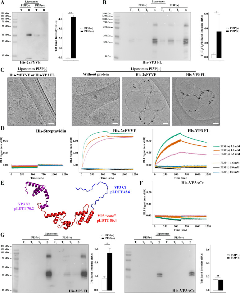
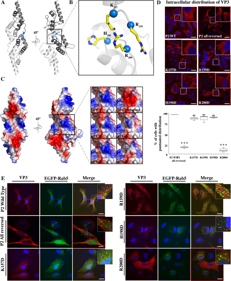
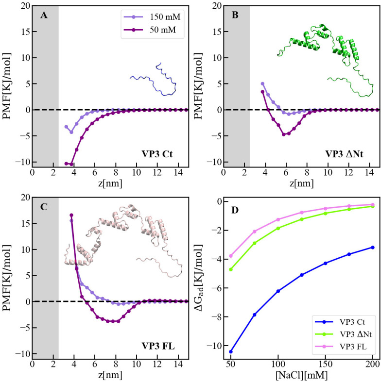
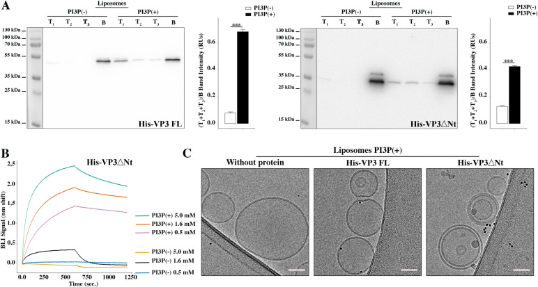
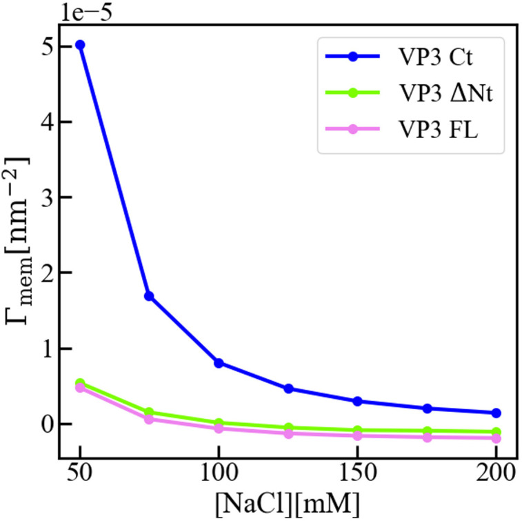
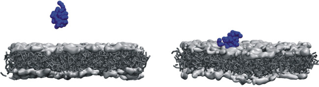
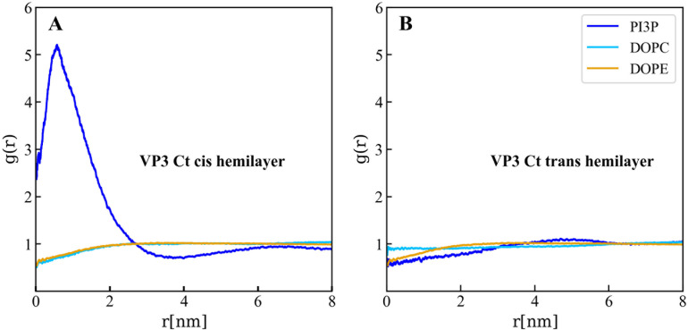
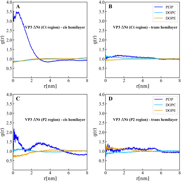
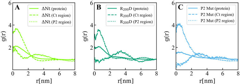
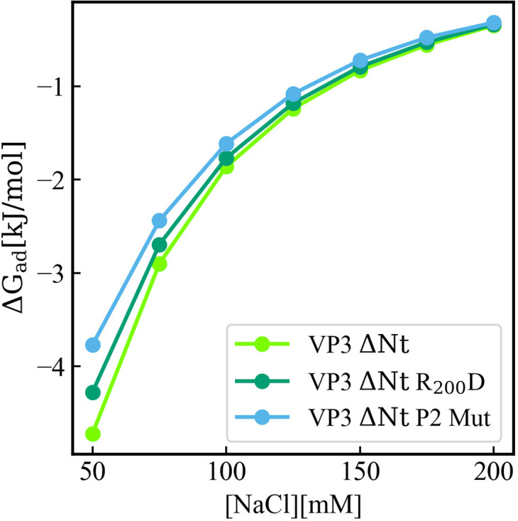
Birnaviruses are a group of double-stranded RNA (dsRNA) viruses infecting birds, fish, and insects. Early endosomes (EE) constitute the platform for viral replication. Here, we study the mechanism of birnaviral targeting of EE membranes. Using the Infectious Bursal Disease Virus (IBDV) as a model, we validate that the viral protein 3 (VP3) binds to phosphatidylinositol-3-phosphate (PI3P) present in EE membranes. We identify the domain of VP3 involved in PI3P-binding, named P2 and localized in the core of VP3, and establish the critical role of the arginine at position 200 (R 200 ), conserved among all known birnaviruses. Mutating R 200 abolishes viral replication. Moreover, we propose a two-stage modular mechanism for VP3 association with EE. Firstly, the carboxy-terminal region of VP3 adsorbs on the membrane, and then the VP3 core reinforces the membrane engagement by specifically binding PI3P through its P2 domain, additionally promoting PI3P accumulation.
Overview
The claims and statistics of this paper haven’t been checked yet.
Run the check — free, no account needed. It reads the open-access text and checks the sources it cites.
Integrity signals
Registry
Cites
Statistics
Figures
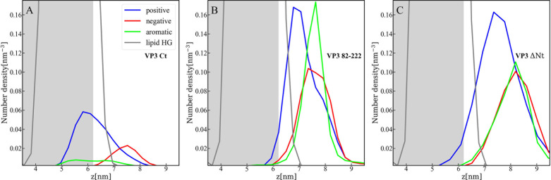
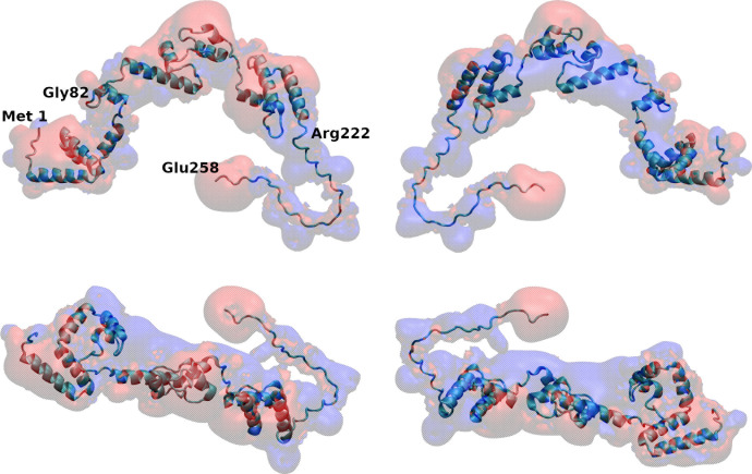
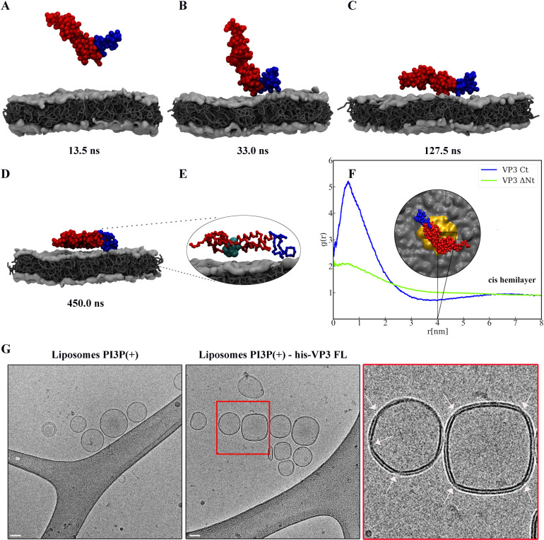
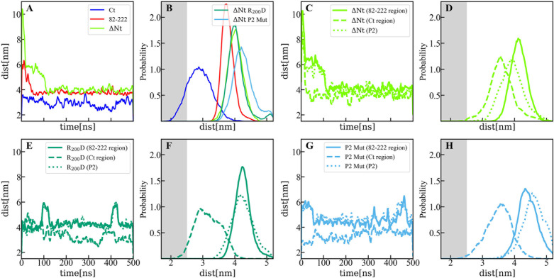
Verification
Claims the paper makes
The claims and statistics of this paper haven’t been checked yet.
Run the check — free, no account needed. It reads the open-access text and checks the sources it cites.
Self-check
Percentages, sums and reported differences re-computed from the paper’s own numbers.
The self-check needs the paper’s full text.
Citations
Cites
Highly accurate protein structure prediction with AlphaFold
Jumper, J., Evans, R., Pritzel, A., Green, T., Figurnov, M., Ronneberger, O., Tunyasuvunakool, K., Bates, R., Žídek, A., Potapenko, A., et al. (2021) · Nature · doi:10.1038/s41586-021-03819-2 · Cited by 43,764
Gaullier, J.-M., Simonsen, A., D'Arrigo, A., Bremnes, B., Stenmark, H., & Aasland, R. (1998) · Nature · doi:10.1038/28767 · Cited by 553
Research works printed but not linked: 74
- Baltimore, 1971, Expression of animal virus genomes, Bacteriological Reviews, 35, 235, 10.1128/br.35.3.235-241.1971 · DOI
- Banani, 2017, Biomolecular condensates: organizers of cellular biochemistry, Nature Reviews. Molecular Cell Biology, 18, 285, 10.1038/nrm.2017.7 · DOI
- Bolte, 2006, A guided tour into subcellular colocalization analysis in light microscopy, Journal of Microscopy, 224, 213, 10.1111/j.1365-2818.2006.01706.x · DOI
- Brodrick, 2023, The formation and function of Birnaviridae virus factories, International Journal of Molecular Sciences, 24, 10.3390/ijms24108471 · DOI
- Burd, 1998, Phosphatidylinositol(3)-phosphate signaling mediated by specific binding to RING FYVE domains, Molecular Cell, 2, 157, 10.1016/s1097-2765(00)80125-2 · DOI
- Casañas, 2008, Structural insights into the multifunctional protein VP3 of birnaviruses, Structure, 16, 29, 10.1016/j.str.2007.10.023 · DOI
- Chiarpotti, 2021, Nanoparticles modified with cell penetrating peptides: assessing adsorption on membranes containing acidic lipids, Colloids and Surfaces. B, Biointerfaces, 197, 10.1016/j.colsurfb.2020.111373 · DOI
- Coulibaly, 2005, The birnavirus crystal structure reveals structural relationships among icosahedral viruses, Cell, 120, 761, 10.1016/j.cell.2005.01.009 · DOI
- Darden, 1993, Particle mesh Ewald: An N⋅log(N) method for Ewald sums in large systems, The Journal of Chemical Physics, 98, 10089, 10.1063/1.464397 · DOI
- de Jong, 2013, Improved parameters for the martini coarse-grained protein force field, Journal of Chemical Theory and Computation, 9, 687, 10.1021/ct300646g · DOI · Paper page
- Delgui, 2013, The endosomal pathway and the Golgi complex are involved in the infectious bursal disease virus life cycle, Journal of Virology, 87, 8993, 10.1128/JVI.03152-12 · DOI
- Delmas, 2019, ICTV virus taxonomy profile: birnaviridae, The Journal of General Virology, 100, 5, 10.1099/jgv.0.001185 · DOI
- Dolinsky, 2004, PDB2PQR: an automated pipeline for the setup of Poisson-Boltzmann electrostatics calculations, Nucleic Acids Research, 32, W665, 10.1093/nar/gkh381 · DOI
- Fernandes, 2011, Phosphoinositides direct equine infectious anemia virus gag trafficking and release, Traffic, 12, 438, 10.1111/j.1600-0854.2010.01153.x · DOI
- Ghanem, 2012, Significantly improved rescue of rabies virus from cDNA plasmids, European Journal of Cell Biology, 91, 10, 10.1016/j.ejcb.2011.01.008 · DOI
- Gibson, 2009, Enzymatic assembly of DNA molecules up to several hundred kilobases, Nature Methods, 6, 343, 10.1038/nmeth.1318 · DOI
- Gillooly, 2000, Localization of phosphatidylinositol 3-phosphate in yeast and mammalian cells, The EMBO Journal, 19, 4577, 10.1093/emboj/19.17.4577 · DOI · Paper page
- Gimenez, 2018, Infectious bursal disease virus hijacks endosomal membranes as the scaffolding structure for viral replication, Journal of Virology, 92, 10.1128/JVI.01964-17 · DOI
- Gimenez, 2021, Phosphatidylinositol 3-phosphate mediates the establishment of infectious bursal disease virus replication complexes in association with early endosomes, Journal of Virology, 95, 10.1128/JVI.02313-20 · DOI
- Gorbalenya, 2002, The palm subdomain-based active site is internally permuted in viral RNA-dependent RNA polymerases of an ancient lineage, Journal of Molecular Biology, 324, 47, 10.1016/s0022-2836(02)01033-1 · DOI
- Gorvel, 1991, rab5 controls early endosome fusion in vitro, Cell, 64, 915, 10.1016/0092-8674(91)90316-q · DOI
- Johnson, 1999, Analyzing protein circular dichroism spectra for accurate secondary structures, Proteins, 35, 307, 10.1002/(SICI)1097-0134(19990515)35:3 · DOI
- Kairys, 2019, Binding affinity in drug design: experimental and computational techniques, Expert Opinion on Drug Discovery, 14, 755, 10.1080/17460441.2019.1623202 · DOI
- Kolli, 2015, Structure-function analysis of vaccinia virus h7 protein reveals a novel phosphoinositide binding fold essential for poxvirus replication, Journal of Virology, 89, 2209, 10.1128/JVI.03073-14/ASSET/F1575F5D-784E-4FD0-9DDB-A2B374935CCA/ASSETS/GRAPHIC/ZJV9990900510007.JPEG · DOI
- Komada, 1995, Growth factor-induced tyrosine phosphorylation of hrs, a novel 115-kilodalton protein with a structurally conserved putative zinc finger domain, Molecular and Cellular Biology, 15, 6213, 10.1128/MCB.15.11.6213 · DOI · Paper page
- Koonin, 2020, Global organization and proposed megataxonomy of the virus world, Microbiology and Molecular Biology Reviews, 84, 10.1128/MMBR.00061-19 · DOI
- Lees, 2006, A reference database for circular dichroism spectroscopy covering fold and secondary structure space, Bioinformatics, 22, 1955, 10.1093/bioinformatics/btl327 · DOI
- Lombardo, 1999, VP1, the putative RNA-dependent RNA polymerase of infectious bursal disease virus, forms complexes with the capsid protein VP3, leading to efficient encapsidation into virus-like particles, Journal of Virology, 73, 6973, 10.1128/JVI.73.8.6973-6983.1999 · DOI
- Luque, 2009, Infectious bursal disease virus is an icosahedral polyploid dsRNA virus, PNAS, 106, 2148, 10.1073/pnas.0808498106 · DOI
- Luque, 2009, Infectious Bursal disease virus: ribonucleoprotein complexes of a double-stranded RNA virus, Journal of Molecular Biology, 386, 891, 10.1016/j.jmb.2008.11.029 · DOI
- Maraver, 2003, The oligomerization domain of VP3, the scaffolding protein of infectious bursal disease virus, plays a critical role in capsid assembly, Journal of Virology, 77, 6438, 10.1128/JVI.77.11.6438-6449.2003 · DOI
- Marrink, 2007, The MARTINI force field: coarse grained model for biomolecular simulations, The Journal of Physical Chemistry. B, 111, 7812, 10.1021/jp071097f · DOI
- Matsuda, 2004, Electroporation and RNA interference in the rodent retina in vivo and in vitro, PNAS, 101, 16, 10.1073/pnas.2235688100 · DOI
- McNulty, 2010, Multiple phosphatidylinositol 3-kinases regulate vaccinia virus morphogenesis, PLOS ONE, 5, 10.1371/journal.pone.0010884 · DOI
- Miles, 2018, CDtoolX, a downloadable software package for processing and analyses of circular dichroism spectroscopic data, Protein Science, 27, 1717, 10.1002/pro.3474 · DOI
- Nascimbeni, 2017, Local detection of PtdIns3P at autophagosome biogenesis membrane platforms, Autophagy, 13, 1602, 10.1080/15548627.2017.1341465 · DOI
- Olson, 1979, Preparation of liposomes of defined size distribution by extrusion through polycarbonate membranes, Biochimica et Biophysica Acta, 557, 9, 10.1016/0005-2736(79)90085-3 · DOI
- Parrinello, 1981, Polymorphic transitions in single crystals: a new molecular dynamics method, Journal of Applied Physics, 52, 7182, 10.1063/1.328693 · DOI
- Periole, 2009, Combining an elastic network with a coarse-grained molecular force field: structure, dynamics, and intermolecular recognition, Journal of Chemical Theory and Computation, 5, 2531, 10.1021/CT9002114/SUPPL_FILE/CT9002114_SI_001.PDF · DOI
- Poma, 2017, Combining the MARTINI and structure-based coarse-grained approaches for the molecular dynamics studies of conformational transitions in proteins, Journal of Chemical Theory and Computation, 13, 1366, 10.1021/ACS.JCTC.6B00986/ASSET/IMAGES/LARGE/CT-2016-00986K_0005.JPEG · DOI
- Potgieter, 2009, Improved strategies for sequence-independent amplification and sequencing of viral double-stranded RNA genomes, The Journal of General Virology, 90, 1423, 10.1099/vir.0.009381-0 · DOI
- Provencher, 1981, Estimation of globular protein secondary structure from circular dichroism, Biochemistry, 20, 33, 10.1021/bi00504a006 · DOI
- Puffer, 1998, Equine infectious anemia virus Gag polyprotein late domain specifically recruits cellular AP-2 adapter protein complexes during virion assembly, Journal of Virology, 72, 10218, 10.1128/JVI.72.12.10218-10221.1998 · DOI
- Qi, 2007, An improved method for infectious bursal disease virus rescue using RNA polymerase II system, Journal of Virological Methods, 142, 81, 10.1016/j.jviromet.2007.01.021 · DOI
- Ramírez, 2019, Adsorption and insertion of polyarginine peptides into membrane pores: The trade-off between electrostatics, acid-base chemistry and pore formation energy, Journal of Colloid and Interface Science, 552, 701, 10.1016/j.jcis.2019.05.087 · DOI
- Reddy, 2005, 5 nm gold-Ni-NTA binds his tags, Microscopy and Microanalysis, 11, 1118, 10.1017/S1431927605507712 · DOI
- Reddy, 2022, Birnaviridae virus factories show features of liquid-liquid phase separation and are distinct from paracrystalline arrays of virions observed by electron microscopy, Journal of Virology, 96, 10.1128/jvi.02024-21 · DOI
- Rydzanicz, 2005, Assembly PCR oligo maker: a tool for designing oligodeoxynucleotides for constructing long DNA molecules for RNA production, Nucleic Acids Research, 33, 10.1093/nar/gki380 · DOI
- Saugar, 2010, Electrostatic interactions between capsid and scaffolding proteins mediate the structural polymorphism of a double-stranded RNA virus, The Journal of Biological Chemistry, 285, 3643, 10.1074/jbc.M109.075994 · DOI
- Schindelin, 2012, Fiji: an open-source platform for biological-image analysis, Nature Methods, 9, 676, 10.1038/nmeth.2019 · DOI · Paper page
- Shah, 2014, Bio-layer interferometry for measuring kinetics of protein-protein interactions and allosteric ligand effects, Journal of Visualized Experiments, 01, 10.3791/51383 · DOI
- Sievers, 2011, Fast, scalable generation of high-quality protein multiple sequence alignments using Clustal Omega, Molecular Systems Biology, 7, 10.1038/msb.2011.75 · DOI
- Simonsen, 1998, EEA1 links PI(3)K function to Rab5 regulation of endosome fusion, Nature, 394, 494, 10.1038/28879 · DOI · Paper page
- Sreerama, 2000, Estimation of protein secondary structure from circular dichroism spectra: comparison of CONTIN, SELCON, and CDSSTR methods with an expanded reference set, Analytical Biochemistry, 287, 252, 10.1006/abio.2000.4880 · DOI
- Stenmark, 1996, Endosomal localization of the autoantigen EEA1 is mediated by a zinc-binding FYVE finger, The Journal of Biological Chemistry, 271, 24048, 10.1074/jbc.271.39.24048 · DOI
- Tanzi, 2003, Equine infectious anemia virus utilizes host vesicular protein sorting machinery during particle release, Journal of Virology, 77, 8440, 10.1128/jvi.77.15.8440-8447.2003 · DOI
- Thakur, 2024, EMBL’s European Bioinformatics Institute (EMBL-EBI) in 2023, Nucleic Acids Research, 52, D10, 10.1093/nar/gkad1088 · DOI
- Valli, 2012, The VP3 factor from viruses of birnaviridae family suppresses rna silencing by binding both long and small rna duplexes, PLOS ONE, 7, 10.1371/journal.pone.0045957 · DOI
- Walker, 2021, Changes to virus taxonomy and to the international code of virus classification and nomenclature ratified by the international committee on taxonomy of viruses (2021), Archives of Virology, 166, 2633, 10.1007/s00705-021-05156-1 · DOI
- Wassenaar, 2015, Computational lipidomics with insane: a versatile tool for generating custom membranes for molecular simulations, Journal of Chemical Theory and Computation, 11, 2144, 10.1021/acs.jctc.5b00209 · DOI · Paper page
- Whitmore, 2004, DICHROWEB, an online server for protein secondary structure analyses from circular dichroism spectroscopic data, Nucleic Acids Research, 32, 10.1093/nar/gkh371 · DOI
- Whitmore, 2008, Protein secondary structure analyses from circular dichroism spectroscopy: methods and reference databases, Biopolymers, 89, 392, 10.1002/bip.20853 · DOI
- Wickham, 2016, Ggplot2: Elegant Graphics for Data Analysis, 10.1007/978-3-319-24277-4 · DOI
- Wills, 1991, Form, function, and use of retroviral gag proteins, AIDS, 5, 639, 10.1097/00002030-199106000-00002 · DOI · Paper page
- Yanai, 2006, Development of a novel Borna disease virus reverse genetics system using RNA polymerase II promoter and SV40 nuclear import signal, Microbes and Infection, 8, 1522, 10.1016/j.micinf.2006.01.010 · DOI
- Zhou, 2022, ggmsa: a visual exploration tool for multiple sequence alignment and associated data, Briefings in Bioinformatics, 23, 10.1093/bib/bbac222 · DOI
- Da Costa B, Soignier S, Chevalier C, Henry C, Thory C, Huet JC, Delmas B. 2018. Blotched snakehead virus is a new aquatic birnavirus that is slightly more related to avibirnavirus than to aquabirnavirus. NCBI Protein. https://www.ncbi.nlm.nih.gov/protein/YP_052872.1. YP_052872.1.
- Mohr PG, Moody NJ, Williams LM, Hoad J, Crane MS. 2018. Molecular characterization of Tasmanian aquabirnaviruses from 1998 to 2013. NCBI Protein. https://www.ncbi.nlm.nih.gov/protein/YP_009255397.1. YP_009255397.1.
- Blake S, Ma JY, Caporale DA, Jairath S, Nicholson BL. 2021. Phylogenetic relationships of aquatic birnaviruses based on deduced amino acid sequences of genome segment A cDNA. NCBI Protein. https://www.ncbi.nlm.nih.gov/protein/YP_010084301.1. YP_010084301.1.
- Suzuki S, Kimura M, Kusuda R. 2018. The complete nucleotide sequence of the polyprotein and VP5 gene of a marine birnavirus. NCBI Protein. https://www.ncbi.nlm.nih.gov/protein/NP_690805.1. NP_690805.1.
- Duncan R, Dobos P. 2018. The nucleotide sequence of infectious pancreatic necrosis virus (IPNV) dsRNA segment A reveals one large ORF encoding a precursor polyprotein. NCBI Protein. https://www.ncbi.nlm.nih.gov/protein/NP_047196.1. NP_047196.1.
- Mohr PG, Moody NJ, Williams LM, Hoad J, Crane MS. 2018. Molecular characterization of Tasmanian aquabirnaviruses from 1998 to 2013. NCBI Protein. https://www.ncbi.nlm.nih.gov/protein/YP_009177608.1. YP_009177608.1.
- Nobiron I, Galloux M, Henry C, Torhy C, Boudinot P, Lejal N, Da Costa B, Delmas B. 2018. Genome and polypeptides characterization of Tellina virus 1 reveals a fifth genetic cluster in the Birnaviridae family. NCBI Protein. https://www.ncbi.nlm.nih.gov/protein/YP_009509080.1. YP_009509080.
- Chen J, Toh X, Ong J, Wang Y, Teo X, Lee B, Wong P, Chee S, Wee D, Wang A, Ng Y, Tan BM, Khor D, Chong S. 2022. Detection and characterisation of a novel marine Birnavirus isolated from Asian Seabass in Singapore. NCBI Protein. https://www.ncbi.nlm.nih.gov/protein/YP_010086267.1. YP_010086267.1.
Metadata
- Authors
- Zanetti, F. A., Fernandez, I., Baquero, E., Guardado-Calvo, P., Ferrino-Iriarte, A., Dubois, S., Morel, E., Alfonso, V., Aguilera, M. O., Celayes, M. E., Polo, L. M., Suhaiman, L., Galassi, V. V., Chiarpotti, M. V., Allende-Ballestero, C., Rodriguez, J. M., Castón, J. R., Lijavetzky, D., Taboga, O., Colombo, M. I., Del Pópolo, M., Rey, F. A., & Delgui, L. R.
- Affiliations
- Consejo Nacional de Investigaciones Científicas y Técnicas (AR)
- Institut Pasteur (FR)
- Institut Pasteur, Université Paris Cité, Structural Virology Unit
- Centre National de la Recherche Scientifique (FR)
- Université Paris Cité, INSERM UMR-S1151, CNRS UMR-S8253, Institut Necker Enfants Malades
- Instituto Interdisciplinario de Ciencias Básicas (ICB), Consejo Nacional de Investigaciones Científicas y Técnicas (CONICET)
- Consejo Superior de Investigaciones Científicas (ES)
- Department of Structure of Macromolecules, Centro Nacional de Biotecnología (CNB-CSIC)
- Instituto de Histología y Embriología de Mendoza, Universidad Nacional de Cuyo (UNCuyo), Consejo Nacional de Investigaciones Científicas y Técnicas (CONICET), Centro Universitario
- Corresponding author
- Delgui, Laura Ruth
- ORCID
- Venue
- eLife
- Publisher
- eLife Sciences Publications, Ltd
- Published
- 2025-03-06
- Volume
- 13
- Pages
- RP97261
- ISSN
- 2050-084X (ISSN-L)
- PMC
- PMC11884790
- OpenAlex
- W4395667216
- Cited by
- 4
- References
- 2
- Citations by year
- 2026: 3 · 2025: 1
- Type
- posted content
- Language
- en
- Open access
- gold
- doi.org
- Licence
- Locations
- eLife (publishedVersion, cc-by)
- PubMed Central (publishedVersion, cc-by)
- PubMed (publishedVersion)
- DIGITAL.CSIC (Spanish National Research Council (CSIC)) (acceptedVersion)
- HAL (submittedVersion, cc-by)
- DOAJ (DOAJ: Directory of Open Access Journals) (submittedVersion, cc-by-sa)
- Funders
- Ministry of Science, Technology and Innovation (PICT 2016-0528, PICT 2019-01324, PICT 2015-2210, PICT 2019-01889, PICT 2020-3795)
- National Scientific and Technical Research Council (PIP 2015-2017 11220150100114CO, PIP 2021-2023 11220200103195CO, PIP 11220200103223CO, 2021–2023 11220200103139CO)
- National University of Cuyo (2013-2015 M006, 2016-2018 M029, 2019-2021 M071, 2022-2024 M012, Teaching Staff Mobility Program in 2018-2019)
- Ministry of Education (BEC.AR 2018)
- International Union of Biochemistry and Molecular Biology (2020 Mid-Career Research Fellowship)
- Spanish Ministry of Science and Innovation (PID2020-113287RB-I00)
- Comunidad Autónoma de Madrid (P2018/NMT-4389)
- Topics
- Life Sciences › Biochemistry, Genetics and Molecular Biology › Cell Biology › Cellular transport and secretion
- Life Sciences › Agricultural and Biological Sciences › Animal Science and Zoology › Animal Virus Infections Studies
- Health Sciences › Medicine › Epidemiology › Virology and Viral Diseases
- Keywords
- birnavirus, VP3, phosphatidylinositol 3-phosphate, early endosomes, membrane targeting, infectious bursal disease virus, viral replication, P2 domain, double-stranded RNA virus, membrane adsorption
- MeSH terms
- Animals, Humans, Infectious bursal disease virus, Intracellular Membranes, Protein Binding, Endosomes, Virus Replication, Viral Structural Proteins, Phosphatidylinositol Phosphates
About these checks
Registry, retraction, Cites and statistics results are computed, not judged, with one exception: a printed reference that no index lists is read once by a model before it counts as a citation error. Claims are extracted by a model and kept only when their quote appears verbatim in the text; each cited source is then checked, with a model’s help, for whether it says what the claim relies on. Those verdicts can be wrong — read the claim’s quote and the cited source before relying on one.
Statistics and arithmetic are recomputed, not judged.
Confidence bands are shown only for fields calibrated on our annotated test set. None is calibrated yet, so no band is shown.
What counts as a claim ›How we measure confidence ›How we check this ›
Are you an author, or do you see a problem with this page? Request a review
