

The DOI matches its Crossref record; 63 of the 63 references it cites were found in a registry or an index; 6 of the 75 sources its claims cite were checked for support.
Positron Emission Tomography Imaging With [ 18 F]flortaucipir and Postmortem Assessment of Alzheimer Disease Neuropathologic Changes
Open accessAbstract
Importance: Positron emission tomography (PET) may increase the diagnostic accuracy and confirm the underlying neuropathologic changes of Alzheimer disease (AD). Objective: To determine the accuracy of antemortem [ 18 F]flortaucipir PET images for predicting the presence of AD-type tau pathology at autopsy. Design, Setting, and Participants: This diagnostic study (A16 primary cohort) was conducted from October 2015 to June 2018 at 28 study sites (27 in US sites and 1 in Australia). Individuals with a terminal illness who were older than 50 years and had a projected life expectancy of less than 6 months were enrolled. All participants underwent [ 18 F]flortaucipir PET imaging, and scans were interpreted by 5 independent nuclear medicine physicians or radiologists. Supplemental autopsy [ 18 F]flortaucipir images and pathological samples were also collected from 16 historically collected cases. A second study (FR01 validation study) was conducted from March 26 to April 26, 2019, in which 5 new readers assessed the original PET images for comparison to autopsy. Main Outcomes and Measures: [ 18 F]flortaucipir PET images were visually assessed and compared with immunohistochemical tau pathology. An AD tau pattern of flortaucipir retention was assessed for correspondence with a postmortem B3-level (Braak stage V or VI) pathological pattern of tau accumulation and to the presence of amyloid-β plaques sufficient to meet the criteria for high levels of AD neuropathological change. Success was defined as having at least 3 of the 5 readers above the lower bounds of the 95% CI for both sensitivity and specificity of 50% or greater. Results: A total of 156 patients were enrolled in the A16 study and underwent [ 18 F]flortaucipir PET imaging. Of these, 73 died during the study, and valid autopsies were performed for 67 of these patients. Three autopsies were evaluated as test cases and removed from the primary cohort (n = 64). Of the 64 primary cohort patients, 34 (53%) were women and 62 (97%) were white; mean (SD) age was 82.5 (9.6) years; and 49 (77%) had dementia, 1 (2%) had mild cognitive impairment, and 14 (22%) had normal cognition. Prespecified success criteria were met for the A16 primary cohort. The flortaucipir PET scans predicted a B3 level of tau pathology, with sensitivity ranging from 92.3% (95% CI, 79.7%-97.3%) to 100.0% (95% CI, 91.0%-100.0%) and specificity ranging from 52.0% (95% CI, 33.5%-70.0%) to 92.0% (95% CI, 75.0%-97.8%). A high level of AD neuropathological change was predicted with sensitivity of 94.7% (95% CI, 82.7%-98.5%) to 100.0% (95% CI, 90.8%-100.0%) and specificity of 50.0% (95% CI, 32.1%-67.9%) to 92.3% (95% CI, 75.9%-97.9%). The FR01 validation study also met prespecified success criteria. Addition of the supplemental autopsy data set and 3 test cases, which comprised a total of 82 patients and autopsies for both the A16 and FR01 studies, resulted in improved specificity and comparable overall accuracy. Among the 156 enrolled participants, 14 (9%) experienced at least 1 treatment-emergent adverse event. Conclusions and Relevance: This study’s findings suggest that PET imaging with [ 18 F]flortaucipir could be used to identify the density and distribution of AD-type tau pathology and the presence of high levels of AD neuropathological change, supporting a neuropathological diagnosis of AD.
Overview
AI snapshot
Written from the paper’s full text.
TL;DR
In a prospective autopsy-validated diagnostic study, [18F]flortaucipir PET visual reads detected B3-level tau neurofibrillary tangles and high Alzheimer disease neuropathologic change with high sensitivity and moderate-to-high specificity. A second independent reader validation study confirmed the findings, and quantitative SUVR analysis also showed high specificity.
Key findings
In the A16 primary cohort (n = 64), flortaucipir PET met prespecified success criteria for detecting B3 NFT score: sensitivity ranged from 92.3% (95% CI, 79.7%-97.3%) to 100.0% (95% CI, 91.0%-100.0%) and specificity from 52.0% (95% CI, 33.5%-70.0%) to 92.0% (95% CI, 75.0%-97.8%) across 5 readers; majority read sensitivity was 92.3% and specificity 80.0%. For high ADNC, sensitivity ranged from 94.7% (95% CI, 82.7%-98.5%) to 100.0% (95% CI, 90.8%-100.0%) and specificity from 50.0% (95% CI, 32.1%-67.9%) to 92.3% (95% CI, 75.9%-97.9%); majority read sensitivity was 94.7% and specificity 80.8%.
- In the full autopsy data set (n = 82), B3 sensitivity ranged from 89.1% (95% CI, 77.0%-95.3%) to 93.5% (95% CI, 82.5%-97.8%) and specificity from 66.7% (95% CI, 50.3%-79.8%) to 94.4% (95% CI, 81.9%-98.5%); high ADNC sensitivity ranged from 95.1% (95% CI, 83.9%-98.7%) to 97.6% (95% CI, 87.4%-99.6%) and specificity from 65.9% (95% CI, 50.5%-78.4%) to 90.2% (95% CI, 77.5%-96.1%). Majority read results were B3 sensitivity 89.1% and specificity 86.1%, and high ADNC sensitivity 95.1% and specificity 82.9%.
- The FR01 validation study also met prespecified success criteria. Interrater reliability was high: A16 all read scans (n = 105) showed 89.9% agreement (Fleiss κ, 0.80; P < .001), A16 primary cohort scans (n = 64) showed 88.4% agreement (Fleiss κ, 0.74; P < .001), and FR01 (n = 82) had Fleiss κ 0.82 (P < .001).
- Quantitative SUVR analysis in 60 of 67 scans meeting quality criteria established an optimal cut point greater than 1.113, yielding sensitivity of 84.2% for B3 NFT score and 86.5% for high ADNC, with 100% specificity for both. Safety: 14 of 156 enrolled participants (9%) had at least 1 treatment-emergent adverse event; agitation (n = 3) and headache (n = 2) were most common, and 3 participants (2%) had serious adverse events within 48 hours, including 2 deaths reported as not associated with study drug or procedure.
Methods
This multicenter diagnostic study (A16 primary cohort; NCT02516046) enrolled participants older than 50 years with terminal illness and projected life expectancy less than 6 months, ranging from clinically normal cognition to dementia, at 28 sites (27 US, 1 Australia) from October 2015 through June 2018. The A16 primary cohort included 64 participants with valid evaluable flortaucipir images and autopsy; the full autopsy data set included 82 cases (64 primary cohort, 2 evaluable test cases, 16 supplemental historically collected autopsy cases), and a separate FR01 validation study (NCT03901092) reinterpreted 82 images.
- PET imaging was acquired for 20 minutes (4 × 5-minute frames) beginning approximately 80 minutes after intravenous 370 (±10%) MBq [18F]flortaucipir; scans were visually rated by 5 blinded nuclear medicine physicians or radiologists as negative, moderate, or advanced AD tau pattern, and quantitative SUVRs used a PERSI white matter reference region and a weighted neocortical target.
- Neuropathological assessment was performed by 2 blinded authors using NIA–AA guidelines, with AT8 immunohistochemistry for Braak NFT staging and 6E10 for amyloid plaque; primary comparisons were flortaucipir PET classification against B3 NFT score (Braak V-VI) and high ADNC level. Analyses calculated sensitivity, specificity, accuracy, predictive values, and likelihood ratios per reader and for majority read, with Wilson 95% CIs, Fleiss and simple κ statistics, and ROC analysis for the SUVR cut point.
Limitations
The paper states that the A16 cohort was older than typical symptomatic AD trial populations, had more advanced clinical disease with only 1 case of mild cognitive impairment, and lacked racial/ethnic diversity; visual reads showed best accuracy for the most advanced NFT tau load and ADNC, and earlier AD stages may meet intermediate pathological criteria or have neuritic plaque without substantial tau. The paper also states that up to 2 of 5 readers did not meet statistical specificity criteria for primary outcomes, largely from overcalling small noncontiguous temporal lobe activity, and that read errors may relate to imprecise cerebellar reference region drawing; subcortical [18F]flortaucipir retention appeared to represent off-target binding of unknown source.
- The paper states that larger sample sizes are needed to confirm high specificity for distinguishing AD from non-AD tau pathologies.
- Evident from the text: the primary cohort lacked structural imaging and used PET-to-PET registration; the full autopsy data set combined primary, test-case, and historically collected supplemental cases, and cognitively normal participants could remain eligible for autopsy regardless of time from scan to autopsy, which may introduce heterogeneity.
Model-written summary. Check it against the paper before relying on it.
Integrity signals
Registry
Cites
Statistics
Figures
Verification
Claims the paper makes
40 Own result · 28 Not checked yet · 16 Own interpretation · 10 Own method · 4 No basis given · 2 Supported · 1 Supported so far
The sources with a verdict support this claim; the rest weren’t checked yet, have no full text we could read, or their verdicts aren’t rated yet.
In the paper
“Positron emission tomography (PET) ligands have been shown to provide a minimally invasive estimate of the neuropathological features of AD, such as Aβ neuritic plaque deposition.15,16,17,18”
§ Positron Emission Tomography Imaging With [18F]flortaucipir and Postmortem Assessment of Alzheimer Disease Neuropathologic Changes > Introduction
What the sources say
- Supportedcites [15] Wong DF, Rosenberg PB, Zhou Y,. In vivo imaging of amyloid deposition in Alzheimer disease using the radioligand 18F-AV-45 (florbetapir [corrected] F 18). J Nucl Med. 2010; 51(6): 913- 920. doi: 10.2967/jnumed.109.069088 20501908 PMC3101877 source
The Introduction (page not specified) describes PET tracers as noninvasive markers that bind aggregated Aβ in amyloid plaques and can assess relative brain amyloid pathology. The Results (page not specified) report greater cortical tracer uptake in patients with Alzheimer disease than in healthy controls, supporting the claim that PET ligands provide an estimate of this pathological feature. (LLM, conf=0.86)
- Not checked yetcites [16] Clark CM, Pontecorvo MJ, Beach TG,; AV-45-A16 Study Group. Cerebral PET with florbetapir compared with neuropathology at autopsy for detection of neuritic amyloid-β plaques: a prospective cohort study. Lancet Neurol. 2012; 11(8): 669- 678. doi: 10.1016/S1474-4422(12)70142-4 22749065 source
- Supportedcites [17] Curtis C, Gamez JE, Singh U,. Phase 3 trial of flutemetamol labeled with radioactive fluorine 18 imaging and neuritic plaque density. JAMA Neurol. 2015; 72(3): 287- 294. doi: 10.1001/jamaneurol.2014.4144 25622185 source
The Abstract reports that PET imaging with flutemetamol detected brain β-amyloid in vivo and was assessed against neuritic plaque levels determined at autopsy. It reports high sensitivity and specificity in the study’s end-of-life population, supporting the claim that a PET ligand can provide an in-vivo estimate of this neuropathological feature. (LLM, conf=0.90)
- Supportedcites [18] Sabri O, Sabbagh MN, Seibyl J,; Florbetaben Phase 3 Study Group. Florbetaben PET imaging to detect amyloid beta plaques in Alzheimer’s disease: phase 3 study. Alzheimers Dement. 2015; 11(8): 964- 974. doi: 10.1016/j.jalz.2015.02.004 25824567 source
The Additional information section reports high diagnostic efficacy for florbetaben PET in detecting neuritic plaques and confirms its efficacy for in vivo detection of β-amyloid deposits. The study compared PET findings with post-mortem histopathology, supporting the description of PET as a minimally invasive estimate of this neuropathological feature. (LLM, conf=0.92)
boostedcorrelational- Asserted: passes — The authors put it forward as true.
- Checkable: passes — It can be checked against evidence.
- Load-bearing: passes — The paper’s conclusions depend on it.
- Based: passes — The paper gives a reason why it holds.
- Supported
Every source the paper cites for this claim was checked and supports it.
In the paper
“These results are consistent with previously observed correspondence between elevated flortaucipir PET signal and elevated signal on amyloid PET.23,45”
§ Positron Emission Tomography Imaging With [18F]flortaucipir and Postmortem Assessment of Alzheimer Disease Neuropathologic Changes > Discussion
What the sources say
- Supportedcites [23] Pontecorvo MJ, Devous MD Sr, Navitsky M,; 18F-AV-1451-A05 investigators. Relationships between flortaucipir PET tau binding and amyloid burden, clinical diagnosis, age and cognition. Brain. 2017; 140(3): 748- 763. doi: 10.1093/brain/aww334 28077397 PMC5382945 source
The Results section reports a significant correlation between flortaucipir and florbetapir PET signals (r = 0.64, P < 0.0001). It also says the probability of elevated flortaucipir signal increased with florbetapir signal, while noting that amyloid-positive scans did not always have elevated flortaucipir signal. (LLM, conf=0.93)
- Supportedcites [45] Johnson KA, Schultz A, Betensky RA,. Tau positron emission tomographic imaging in aging and early Alzheimer disease. Ann Neurol. 2016; 79(1): 110- 119. doi: 10.1002/ana.24546 26505746 PMC4738026 source
The Results section reports that higher inferior temporal 18F T807 (flortaucipir) binding was associated with higher cortical PiB amyloid PET retention. The Discussion adds that neocortical 18F T807 binding above SUVR 1.3 occurred only in individuals with elevated amyloid PET signal, while cautioning that larger samples are needed to establish how robust that finding is. (LLM, conf=0.93)
plaincorrelational- Asserted: passes — The authors put it forward as true.
- Checkable: passes — It can be checked against evidence.
- Load-bearing: unclear — It is unclear whether the paper’s conclusions depend on it.
- Based: passes — The paper gives a reason why it holds.
- Supported
Every source the paper cites for this claim was checked and supports it.
In the paper
“In support of this finding and the quantitative data presented herein, a recent study of 26 cases comparing flortaucipir PET image and autopsy findings demonstrated a sensitivity of 87% and specificity of 82% for identifying AD-spectrum pathological diagnoses, using quantitative analysis of a mesial, inferior, or middle temporal lobe region of interest.25”
§ Positron Emission Tomography Imaging With [18F]flortaucipir and Postmortem Assessment of Alzheimer Disease Neuropathologic Changes > Discussion
What the sources say
- Supportedcites [25] Lowe VJ, Lundt ES, Albertson SM,. Tau-positron emission tomography correlates with neuropathology findings. Alzheimers Dement. 2020; 16(3): 561- 571. doi: 10.1016/j.jalz.2019.09.079 31784374 PMC7067654 source
Split into 2 atoms — 2 confirmed, 0 partially supported, 0 disputed, 0 misattributed, 0 unverifiable. Expand the claim to see per-atom verdicts.
boostedcorrelationaln = 26- Asserted: passes — The authors put it forward as true.
- Checkable: passes — It can be checked against evidence.
- Load-bearing: passes — The paper’s conclusions depend on it.
- Based: passes — The paper gives a reason why it holds.
- Supported
A result the paper reports from its own data — it cites no other source for it, so there is none to check. The quote shows it is in the paper.
In the paper
“The A16 primary cohort analysis of 64 patients met prespecified success criteria, with flortaucipir PET imaging demonstrating statistically significant sensitivity and specificity for detecting both NFT score of B3 and high level of ADNC, as determined by the interpretations of at least 3 of the 5 physican readers of the PET scans (Table 2; Figure 2).”
§ Positron Emission Tomography Imaging With [18F]flortaucipir and Postmortem Assessment of Alzheimer Disease Neuropathologic Changes > Results > Primary Cohort Outcomes
boostedcorrelationaln = 64- Asserted: passes — The authors put it forward as true.
- Checkable: passes — It can be checked against evidence.
- Load-bearing: passes — The paper’s conclusions depend on it.
- Based: passes — The paper gives a reason why it holds.
- Rests on an assumption: Tau pathology does not change substantially between antemortem flortaucipir positron emission tomography imaging and postmortem assessment.
A result the paper reports from its own data — it cites no other source for it, so there is none to check. The quote shows it is in the paper.
In the paper
“Among the 156 enrolled participants, 14 (9%) experienced at least 1 treatment-emergent adverse event (eTable 2 in the Supplement).”
§ Positron Emission Tomography Imaging With [18F]flortaucipir and Postmortem Assessment of Alzheimer Disease Neuropathologic Changes > Results > Safety Assessment
plainn = 156- Asserted: passes — The authors put it forward as true.
- Checkable: passes — It can be checked against evidence.
- Load-bearing: unclear — It is unclear whether the paper’s conclusions depend on it.
- Based: passes — The paper gives a reason why it holds.
A result the paper reports from its own data — it cites no other source for it, so there is none to check. The quote shows it is in the paper.
In the paper
“The A16 primary cohort analysis of 64 patients met prespecified success criteria, with flortaucipir PET imaging demonstrating statistically significant sensitivity and specificity for detecting both NFT score of B3 and high level of ADNC, as determined by the interpretations of at least 3 of the 5 physican readers of the PET scans (Table 2; Figure 2).”
§ Positron Emission Tomography Imaging With [18F]flortaucipir and Postmortem Assessment of Alzheimer Disease Neuropathologic Changes > Results > Primary Cohort Outcomes
plainn = 64- Asserted: passes — The authors put it forward as true.
- Checkable: passes — It can be checked against evidence.
- Load-bearing: passes — The paper’s conclusions depend on it.
- Based: passes — The paper gives a reason why it holds.
- Rests on an assumption: Tau pathology does not change substantially between antemortem flortaucipir positron emission tomography imaging and postmortem assessment.
A result the paper reports from its own data — it cites no other source for it, so there is none to check. The quote shows it is in the paper.
In the paper
“The A16 primary cohort analysis of 64 patients met prespecified success criteria, with flortaucipir PET imaging demonstrating statistically significant sensitivity and specificity for detecting both NFT score of B3 and high level of ADNC, as determined by the interpretations of at least 3 of the 5 physican readers of the PET scans (Table 2; Figure 2).”
§ Positron Emission Tomography Imaging With [18F]flortaucipir and Postmortem Assessment of Alzheimer Disease Neuropathologic Changes > Results > Primary Cohort Outcomes
boostedcorrelationaln = 64- Asserted: passes — The authors put it forward as true.
- Checkable: passes — It can be checked against evidence.
- Load-bearing: passes — The paper’s conclusions depend on it.
- Based: passes — The paper gives a reason why it holds.
- Rests on an assumption: Tau pathology does not change substantially between antemortem flortaucipir positron emission tomography imaging and postmortem assessment.
A result the paper reports from its own data — it cites no other source for it, so there is none to check. The quote shows it is in the paper.
In the paper
“In the FR01 study (n = 82), the Fleiss κ value was 0.82 (P < .001).”
§ Positron Emission Tomography Imaging With [18F]flortaucipir and Postmortem Assessment of Alzheimer Disease Neuropathologic Changes > Results > Primary Cohort Outcomes
plainn = 82 ·- Asserted: passes — The authors put it forward as true.
- Checkable: passes — It can be checked against evidence.
- Load-bearing: passes — The paper’s conclusions depend on it.
- Based: passes — The paper gives a reason why it holds.
A result the paper reports from its own data — it cites no other source for it, so there is none to check. The quote shows it is in the paper.
In the paper
“Of these, 19 had less than high levels of ADNC at autopsy.”
§ Positron Emission Tomography Imaging With [18F]flortaucipir and Postmortem Assessment of Alzheimer Disease Neuropathologic Changes > Results > Primary Cohort Outcomes
plainn = 26- Asserted: passes — The authors put it forward as true.
- Checkable: passes — It can be checked against evidence.
- Load-bearing: passes — The paper’s conclusions depend on it.
- Based: passes — The paper gives a reason why it holds.
A result the paper reports from its own data — it cites no other source for it, so there is none to check. The quote shows it is in the paper.
In the paper
“The majority read analysis for the A16 primary cohort showed similar results for B3 level sensitivity (92.3%; 95% CI, 79.7%-97.3%) and specificity (80.0%; 95% CI, 60.9%-91.1%), and for high ADNC level sensitivity (94.7%; 95% CI, 82.7%-98.5%) and specificity (80.8%; 95% CI, 62.1%-91.5%) (Table 2).”
§ Positron Emission Tomography Imaging With [18F]flortaucipir and Postmortem Assessment of Alzheimer Disease Neuropathologic Changes > Results > Primary Cohort Outcomes
plaincorrelationaln = 64- Asserted: passes — The authors put it forward as true.
- Checkable: passes — It can be checked against evidence.
- Load-bearing: passes — The paper’s conclusions depend on it.
- Based: passes — The paper gives a reason why it holds.
- Rests on an assumption: Tau pathology does not change substantially between antemortem flortaucipir positron emission tomography imaging and postmortem assessment.
- Probably not a substantial claim. 30
- It may state two separate claims as one. 32
A result the paper reports from its own data — it cites no other source for it, so there is none to check. The quote shows it is in the paper.
In the paper
“After removal of the 3 test cases, the remaining 64 participants were included in the A16 primary cohort, with autopsy occurring a mean (SD) 16.2 (15.0) hours from the recorded time of death and 2.6 (2.14) months after [18F]flortaucipir imaging.”
§ Positron Emission Tomography Imaging With [18F]flortaucipir and Postmortem Assessment of Alzheimer Disease Neuropathologic Changes > Results > Clinical Demographics
plainn = 64- Asserted: passes — The authors put it forward as true.
- Checkable: passes — It can be checked against evidence.
- Load-bearing: fails — The paper’s conclusions may not depend on it.
- Based: passes — The paper gives a reason why it holds.
Probably not a substantial claim. The paper’s conclusions may not depend on it. It may state two separate claims as one.
A result the paper reports from its own data — it cites no other source for it, so there is none to check. The quote shows it is in the paper.
In the paper
“Before study completion, 73 participants died within 9 months of imaging, of whom 67 (92%) had a valid study autopsy (eFigure 2 in the Supplement).”
§ Positron Emission Tomography Imaging With [18F]flortaucipir and Postmortem Assessment of Alzheimer Disease Neuropathologic Changes > Results > Clinical Demographics
plainn = 73- Asserted: passes — The authors put it forward as true.
- Checkable: passes — It can be checked against evidence.
- Load-bearing: unclear — It is unclear whether the paper’s conclusions depend on it.
- Based: passes — The paper gives a reason why it holds.
It may state two separate claims as one.
A result the paper reports from its own data — it cites no other source for it, so there is none to check. The quote shows it is in the paper.
In the paper
“The majority read analysis for the A16 primary cohort showed similar results for B3 level sensitivity (92.3%; 95% CI, 79.7%-97.3%) and specificity (80.0%; 95% CI, 60.9%-91.1%), and for high ADNC level sensitivity (94.7%; 95% CI, 82.7%-98.5%) and specificity (80.8%; 95% CI, 62.1%-91.5%) (Table 2).”
§ Positron Emission Tomography Imaging With [18F]flortaucipir and Postmortem Assessment of Alzheimer Disease Neuropathologic Changes > Results > Primary Cohort Outcomes
plainn = 64- Asserted: passes — The authors put it forward as true.
- Checkable: passes — It can be checked against evidence.
- Load-bearing: passes — The paper’s conclusions depend on it.
- Based: passes — The paper gives a reason why it holds.
It may state two separate claims as one.
- Rests on an assumption: Tau pathology does not change substantially between antemortem flortaucipir positron emission tomography imaging and postmortem assessment.
A result the paper reports from its own data — it cites no other source for it, so there is none to check. The quote shows it is in the paper.
In the paper
“A total of 156 participants were enrolled in this diagnostic study, underwent flortaucipir imaging, and were included in a safety assessment (Table 1).”
§ Positron Emission Tomography Imaging With [18F]flortaucipir and Postmortem Assessment of Alzheimer Disease Neuropathologic Changes > Results > Clinical Demographics
plainn = 156- Asserted: passes — The authors put it forward as true.
- Checkable: passes — It can be checked against evidence.
- Load-bearing: fails — The paper’s conclusions may not depend on it.
- Based: passes — The paper gives a reason why it holds.
Probably not a substantial claim. The paper’s conclusions may not depend on it. It may state two separate claims as one.
A result the paper reports from its own data — it cites no other source for it, so there is none to check. The quote shows it is in the paper.
In the paper
“Three participants (2%) experienced serious adverse events within 48 hours of the flortaucipir scan: death from acute kidney failure, death from malignant neoplasm, and nonfatal myocardial infarction during hemodialysis.”
§ Positron Emission Tomography Imaging With [18F]flortaucipir and Postmortem Assessment of Alzheimer Disease Neuropathologic Changes > Results > Safety Assessment
plainn = 3- Asserted: passes — The authors put it forward as true.
- Checkable: passes — It can be checked against evidence.
- Load-bearing: unclear — It is unclear whether the paper’s conclusions depend on it.
- Based: passes — The paper gives a reason why it holds.
It may state two separate claims as one.
A result the paper reports from its own data — it cites no other source for it, so there is none to check. The quote shows it is in the paper.
In the paper
“For the B3 level, sensitivity ranged from 92.3% (95% CI, 79.7%-97.3%) to 100.0% (95% CI, 91.0%-100.0%), and specificity ranged from 52.0% (95% CI, 33.5%-70.0%) to 92.0% (95% CI, 75.0%-97.8%).”
§ Positron Emission Tomography Imaging With [18F]flortaucipir and Postmortem Assessment of Alzheimer Disease Neuropathologic Changes > Results > Primary Cohort Outcomes
plainn = 64- Asserted: passes — The authors put it forward as true.
- Checkable: passes — It can be checked against evidence.
- Load-bearing: passes — The paper’s conclusions depend on it.
- Based: passes — The paper gives a reason why it holds.
It may state two separate claims as one.
- Rests on an assumption: Tau pathology does not change substantially between antemortem flortaucipir positron emission tomography imaging and postmortem assessment.
A result the paper reports from its own data — it cites no other source for it, so there is none to check. The quote shows it is in the paper.
In the paper
“Agitation (n = 3) and headache (n = 2) were the most common of these events.”
§ Positron Emission Tomography Imaging With [18F]flortaucipir and Postmortem Assessment of Alzheimer Disease Neuropathologic Changes > Results > Safety Assessment
plainn = 3- Asserted: passes — The authors put it forward as true.
- Checkable: passes — It can be checked against evidence.
- Load-bearing: fails — The paper’s conclusions may not depend on it.
- Based: passes — The paper gives a reason why it holds.
Probably not a substantial claim. The paper’s conclusions may not depend on it.
A result the paper reports from its own data — it cites no other source for it, so there is none to check. The quote shows it is in the paper.
In the paper
“Of the 64 primary cohort patients, 49 (77%) had dementia, 1 (2%) had mild cognitive impairment, and 14 (22%) had normal cognition.”
§ Positron Emission Tomography Imaging With [18F]flortaucipir and Postmortem Assessment of Alzheimer Disease Neuropathologic Changes > Results > Clinical Demographics
plainn = 64- Asserted: passes — The authors put it forward as true.
- Checkable: passes — It can be checked against evidence.
- Load-bearing: fails — The paper’s conclusions may not depend on it.
- Based: passes — The paper gives a reason why it holds.
Probably not a substantial claim. The paper’s conclusions may not depend on it. It may state two separate claims as one.
A result the paper reports from its own data — it cites no other source for it, so there is none to check. The quote shows it is in the paper.
In the paper
“Of the 49 patients with dementia, 33 had a clinical diagnosis of AD based on medical history and 16 had non-AD clinical diagnosis at baseline (eTable 1 in the Supplement).”
§ Positron Emission Tomography Imaging With [18F]flortaucipir and Postmortem Assessment of Alzheimer Disease Neuropathologic Changes > Results > Clinical Demographics
plainn = 49- Asserted: passes — The authors put it forward as true.
- Checkable: passes — It can be checked against evidence.
- Load-bearing: fails — The paper’s conclusions may not depend on it.
- Based: passes — The paper gives a reason why it holds.
Probably not a substantial claim. The paper’s conclusions may not depend on it. It may state two separate claims as one.
A result the paper reports from its own data — it cites no other source for it, so there is none to check. The quote shows it is in the paper.
In the paper
“For the high ADNC level, sensitivity was 94.7% (95% CI, 82.7%-98.5%) to 100.0% (95% CI, 90.8%-100.0%) and specificity was 50.0% (95% CI, 32.1%-67.9%) to 92.3% (95% CI, 75.9%-97.9%) (Table 2).”
§ Positron Emission Tomography Imaging With [18F]flortaucipir and Postmortem Assessment of Alzheimer Disease Neuropathologic Changes > Results > Primary Cohort Outcomes
plaincorrelationaln = 64- Asserted: passes — The authors put it forward as true.
- Checkable: passes — It can be checked against evidence.
- Load-bearing: passes — The paper’s conclusions depend on it.
- Based: passes — The paper gives a reason why it holds.
It may state two separate claims as one.
- Rests on an assumption: Tau pathology does not change substantially between antemortem flortaucipir positron emission tomography imaging and postmortem assessment.
A result the paper reports from its own data — it cites no other source for it, so there is none to check. The quote shows it is in the paper.
In the paper
“Both death events were reported by the investigator as severe adverse events that were not associated with the study drug or procedure.”
§ Positron Emission Tomography Imaging With [18F]flortaucipir and Postmortem Assessment of Alzheimer Disease Neuropathologic Changes > Results > Safety Assessment
plainn = 2- Asserted: passes — The authors put it forward as true.
- Checkable: passes — It can be checked against evidence.
- Load-bearing: fails — The paper’s conclusions may not depend on it.
- Based: passes — The paper gives a reason why it holds.
Probably not a substantial claim. The paper’s conclusions may not depend on it. It may state two separate claims as one.
A result the paper reports from its own data — it cites no other source for it, so there is none to check. The quote shows it is in the paper.
In the paper
“For quantitative analysis, 60 (90%) of 67 scans (primary cohort plus test cases) met the scan quality criteria for SUVR calculation; 6 scans (9%) had severe inter- and within-frame motion, and 1 scan (2%) was acquired outside of the allowable postinjection acquisition time window.”
§ Positron Emission Tomography Imaging With [18F]flortaucipir and Postmortem Assessment of Alzheimer Disease Neuropathologic Changes > Results > Primary Cohort Outcomes
plain- Asserted: passes — The authors put it forward as true.
- Checkable: passes — It can be checked against evidence.
- Load-bearing: passes — The paper’s conclusions depend on it.
- Based: passes — The paper gives a reason why it holds.
It may state two separate claims as one.
A result the paper reports from its own data — it cites no other source for it, so there is none to check. The quote shows it is in the paper.
In the paper
“Success criteria were also met for high level of ADNC with sensitivity ranging from 95.1% (95% CI, 83.9%-98.7%) to 97.6% (95% CI 87.4%-99.6%) and specificity ranging from 65.9% (95% CI, 50.5%-78.4%) to 90.2% (95% CI, 77.5%-96.1%) (Table 2).”
§ Positron Emission Tomography Imaging With [18F]flortaucipir and Postmortem Assessment of Alzheimer Disease Neuropathologic Changes > Results > Primary Cohort Outcomes
plaincorrelationaln = 82- Asserted: passes — The authors put it forward as true.
- Checkable: passes — It can be checked against evidence.
- Load-bearing: passes — The paper’s conclusions depend on it.
- Based: passes — The paper gives a reason why it holds.
It may state two separate claims as one.
A result the paper reports from its own data — it cites no other source for it, so there is none to check. The quote shows it is in the paper.
In the paper
“The FR01 validation study also met the prespecified success criteria for both the FR01 primary cohort (n = 64) and full autopsy data set (n = 82) analyses, demonstrating statistically significant sensitivity and specificity (above 95% CI lower limits >50%) of flortaucipir PET imaging for detecting both the NFT score of B3 NFT and high ADNC level by at least 3 of the 5 readers.”
§ Positron Emission Tomography Imaging With [18F]flortaucipir and Postmortem Assessment of Alzheimer Disease Neuropathologic Changes > Results > Primary Cohort Outcomes
boostedcorrelationaln = 82- Asserted: passes — The authors put it forward as true.
- Checkable: passes — It can be checked against evidence.
- Load-bearing: passes — The paper’s conclusions depend on it.
- Based: passes — The paper gives a reason why it holds.
It may state two separate claims as one.
A result the paper reports from its own data — it cites no other source for it, so there is none to check. The quote shows it is in the paper.
In the paper
“The majority read analysis for the A16 full autopsy data set showed similar results for B3 level sensitivity (89.1%; 95% CI, 77.0%-95.3%) and specificity (86.1%; 95% CI, 71.3%-93.9%), and for high ADNC level sensitivity (95.1%; 95% CI, 83.9%-98.7%) and specificity (82.9%; 95% CI, 68.7%-91.5%) (Table 2).”
§ Positron Emission Tomography Imaging With [18F]flortaucipir and Postmortem Assessment of Alzheimer Disease Neuropathologic Changes > Results > Primary Cohort Outcomes
plainn = 82- Asserted: passes — The authors put it forward as true.
- Checkable: passes — It can be checked against evidence.
- Load-bearing: passes — The paper’s conclusions depend on it.
- Based: passes — The paper gives a reason why it holds.
It may state two separate claims as one.
A result the paper reports from its own data — it cites no other source for it, so there is none to check. The quote shows it is in the paper.
In the paper
“For the A16 study, all read scans (n = 105 of enrolled patients) showed 89.9% agreement (Fleiss κ, 0.80; P < .001); the primary cohort scans (n = 64) showed 88.4% agreement (Fleiss κ, 0.74; P < .001).”
§ Positron Emission Tomography Imaging With [18F]flortaucipir and Postmortem Assessment of Alzheimer Disease Neuropathologic Changes > Results > Primary Cohort Outcomes
plainn = 105 ·- Asserted: passes — The authors put it forward as true.
- Checkable: passes — It can be checked against evidence.
- Load-bearing: unclear — It is unclear whether the paper’s conclusions depend on it.
- Based: passes — The paper gives a reason why it holds.
It may state two separate claims as one.
A result the paper reports from its own data — it cites no other source for it, so there is none to check. The quote shows it is in the paper.
In the paper
“For the full autopsy data set (n = 82), all 5 readers met success criteria with lower confidence limits of sensitivity and specificity greater than 50% for the NFT score of B3 with sensitivity ranging from 89.1% (95% CI, 77.0%-95.3%) to 93.5% (95% CI, 82.5%-97.8%) and specificity ranging from 66.7% (95% CI, 50.3%-79.8%) to 94.4% (95% CI, 81.9%-98.5%).”
§ Positron Emission Tomography Imaging With [18F]flortaucipir and Postmortem Assessment of Alzheimer Disease Neuropathologic Changes > Results > Primary Cohort Outcomes
plainn = 82- Asserted: passes — The authors put it forward as true.
- Checkable: passes — It can be checked against evidence.
- Load-bearing: passes — The paper’s conclusions depend on it.
- Based: passes — The paper gives a reason why it holds.
It may state two separate claims as one.
A result the paper reports from its own data — it cites no other source for it, so there is none to check. The quote shows it is in the paper.
In the paper
“This quantitative cut point yielded a sensitivity of 84.2% for an NFT score of B3 and 86.5% for a high level of ADNC, with 100% specificity for both autopsy measures (eFigure 3 in the Supplement).”
§ Positron Emission Tomography Imaging With [18F]flortaucipir and Postmortem Assessment of Alzheimer Disease Neuropathologic Changes > Results > Primary Cohort Outcomes
plaincorrelational- Asserted: passes — The authors put it forward as true.
- Checkable: passes — It can be checked against evidence.
- Load-bearing: passes — The paper’s conclusions depend on it.
- Based: passes — The paper gives a reason why it holds.
It may state two separate claims as one.
The paper states this without a source or data behind it here, so there is nothing to check it against. The quote shows it is in the paper.
In the paper
“Although [18F]flortaucipir retention in the neocortex appeared to match the distribution of aggregated tau in AD at autopsy, [18F]flortaucipir retention in subcortical structures also appeared to represent off-target binding.”
§ Positron Emission Tomography Imaging With [18F]flortaucipir and Postmortem Assessment of Alzheimer Disease Neuropathologic Changes > Discussion > Limitations
hedgedcorrelational- Asserted: passes — The authors put it forward as true.
- Checkable: passes — It can be checked against evidence.
- Load-bearing: unclear — It is unclear whether the paper’s conclusions depend on it.
- Based: passes — The paper gives a reason why it holds.
It may state two separate claims as one.
None of the sources cited for this claim have been checked yet.
In the paper
“Neuropathological criteria for the diagnosis of AD were established by the National Institute on Aging–Alzheimer Association (NIA–AA)4 and were updated in 2012 to reflect the current thinking on AD pathological assessment.5,6”
§ Positron Emission Tomography Imaging With [18F]flortaucipir and Postmortem Assessment of Alzheimer Disease Neuropathologic Changes > Introduction
What the sources say
- Not checked yetcites [4] Hyman BT. The neuropathological diagnosis of Alzheimer’s disease: clinical-pathological studies. Neurobiol Aging. 1997; 18(4)(suppl): S27- S32. doi: 10.1016/S0197-4580(97)00066-3 9330982
- Not checked yetcites [5] Hyman BT, Phelps CH, Beach TG,. National Institute on Aging-Alzheimer’s Association guidelines for the neuropathologic assessment of Alzheimer’s disease. Alzheimers Dement. 2012; 8(1): 1- 13. doi: 10.1016/j.jalz.2011.10.007 22265587 PMC3266529 source
- Not checked yetcites [6] Jack CR Jr, Bennett DA, Blennow K,; Contributors. NIA-AA research framework: toward a biological definition of Alzheimer’s disease. Alzheimers Dement. 2018; 14(4): 535- 562. doi: 10.1016/j.jalz.2018.02.018 29653606 PMC5958625 source
plain- Asserted: passes — The authors put it forward as true.
- Checkable: passes — It can be checked against evidence.
- Load-bearing: fails — The paper’s conclusions may not depend on it.
- Based: passes — The paper gives a reason why it holds.
Probably not a substantial claim. The paper’s conclusions may not depend on it. It may state two separate claims as one.
In the paper
“Pathological accumulation of aggregated hyperphosphorylated tau protein in neurons and glia underlies a wide range of neurodegenerative disorders.11”
§ Positron Emission Tomography Imaging With [18F]flortaucipir and Postmortem Assessment of Alzheimer Disease Neuropathologic Changes > Introduction
What the sources say
- Not checked yetcites [11] Querfurth HW, LaFerla FM. Alzheimer’s disease. N Engl J Med. 2010; 362(4): 329- 344. doi: 10.1056/NEJMra0909142 20107219 source
plaindirect- Asserted: passes — The authors put it forward as true.
- Checkable: passes — It can be checked against evidence.
- Load-bearing: passes — The paper’s conclusions depend on it.
- Based: passes — The paper gives a reason why it holds.
Probably not a substantial claim.
None of the sources cited for this claim have been checked yet.
In the paper
“With the advent of biomarkers that enable in vivo identification of underlying AD pathological conditions, and the acceptance that AD begins years before the emergence of cognitive impairment, AD is now commonly accepted as a clinicopathological entity7,8 that can be diagnosed without histopathological examination, in the presence of appropriate biomarker evidence of an underlying condition.6,8,9”
§ Positron Emission Tomography Imaging With [18F]flortaucipir and Postmortem Assessment of Alzheimer Disease Neuropathologic Changes > Introduction
What the sources say
- Not checked yetcites [7] Dubois B, Feldman HH, Jacova C,. Revising the definition of Alzheimer’s disease: a new lexicon. Lancet Neurol. 2010; 9(11): 1118- 1127. doi: 10.1016/S1474-4422(10)70223-4 20934914
- Not checked yetcites [8] McKhann GM, Knopman DS, Chertkow H,. The diagnosis of dementia due to Alzheimer’s disease: recommendations from the National Institute on Aging-Alzheimer’s Association workgroups on diagnostic guidelines for Alzheimer’s disease. Alzheimers Dement. 2011; 7(3): 263- 269. doi: 10.1016/j.jalz.2011.03.005 21514250 PMC3312024 source
- Not checked yetcites [6] Jack CR Jr, Bennett DA, Blennow K,; Contributors. NIA-AA research framework: toward a biological definition of Alzheimer’s disease. Alzheimers Dement. 2018; 14(4): 535- 562. doi: 10.1016/j.jalz.2018.02.018 29653606 PMC5958625 source
- Not checked yetcites [9] Knopman DS, Petersen RC, Jack CR Jr. A brief history of “Alzheimer disease”: multiple meanings separated by a common name. Neurology. 2019; 92(22): 1053- 1059. doi: 10.1212/WNL.0000000000007583 31028129 PMC6556090
plain- Asserted: passes — The authors put it forward as true.
- Checkable: passes — It can be checked against evidence.
- Load-bearing: passes — The paper’s conclusions depend on it.
- Based: passes — The paper gives a reason why it holds.
Probably not a substantial claim.
In the paper
“Tau is an intracellular protein that binds to and stabilizes axonal microtubules in neurons, thereby regulating intracellular transport.10”
§ Positron Emission Tomography Imaging With [18F]flortaucipir and Postmortem Assessment of Alzheimer Disease Neuropathologic Changes > Introduction
What the sources say
- Not checked yetcites [10] Avila J, Lucas JJ, Perez M, Hernandez F. Role of tau protein in both physiological and pathological conditions. Physiol Rev. 2004; 84(2): 361- 384. doi: 10.1152/physrev.00024.2003 15044677
plaindirect- Asserted: passes — The authors put it forward as true.
- Checkable: passes — It can be checked against evidence.
- Load-bearing: fails — The paper’s conclusions may not depend on it.
- Based: passes — The paper gives a reason why it holds.
Probably not a substantial claim. The paper’s conclusions may not depend on it.
None of the sources cited for this claim have been checked yet.
In the paper
“Alzheimer disease (AD) is characterized by aggregated tau–containing neurofibrillary tangles (NFTs)1 and by amyloid plaque composed largely of aggregated amyloid-β (Aβ) fragments.2,3”
§ Positron Emission Tomography Imaging With [18F]flortaucipir and Postmortem Assessment of Alzheimer Disease Neuropathologic Changes > Introduction
What the sources say
- Not checked yetcites [1] Goedert M, Jakes R. Mutations causing neurodegenerative tauopathies. Biochim Biophys Acta. 2005; 1739(2-3): 240- 250. doi: 10.1016/j.bbadis.2004.08.007 15615642
- Not checked yetcites [2] Kidd M. Paired helical filaments in electron microscopy of Alzheimer’s disease. Nature. 1963; 197: 192- 193. doi: 10.1038/197192b0 14032480
- Not checked yetcites [3] Masters CL, Multhaup G, Simms G, Pottgiesser J, Martins RN, Beyreuther K. Neuronal origin of a cerebral amyloid: neurofibrillary tangles of Alzheimer’s disease contain the same protein as the amyloid of plaque cores and blood vessels. EMBO J. 1985; 4(11): 2757- 2763. doi: 10.1002/j.1460-2075.1985.tb04000.x 4065091 PMC554575
plain- Asserted: passes — The authors put it forward as true.
- Checkable: passes — It can be checked against evidence.
- Load-bearing: fails — The paper’s conclusions may not depend on it.
- Based: passes — The paper gives a reason why it holds.
Probably not a substantial claim. The paper’s conclusions may not depend on it. It may state two separate claims as one.
None of the sources cited for this claim have been checked yet.
In the paper
“For example, large autopsy studies have demonstrated a high sensitivity and specificity for amyloid PET to distinguish individuals with subsequent autopsy findings of no or sparse neuritic plaque from individuals with moderate to frequent plaque.16,17,18”
§ Positron Emission Tomography Imaging With [18F]flortaucipir and Postmortem Assessment of Alzheimer Disease Neuropathologic Changes > Introduction
What the sources say
- Not checked yetcites [16] Clark CM, Pontecorvo MJ, Beach TG,; AV-45-A16 Study Group. Cerebral PET with florbetapir compared with neuropathology at autopsy for detection of neuritic amyloid-β plaques: a prospective cohort study. Lancet Neurol. 2012; 11(8): 669- 678. doi: 10.1016/S1474-4422(12)70142-4 22749065 source
- Not checked yetcites [17] Curtis C, Gamez JE, Singh U,. Phase 3 trial of flutemetamol labeled with radioactive fluorine 18 imaging and neuritic plaque density. JAMA Neurol. 2015; 72(3): 287- 294. doi: 10.1001/jamaneurol.2014.4144 25622185 source
- Not checked yetcites [18] Sabri O, Sabbagh MN, Seibyl J,; Florbetaben Phase 3 Study Group. Florbetaben PET imaging to detect amyloid beta plaques in Alzheimer’s disease: phase 3 study. Alzheimers Dement. 2015; 11(8): 964- 974. doi: 10.1016/j.jalz.2015.02.004 25824567 source
boostedcorrelational- Asserted: passes — The authors put it forward as true.
- Checkable: passes — It can be checked against evidence.
- Load-bearing: passes — The paper’s conclusions depend on it.
- Based: passes — The paper gives a reason why it holds.
Probably not a substantial claim.
What the paper did — it cites no source for this, so there is none to check. The quote shows it is in the paper.
In the paper
“Neuropathological assessment was performed by 2 authors (T.G.B. and T.J.M.) blinded to clinical and imaging results and using NIA–AA diagnostic scoring guidelines.5,40”
§ Positron Emission Tomography Imaging With [18F]flortaucipir and Postmortem Assessment of Alzheimer Disease Neuropathologic Changes > Methods > Neuropathological Assessment
plain- Asserted: passes — The authors put it forward as true.
- Checkable: passes — It can be checked against evidence.
- Load-bearing: passes — The paper’s conclusions depend on it.
- Based: passes — The paper gives a reason why it holds.
Probably not a substantial claim.
In the paper
“In vitro autoradiography studies of brain tissue from symptomatic patients with AD have found that [18F]flortaucipir signal correlates with the level of paired helical filament tau by immunohistochemistry and binds with a dissociation constant in the approximately 0.5 nM range.20”
§ Positron Emission Tomography Imaging With [18F]flortaucipir and Postmortem Assessment of Alzheimer Disease Neuropathologic Changes > Introduction
What the sources say
- Not checked yetcites [20] Xia CF, Arteaga J, Chen G,. [(18)F]T807, a novel tau positron emission tomography imaging agent for Alzheimer’s disease. Alzheimers Dement. 2013; 9(6): 666- 676. doi: 10.1016/j.jalz.2012.11.008 23411393
plaincorrelational- Asserted: passes — The authors put it forward as true.
- Checkable: passes — It can be checked against evidence.
- Load-bearing: passes — The paper’s conclusions depend on it.
- Based: passes — The paper gives a reason why it holds.
It may state two separate claims as one.
None of the sources cited for this claim have been checked yet.
In the paper
“Studies comparing antemortem tau PET imaging with autopsy have begun assessing the ability of flortaucipir to detect underlying NFTs in AD and non-AD dementias.25,26,27,28,29,30,31,32,33”
§ Positron Emission Tomography Imaging With [18F]flortaucipir and Postmortem Assessment of Alzheimer Disease Neuropathologic Changes > Introduction
What the sources say
- Not checked yetcites [25] Lowe VJ, Lundt ES, Albertson SM,. Tau-positron emission tomography correlates with neuropathology findings. Alzheimers Dement. 2020; 16(3): 561- 571. doi: 10.1016/j.jalz.2019.09.079 31784374 PMC7067654 source
- Not checked yetcites [26] Josephs KA, Whitwell JL, Tacik P,. [18F]AV-1451 tau-PET uptake does correlate with quantitatively measured 4R-tau burden in autopsy-confirmed corticobasal degeneration. Acta Neuropathol. 2016; 132(6): 931- 933. doi: 10.1007/s00401-016-1618-1 27645292 PMC5107140 source
- Not checked yetcites [27] Marquié M, Normandin MD, Meltzer AC,. Pathological correlations of [F-18]-AV-1451 imaging in non-Alzheimer tauopathies. Ann Neurol. 2017; 81(1): 117- 128. doi: 10.1002/ana.24844 27997036 PMC5319193 source
- Not checked yetcites [28] Marquié M, Verwer EE, Meltzer AC,. Lessons learned about [F-18]-AV-1451 off-target binding from an autopsy-confirmed Parkinson’s case. Acta Neuropathol Commun. 2017; 5(1): 75. doi: 10.1186/s40478-017-0482-0 29047416 PMC5648451 source
- Not checked yetcites [29] McMillan CT, Irwin DJ, Nasrallah I,. Multimodal evaluation demonstrates in vivo 18F-AV-1451 uptake in autopsy-confirmed corticobasal degeneration. Acta Neuropathol. 2016; 132(6): 935- 937. doi: 10.1007/s00401-016-1640-3 27815633 PMC5154298 source
- Not checked yetcites [30] Smith R, Puschmann A, Schöll M,. 18F-AV-1451 tau PET imaging correlates strongly with tau neuropathology in MAPT mutation carriers. Brain. 2016; 139(pt 9): 2372- 2379. doi: 10.1093/brain/aww163 27357347 PMC4995360 source
- Not checked yetcites [31] Smith R, Schöll M, Honer M, Nilsson CF, Englund E, Hansson O. Tau neuropathology correlates with FDG-PET, but not AV-1451-PET, in progressive supranuclear palsy. Acta Neuropathol. 2017; 133(1): 149- 151. doi: 10.1007/s00401-016-1650-1 27900460 PMC5209394 source
- Not checked yetcites [32] Day GS, Gordon BA, Perrin RJ,. In vivo [18F]-AV-1451 tau-PET imaging in sporadic Creutzfeldt-Jakob disease. Neurology. 2018; 90(10): e896- e906. doi: 10.1212/WNL.0000000000005064 29438042 PMC5863493 source
- Not checked yetcites [33] Smith R, Wibom M, Pawlik D, Englund E, Hansson O. Correlation of in vivo [18F]flortaucipir with postmortem Alzheimer disease tau pathology. JAMA Neurol. 2019; 76(3): 310- 317. doi: 10.1001/jamaneurol.2018.3692 30508025 PMC6439731 source
plaincorrelational- Asserted: passes — The authors put it forward as true.
- Checkable: passes — It can be checked against evidence.
- Load-bearing: passes — The paper’s conclusions depend on it.
- Based: passes — The paper gives a reason why it holds.
Probably not a substantial claim.
What the paper did — it cites no source for this, so there is none to check. The quote shows it is in the paper.
In the paper
“The flortaucipir PET scans were evaluated by 5 readers who were blinded to clinical and neuropathological results.”
§ Positron Emission Tomography Imaging With [18F]flortaucipir and Postmortem Assessment of Alzheimer Disease Neuropathologic Changes > Methods > Imaging Visual Interpretation
plain- Asserted: passes — The authors put it forward as true.
- Checkable: passes — It can be checked against evidence.
- Load-bearing: passes — The paper’s conclusions depend on it.
- Based: passes — The paper gives a reason why it holds.
Probably not a substantial claim.
None of the sources cited for this claim have been checked yet.
In the paper
“Neuropathological assessment was performed by 2 authors (T.G.B. and T.J.M.) blinded to clinical and imaging results and using NIA–AA diagnostic scoring guidelines.5,40”
§ Positron Emission Tomography Imaging With [18F]flortaucipir and Postmortem Assessment of Alzheimer Disease Neuropathologic Changes > Methods > Neuropathological Assessment
What the sources say
- Not checked yetcites [5] Hyman BT, Phelps CH, Beach TG,. National Institute on Aging-Alzheimer’s Association guidelines for the neuropathologic assessment of Alzheimer’s disease. Alzheimers Dement. 2012; 8(1): 1- 13. doi: 10.1016/j.jalz.2011.10.007 22265587 PMC3266529 source
- Not checked yetcites [40] Montine TJ, Phelps CH, Beach TG,; National Institute on Aging; Alzheimer’s Association. National Institute on Aging-Alzheimer’s Association guidelines for the neuropathologic assessment of Alzheimer’s disease: a practical approach. Acta Neuropathol. 2012; 123(1): 1- 11. doi: 10.1007/s00401-011-0910-3 22101365 PMC3268003 source
plain- Asserted: passes — The authors put it forward as true.
- Checkable: passes — It can be checked against evidence.
- Load-bearing: passes — The paper’s conclusions depend on it.
- Based: passes — The paper gives a reason why it holds.
Probably not a substantial claim.
What the paper did — it cites no source for this, so there is none to check. The quote shows it is in the paper.
In the paper
“To assess overall interreader agreement, Fleiss κ statistics were calculated.”
§ Positron Emission Tomography Imaging With [18F]flortaucipir and Postmortem Assessment of Alzheimer Disease Neuropathologic Changes > Methods > Statistical Analysis
plain- Asserted: passes — The authors put it forward as true.
- Checkable: passes — It can be checked against evidence.
- Load-bearing: fails — The paper’s conclusions may not depend on it.
- Based: passes — The paper gives a reason why it holds.
Probably not a substantial claim. The paper’s conclusions may not depend on it.
What the paper did — it cites no source for this, so there is none to check. The quote shows it is in the paper.
In the paper
“Scans that were considered unevaluable (eg, head out of field of view, severe motion, acquisition start time offsets, and low counts) by at least 3 of the 5 readers were not used in any analyses.”
§ Positron Emission Tomography Imaging With [18F]flortaucipir and Postmortem Assessment of Alzheimer Disease Neuropathologic Changes > Methods > Imaging Visual Interpretation
plain- Asserted: passes — The authors put it forward as true.
- Checkable: passes — It can be checked against evidence.
- Load-bearing: fails — The paper’s conclusions may not depend on it.
- Based: passes — The paper gives a reason why it holds.
Probably not a substantial claim. The paper’s conclusions may not depend on it.
What the paper did — it cites no source for this, so there is none to check. The quote shows it is in the paper.
In the paper
“Success was defined as having at least 3 of the 5 readers above the lower bounds of the 95% CI for both sensitivity and specificity of 50% or greater.”
§ Positron Emission Tomography Imaging With [18F]flortaucipir and Postmortem Assessment of Alzheimer Disease Neuropathologic Changes > Methods > Statistical Analysis
plain- Asserted: passes — The authors put it forward as true.
- Checkable: passes — It can be checked against evidence.
- Load-bearing: passes — The paper’s conclusions depend on it.
- Based: passes — The paper gives a reason why it holds.
Probably not a substantial claim.
What the paper did — it cites no source for this, so there is none to check. The quote shows it is in the paper.
In the paper
“In a subsequent validation study (18F-AV-1415-FR01; NCT03901092), hereafter referred to as the FR01 validation study, a second set of 5 nuclear medicine physicians or radiologists reinterpreted all available scan data.”
§ Positron Emission Tomography Imaging With [18F]flortaucipir and Postmortem Assessment of Alzheimer Disease Neuropathologic Changes > Methods > Study Design
plain- Asserted: passes — The authors put it forward as true.
- Checkable: passes — It can be checked against evidence.
- Load-bearing: passes — The paper’s conclusions depend on it.
- Based: passes — The paper gives a reason why it holds.
Probably not a substantial claim.
What the paper did — it cites no source for this, so there is none to check. The quote shows it is in the paper.
In the paper
“The preplanned A16 primary cohort included all enrolled participants who had valid and evaluable flortaucipir images and who had an autopsy (n = 64), and excluded the 3 test cases and 16 supplemental historically collected autopsy cases.”
§ Positron Emission Tomography Imaging With [18F]flortaucipir and Postmortem Assessment of Alzheimer Disease Neuropathologic Changes > Methods > Statistical Analysis
plainn = 64- Asserted: passes — The authors put it forward as true.
- Checkable: passes — It can be checked against evidence.
- Load-bearing: passes — The paper’s conclusions depend on it.
- Based: passes — The paper gives a reason why it holds.
Probably not a substantial claim. It may state two separate claims as one.
What the paper did — it cites no source for this, so there is none to check. The quote shows it is in the paper.
In the paper
“A preplanned exploratory analysis was performed on the full autopsy data set (n = 82), which included the 64 patients in the primary cohort, 2 evaluable autopsies from the 3 test cases, and the 16 supplemental autopsy cases.”
§ Positron Emission Tomography Imaging With [18F]flortaucipir and Postmortem Assessment of Alzheimer Disease Neuropathologic Changes > Methods > Statistical Analysis
plainn = 82- Asserted: passes — The authors put it forward as true.
- Checkable: passes — It can be checked against evidence.
- Load-bearing: fails — The paper’s conclusions may not depend on it.
- Based: passes — The paper gives a reason why it holds.
Probably not a substantial claim. The paper’s conclusions may not depend on it.
What the paper did — it cites no source for this, so there is none to check. The quote shows it is in the paper.
In the paper
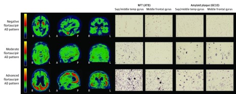
“Five board-certified nuclear medicine physicians or radiologists (eAppendix in the Supplement) independently visually rated flortaucipir PET scans as either having or not having a pattern of flortaucipir retention consistent with AD (Figure 1).”
§ Positron Emission Tomography Imaging With [18F]flortaucipir and Postmortem Assessment of Alzheimer Disease Neuropathologic Changes > Methods > Study Design
plain- Asserted: passes — The authors put it forward as true.
- Checkable: passes — It can be checked against evidence.
- Load-bearing: passes — The paper’s conclusions depend on it.
- Based: passes — The paper gives a reason why it holds.
Probably not a substantial claim.
None of the sources cited for this claim have been checked yet.
In the paper
“Amyloid and tau pathologies may begin independently6,43 but are highly associated with each other in symptomatic stages of AD.44”
§ Positron Emission Tomography Imaging With [18F]flortaucipir and Postmortem Assessment of Alzheimer Disease Neuropathologic Changes > Discussion
What the sources say
- Not checked yetcites [6] Jack CR Jr, Bennett DA, Blennow K,; Contributors. NIA-AA research framework: toward a biological definition of Alzheimer’s disease. Alzheimers Dement. 2018; 14(4): 535- 562. doi: 10.1016/j.jalz.2018.02.018 29653606 PMC5958625 source
- Not checked yetcites [43] Mungas D, Tractenberg R, Schneider JA, Crane PK, Bennett DA. A 2-process model for neuropathology of Alzheimer’s disease. Neurobiol Aging. 2014; 35(2): 301- 308. doi: 10.1016/j.neurobiolaging.2013.08.007 24080173 PMC3977862
- Not checked yetcites [44] Monsell SE, Mock C, Roe CM,. Comparison of symptomatic and asymptomatic persons with Alzheimer disease neuropathology. Neurology. 2013; 80(23): 2121- 2129. doi: 10.1212/WNL.0b013e318295d7a1 23645594 PMC3716351
hedgedcorrelational- Asserted: passes — The authors put it forward as true.
- Checkable: passes — It can be checked against evidence.
- Load-bearing: unclear — It is unclear whether the paper’s conclusions depend on it.
- Based: passes — The paper gives a reason why it holds.
It may state two separate claims as one.
A conclusion the paper draws from its own results — it cites no other source for it, so there is none to check. The quote shows it is in the paper.
In the paper
“The primary study results were further strengthened in secondary exploratory analyses that included 16 supplemental autopsy cases, establishing a high specificity for detecting AD tau.”
§ Positron Emission Tomography Imaging With [18F]flortaucipir and Postmortem Assessment of Alzheimer Disease Neuropathologic Changes > Discussion
boostedcorrelationaln = 16- Asserted: passes — The authors put it forward as true.
- Checkable: passes — It can be checked against evidence.
- Load-bearing: unclear — It is unclear whether the paper’s conclusions depend on it.
- Based: passes — The paper gives a reason why it holds.
It may state two separate claims as one.
A conclusion the paper draws from its own results — it cites no other source for it, so there is none to check. The quote shows it is in the paper.
In the paper
“The A16 study demonstrated statistically significant sensitivity and specificity of PET imaging with [18F]flortaucipir for detecting tau neurofibrillary pathology (NFT score of B3 corresponding with Braak stages V and VI) and high levels of ADNC neuropathologic changes (according to NIA–AA criteria).”
§ Positron Emission Tomography Imaging With [18F]flortaucipir and Postmortem Assessment of Alzheimer Disease Neuropathologic Changes > Discussion
boostedcorrelational- Asserted: passes — The authors put it forward as true.
- Checkable: passes — It can be checked against evidence.
- Load-bearing: passes — The paper’s conclusions depend on it.
- Based: passes — The paper gives a reason why it holds.
It may state two separate claims as one.
In the paper
“Analyses from the National Alzheimer Coordinating Center pathological database have indicated that individuals with a B2 score had clinically normal result at a rate of 25.3% (34 of 134), and 41 (37.6%) of 109 people with Aβ at autopsy had a B2 score.5”
§ Positron Emission Tomography Imaging With [18F]flortaucipir and Postmortem Assessment of Alzheimer Disease Neuropathologic Changes > Discussion
What the sources say
- Not checked yetcites [5] Hyman BT, Phelps CH, Beach TG,. National Institute on Aging-Alzheimer’s Association guidelines for the neuropathologic assessment of Alzheimer’s disease. Alzheimers Dement. 2012; 8(1): 1- 13. doi: 10.1016/j.jalz.2011.10.007 22265587 PMC3266529 source
hedgedcorrelationaln = 109- Asserted: passes — The authors put it forward as true.
- Checkable: passes — It can be checked against evidence.
- Load-bearing: passes — The paper’s conclusions depend on it.
- Based: passes — The paper gives a reason why it holds.
It may state two separate claims as one.
None of the sources cited for this claim have been checked yet.
In the paper
“Although the presence of B3 score may predict the presence of substantial amyloid plaque, amyloid plaque is often present in the absence of B3-level NFTs, particularly in earlier stages of AD.6,48,49”
§ Positron Emission Tomography Imaging With [18F]flortaucipir and Postmortem Assessment of Alzheimer Disease Neuropathologic Changes > Discussion
What the sources say
- Not checked yetcites [6] Jack CR Jr, Bennett DA, Blennow K,; Contributors. NIA-AA research framework: toward a biological definition of Alzheimer’s disease. Alzheimers Dement. 2018; 14(4): 535- 562. doi: 10.1016/j.jalz.2018.02.018 29653606 PMC5958625 source
- Not checked yetcites [48] Bennett DA, Schneider JA, Wilson RS, Bienias JL, Arnold SE. Neurofibrillary tangles mediate the association of amyloid load with clinical Alzheimer disease and level of cognitive function. Arch Neurol. 2004; 61(3): 378- 384. doi: 10.1001/archneur.61.3.378 15023815 source
- Not checked yetcites [49] Bennett DA, Schneider JA, Bienias JL, Evans DA, Wilson RS. Mild cognitive impairment is related to Alzheimer disease pathology and cerebral infarctions. Neurology. 2005; 64(5): 834- 841. doi: 10.1212/01.WNL.0000152982.47274.9E 15753419 source
hedgedcorrelational- Asserted: passes — The authors put it forward as true.
- Checkable: passes — It can be checked against evidence.
- Load-bearing: unclear — It is unclear whether the paper’s conclusions depend on it.
- Based: passes — The paper gives a reason why it holds.
It may state two separate claims as one.
None of the sources cited for this claim have been checked yet.
In the paper
“Further method improvements, including quantitative analysis of mesial temporal structures,25,54,55 may alter the sensitivity of [18F]flortaucipir to detect earlier pathological stages of NFTs.”
§ Positron Emission Tomography Imaging With [18F]flortaucipir and Postmortem Assessment of Alzheimer Disease Neuropathologic Changes > Discussion
What the sources say
- Not checked yetcites [25] Lowe VJ, Lundt ES, Albertson SM,. Tau-positron emission tomography correlates with neuropathology findings. Alzheimers Dement. 2020; 16(3): 561- 571. doi: 10.1016/j.jalz.2019.09.079 31784374 PMC7067654
- Not checked yetcites [54] Southekal S, Kotari V, Devous MD Sr,. Temporal lobe quantitation of flortaucipir PET images may improve detection of intermediate neurofibrillary tangle pathology in autopsy-validated cases. Alzheimers Dement. 2019; 15(7): P1486- P1487. doi: 10.1016/j.jalz.2019.08.008
- Not checked yetcites [55] Kotari V, Navitsky M, Southekal S,. Early tau detection and implications for disease progression. Alzheimers Dement. 2019; 15(7):P 1614- P1615. doi: 10.1016/j.jalz.2019.06.4839 PMC9972744 36855201
hedgedconditional- Asserted: passes — The authors put it forward as true.
- Checkable: passes — It can be checked against evidence.
- Load-bearing: fails — The paper’s conclusions may not depend on it.
- Based: passes — The paper gives a reason why it holds.
Probably not a substantial claim. The paper’s conclusions may not depend on it.
A conclusion the paper draws from its own results — it cites no other source for it, so there is none to check. The quote shows it is in the paper.
In the paper
“These characteristics may have implications for the visual and quantitative interpretation of PET scans and for the associations with underlying pathological features.”
§ Positron Emission Tomography Imaging With [18F]flortaucipir and Postmortem Assessment of Alzheimer Disease Neuropathologic Changes > Discussion > Limitations
hedgedconditional- Asserted: passes — The authors put it forward as true.
- Checkable: passes — It can be checked against evidence.
- Load-bearing: fails — The paper’s conclusions may not depend on it.
- Based: passes — The paper gives a reason why it holds.
Probably not a substantial claim. The paper’s conclusions may not depend on it.
None of the sources cited for this claim have been checked yet.
In the paper
“Cortical [18F]flortaucipir PET signal in patients with non-AD dementias is generally lower than expected than that seen in typical patients with AD and, when present, tends to be greatest in anterior temporal lobes, frontal lobe and striatum/globus pallidus, and in some reports of white matter foci.27,28,30,31,32”
§ Positron Emission Tomography Imaging With [18F]flortaucipir and Postmortem Assessment of Alzheimer Disease Neuropathologic Changes > Discussion
What the sources say
- Not checked yetcites [27] Marquié M, Normandin MD, Meltzer AC,. Pathological correlations of [F-18]-AV-1451 imaging in non-Alzheimer tauopathies. Ann Neurol. 2017; 81(1): 117- 128. doi: 10.1002/ana.24844 27997036 PMC5319193
- Not checked yetcites [28] Marquié M, Verwer EE, Meltzer AC,. Lessons learned about [F-18]-AV-1451 off-target binding from an autopsy-confirmed Parkinson’s case. Acta Neuropathol Commun. 2017; 5(1): 75. doi: 10.1186/s40478-017-0482-0 29047416 PMC5648451
- Not checked yetcites [30] Smith R, Puschmann A, Schöll M,. 18F-AV-1451 tau PET imaging correlates strongly with tau neuropathology in MAPT mutation carriers. Brain. 2016; 139(pt 9): 2372- 2379. doi: 10.1093/brain/aww163 27357347 PMC4995360
plaincorrelational- Asserted: passes — The authors put it forward as true.
- Checkable: passes — It can be checked against evidence.
- Load-bearing: passes — The paper’s conclusions depend on it.
- Based: passes — The paper gives a reason why it holds.
It may state two separate claims as one.
What the paper did — it cites no source for this, so there is none to check. The quote shows it is in the paper.
In the paper
“The A16 study cohort was older than the typical symptomatic patients with AD recruited into clinical trials, had more advanced clinical disease with only 1 case of mild cognitive impairment, and lacked racial/ethnic diversity.”
§ Positron Emission Tomography Imaging With [18F]flortaucipir and Postmortem Assessment of Alzheimer Disease Neuropathologic Changes > Discussion > Limitations
plainSingle case- Asserted: passes — The authors put it forward as true.
- Checkable: passes — It can be checked against evidence.
- Load-bearing: unclear — It is unclear whether the paper’s conclusions depend on it.
- Based: passes — The paper gives a reason why it holds.
It may state two separate claims as one.
A conclusion the paper draws from its own results — it cites no other source for it, so there is none to check. The quote shows it is in the paper.
In the paper
“This level of activity may represent noise or early spread of tau.”
§ Positron Emission Tomography Imaging With [18F]flortaucipir and Postmortem Assessment of Alzheimer Disease Neuropathologic Changes > Discussion > Limitations
hedgedconditional- Asserted: passes — The authors put it forward as true.
- Checkable: fails — It may be too vague to check against evidence.
- Load-bearing: fails — The paper’s conclusions may not depend on it.
- Based: passes — The paper gives a reason why it holds.
Probably not a substantial claim. It may be too vague to check against evidence. The paper’s conclusions may not depend on it. It may state two separate claims as one.
In the paper
“Earlier stages of AD may meet intermediate pathological diagnostic criteria or have neuritic plaque with moderate or frequent CERAD scores in the absence of substantial tau.6”
§ Positron Emission Tomography Imaging With [18F]flortaucipir and Postmortem Assessment of Alzheimer Disease Neuropathologic Changes > Discussion > Limitations
What the sources say
- Not checked yetcites [6] Jack CR Jr, Bennett DA, Blennow K,; Contributors. NIA-AA research framework: toward a biological definition of Alzheimer’s disease. Alzheimers Dement. 2018; 14(4): 535- 562. doi: 10.1016/j.jalz.2018.02.018 29653606 PMC5958625 source
hedged- Asserted: passes — The authors put it forward as true.
- Checkable: passes — It can be checked against evidence.
- Load-bearing: fails — The paper’s conclusions may not depend on it.
- Based: passes — The paper gives a reason why it holds.
Probably not a substantial claim. The paper’s conclusions may not depend on it. It may state two separate claims as one.
A conclusion the paper draws from its own results — it cites no other source for it, so there is none to check. The quote shows it is in the paper.
In the paper
“PET scan read errors may also have been associated with imprecise drawing of the cerebellar reference region, resulting in reduced mean cerebellar counts and relatively increased cortical signal above the threshold.”
§ Positron Emission Tomography Imaging With [18F]flortaucipir and Postmortem Assessment of Alzheimer Disease Neuropathologic Changes > Discussion > Limitations
hedgedcorrelational- Asserted: passes — The authors put it forward as true.
- Checkable: passes — It can be checked against evidence.
- Load-bearing: fails — The paper’s conclusions may not depend on it.
- Based: passes — The paper gives a reason why it holds.
Probably not a substantial claim. The paper’s conclusions may not depend on it.
A conclusion the paper draws from its own results — it cites no other source for it, so there is none to check. The quote shows it is in the paper.
In the paper
“The source of this binding is unknown, but given these locations, it can typically be distinguished from neocortical binding in regions associated with AD pathology.”
§ Positron Emission Tomography Imaging With [18F]flortaucipir and Postmortem Assessment of Alzheimer Disease Neuropathologic Changes > Discussion > Limitations
plaincorrelational- Asserted: passes — The authors put it forward as true.
- Checkable: passes — It can be checked against evidence.
- Load-bearing: fails — The paper’s conclusions may not depend on it.
- Based: passes — The paper gives a reason why it holds.
Probably not a substantial claim. The paper’s conclusions may not depend on it. It may state two separate claims as one.
A conclusion the paper draws from its own results — it cites no other source for it, so there is none to check. The quote shows it is in the paper.
In the paper
“These types of scan read errors are potentially mitigated through improved reader training or procedural automation.”
§ Positron Emission Tomography Imaging With [18F]flortaucipir and Postmortem Assessment of Alzheimer Disease Neuropathologic Changes > Discussion > Limitations
hedgedconditional- Asserted: passes — The authors put it forward as true.
- Checkable: fails — It may be too vague to check against evidence.
- Load-bearing: fails — The paper’s conclusions may not depend on it.
- Based: fails — The paper may give no reason why it holds.
Probably not a substantial claim. It may be too vague to check against evidence. The paper’s conclusions may not depend on it. The paper may give no reason why it holds.
The paper relies on this without support.
Tau pathology does not change substantially between antemortem flortaucipir positron emission tomography imaging and postmortem assessment.
“Participants with cognitive impairment who did not come to autopsy within 9 months after the flortaucipir scan were either removed from the study or required to undergo a repeat flortaucipir scan for comparison with the neuropathological result. Cognitively normal patients remained eligible for autopsy, regardless of the time from scan to autopsy.”
“After removal of the 3 test cases, the remaining 64 participants were included in the A16 primary cohort, with autopsy occurring a mean (SD) 16.2 (15.0) hours from the recorded time of death and 2.6 (2.14) months after [18F]flortaucipir imaging.”
Claims other papers make about this one
Each is checked against this paper’s own text: does it say what the citing paper says it does?
The source backs part of the claim, not all of it — or backs it for a different population, condition or comparison.
In the citing paper
“Studies combining antemortem imaging with neuropathological validation have shown that 18F-flortaucipir PET is highly sensitive for presence of high levels of AD neuropathological change with visual rating positivity corresponding to Braak levels IV or greater [184, 185].”
From The use of neuroimaging techniques in the early and differential diagnosis of dementia (2023) · cites this paper as [184]
Experimental — accuracy not yet measuredIn the citing paper
“The current in-vivo detection of tau positivity using 18F-flortaucipir PET seems to correspond only to widespread tau pathology in the brain (ie, Braak stages ≥IV),69 whereas CSF phosphorylated tau elevation can reflect earlier stages of tauopathy.70”
From Clinical diagnosis of Alzheimer’s disease: Recommendations of the International Working Group (2021) · cites this paper as [69]
Citations
Cites
Folstein, M. F., Folstein, S. E., & McHugh, P. R. (1975) · Journal of Psychiatric Research · doi:10.1016/0022-3956(75)90026-6 · Cited by 88,282
Hirono, N., Mori, E., Ikejiri, Y., Imamura, T., Shimomura, T., Hashimoto, M., Yamashita, H., & Ikeda, M. (1997) · PubMed · Cited by 21,012
Kok, J. S., Berg, I. J., & Scherder, E. J. A. (2013) · Dementia and Geriatric Cognitive Disorders Extra · doi:10.1159/000353441 · Cited by 20,589
Older Adults' Reasons for Using Technology while Aging in Place
Peek, S. T. M., Luijkx, K. G., Rijnaard, M. D., Nieboer, M. E., van der Voort, C. S., Aarts, S., van Hoof, J., Vrijhoef, H. J., & Wouters, E. (2015) · Gerontology · doi:10.1159/000430949 · Cited by 19,852
Sensitivity and False Alarm Rate of a Fall Sensor in Long-Term Fall Detection in the Elderly
Kangas, M., Korpelainen, R., Vikman, I., Nyberg, L., & Jämsä, T. J. (2014) · Gerontology · doi:10.1159/000362720 · Cited by 19,007
McKhann, G. M., Knopman, D. S., Chertkow, H., Hyman, B. T., Jack, C. R., Kawas, C. H., Klunk, W. E., Koroshetz, W. J., Manly, J. J., Mayeux, R., et al. (2011) · Alzheimer's & Dementia · doi:10.1016/j.jalz.2011.03.005 · Cited by 18,657
Cook, R. J., Dickens, B. M., & Fathalla, M. F. (2003) · Oxford University Press eBooks · doi:10.1093/acprof:oso/9780199241323.003.0025 · Cited by 10,825
NIA‐AA Research Framework: Toward a biological definition of Alzheimer's disease
Jack, C. R., Bennett, D. A., Blennow, K., Carrillo, M. C., Dunn, B., Haeberlein, S. B., Holtzman, D. M., Jagust, W., Jessen, F., Karlawish, J., et al. (2018) · Alzheimer's & Dementia · doi:10.1016/j.jalz.2018.02.018 · Cited by 10,537
Self-citationMirra, S. S., Heyman, A., McKeel, D., Sumi, S. M., Crain, B. J., Brownlee, L. M., Vogel, F. S., Hughes, J. P., Belle, G. V., Berg, L., et al. (1991) · Neurology · doi:10.1212/wnl.41.4.479 · Cited by 4,961
Research works printed but not linked: 1
- Fleisher, 2018, Tau PET imaging as a screening tool for clinical trials of disease modifying therapies., J Prev Alzheimers Dis, 5, S17
Cited by
Sorted by citations
Items 301–400 of 448
What is new in nuclear medicine imaging for dementia
Saka, E., Akdemir, Ü. Ö., & Atay, L. Ö. (2022) · Nöro Psikiyatri Arşivi · doi:10.29399/npa.28155 · Cited by 4
Watanabe, H., Kishimoto, T., Kaide, S., Tarumizu, Y., Tatsumi, H., Iikuni, S., & Ono, M. (2021) · ACS Medicinal Chemistry Letters · doi:10.1021/acsmedchemlett.1c00071 · Cited by 4
PDE4D inhibitors: Opening a new era of PET diagnostics for Alzheimer's disease
SHI, L. ‐., Wang, X., Si, H., & Song, W. (2024) · Neurochemistry International · doi:10.1016/j.neuint.2024.105903 · Cited by 3
Nasrallah, I. M., Kuo, P. H., Nordberg, A. K., Bohnen, N. I., & Ponisio, M. R. (2025) · American Journal of Roentgenology · doi:10.2214/ajr.24.32325 · Cited by 4
Huang, S., Wu, J., & He, L. (2025) · Frontiers in Neuroscience · doi:10.3389/fnins.2025.1683204 · Cited by 4
The roles of biomarkers in Alzheimer's disease clinical trials
Cummings, J. L., Sharma, S., DeAndrea, G., Osse, A. M. L., & Ortiz, A. A. (2025) · Neurotherapeutics · doi:10.1016/j.neurot.2025.e00811 · Cited by 4
“Microtubule Binding Region tau 243 (MTBRtau-243) and tau PET reflect the presence of neurofibrillary tangles in the brain [67,68].”
Li, J., Li, A., Ye, C., Zhang, L., Jiang, M., Lan, G., González‐Ortiz, F., Yang, J., Zhu, Y., Cai, Y., et al. (2026) · Molecular Neurodegeneration · doi:10.1186/s13024-026-00926-4 · Cited by 4
“The acquisitions of [ 18 F]-D3FSP (FSP) Aβ PET [30] and [ 18 F]-flortaucipir (FTP) tau PET [31] were described in our previous study [26].”
Mathoux, G., Harput, E., Peretti, D. E., Boccalini, C., & Garibotto, V. (2026) · Current Opinion in Neurology · doi:10.1097/wco.0000000000001471 · Cited by 3
Alzheimer's disease Stage 1 and 2: Biology, diagnostics, and treatment
Schindler, S. E., Hansson, O., Algeciras‐Schimnich, A., Arias, J. J., Beasley, B. W., Farrar, G., Fowler, N. R., Hartz, S. M., Jack, C. R., Jönsson, L., et al. (2026) · Alzheimer s & Dementia Translational Research & Clinical Interventions · doi:10.1002/trc2.70216 · Cited by 4
“13 The 5-year risk of progression to mild cognitive impairment (MCI) or dementia was 57% in A β -positive/tau-positive individuals, 17% in A β -positive/tau-negative individuals, and only 6% in A β -negative/tau-negative individuals.”
Neuropathological correlates of age and sex differences in 18F-flortaucipir PET
Coomans, E. M., Smith, R. A., Pawlik, D., Hauer, K. O., Palmqvist, S., Stomrud, E., Mattsson, N., Tremblay, C., Serrano, G. E., Beach, T. G., et al. (2026) · Brain · doi:10.1093/brain/awag136 · Cited by 3
Self-citationNeuroimaging in the era of biologically defined Alzheimer's disease and disease-modifying therapy
Benzinger, T. L. S., Cogswell, P. M., Arani, A., Chaney, A. M., Hutson, K., Glasser, M. F., Graff‐Radford, J., McConathy, J., Raji, C. A., Teunissen, C. E., et al. (2026) · The Lancet Neurology · doi:10.1016/s1474-4422(26)00285-1 · Cited by 4
New Targets for Positron Emission Tomography Imaging in Parkinson´s Disease
Yang, Y., Zhao, Y.-X., Li, X., & Zuo, C. (2025) · Seminars in Nuclear Medicine · doi:10.1053/j.semnuclmed.2025.05.004 · Cited by 3
Smith, R. A., Shaw, L. M. J., Palmqvist, S., Mattsson, N., Klein, G., Tonietto, M., Initiative, A. D. N., Quijano‐Rubio, C., Rank, C. M., Andreadou, M., et al. (2025) · Neurology and Therapy · doi:10.1007/s40120-025-00798-8 · Cited by 4
“…accompanied by increases in the posterolateral temporal, parietal, or occipital region; AD− indicates a negative AD tau pat‑ tern with no increased neocortical activity or increased neocortical activity isolated to the mesial temporal, anterolateral temporal, and/or frontal regions [19, 37, 38].”
Bharathi, P. G., Abbaszadeh, S., Moskal, P., & Alavi, A. A. (2025) · Bio-Algorithms and Med-Systems · doi:10.5604/01.3001.0055.2175 · Cited by 3
Recent developments in geriatric psychopharmacology
Aftab, A., Lam, J. A., Liu, F., Ghosh, A., & Sajatovic, M. (2021) · Expert Review of Clinical Pharmacology · doi:10.1080/17512433.2021.1882848 · Cited by 3
“The first study was conducted in terminally ill individuals who had agreed to undergo tau PET imaging and had agreed for their brains to be examined postmortem [24].”
BackgroundMorgan, R., Prosapio, J., Kara, S., Sonty, S., Youssef, P., & Nedd, K. (2021) · Interdisciplinary Neurosurgery · doi:10.1016/j.inat.2021.101290 · Cited by 3
Tau-PET Bildgebung der Demenzerkrankungen
Bischof, G. N. (2022) · Angewandte Nuklearmedizin · doi:10.1055/a-1712-6020 · Cited by 2
“Dieser ist bisher auch der einzige Tracer, für den in einem ante mortem und post mortem Vergleich der Zusammenhang zwischen in vivo gemessener Tau Last und exvivo ermittelter Tau Anreicherung wissenschaftlich gezeigt werden konnte [16].”
BackgroundInfluentialStrategies for translating proteomics discoveries into drug discovery for dementia
Halder, A., & Drummond, E. (2023) · Neural Regeneration Research · doi:10.4103/1673-5374.373681 · Cited by 4
“…testing for mutations in GRN and C9orf72 , there are currently no fluid biomarkers that permit diagnosis of sporadic FTD. Well-recognized AD biomarkers include amyloid and tau cerebrospinal fluid (CSF) levels and positron emission tomography (PET) scans (Fleisher et al., 2020; Dubois et al., 2021).”
Alzheimer’s Disease Pathology Outside of the Cerebrum Is Related to a Higher Odds of Dementia
Buchman, A. S., Leurgans, S. E., Kim, N., Agrawal, S., Oveisgharan, S., Zammit, A. R., VanderHorst, V., Nag, S., & Bennett, D. A. (2023) · Journal of Alzheimer s Disease · doi:10.3233/jad-230223 · Cited by 3
Age of onset predicted by Aβ profiling in a novel PSEN1 (I180F) mutation
Robbie, L., Fernández, S. G., Montoya, L., Sagare, A. P., Barrera, L., Sheikh‐Bahaei, N., Gutiérrez, L. C., & Ringman, J. M. (2023) · Neuroscience Letters · doi:10.1016/j.neulet.2023.137591 · Cited by 2
Samudra, N. P., Lerner, H. M., Yack, L., Walsh, C. M., Kirsch, H. E., Kudo, K., Yballa, C., Joie, R. L., Gorno-Tempini, M. L., Spina, S., et al. (2024) · Annals of Clinical and Translational Neurology · doi:10.1002/acn3.51974 · Cited by 3
Self-citationEffect Modifiers of the Association of Blood Pressure With Brain Amyloid and Tau Pathology
Cai, W., Neitzel, J., Glodzik, L., Blacker, D., & Ma, Y. (2025) · Neurology · doi:10.1212/wnl.0000000000213441 · Cited by 3
PET Imaging of Amyloid and Tau in Alzheimer’s Disease
Okamura, N., & Harada, R. (2022) · Springer eBooks · doi:10.1007/978-981-16-7977-3_19 · Cited by 3
Enhancing the Diagnostic Accuracy of Amyloid PET: The Impact of MR-Guided PET ReconstructionPreprint
Khalighi, M. M., Young, C. B., Weiss, S., Zeineh, M., Davidzon, G., Mormino, E. C., & Zaharchuk, G. (2025) · medRxiv · doi:10.1101/2025.01.04.25319996 · Cited by 4
“Tomography (PET) imaging has emerged as a valuable tool for visualizing and quantifying these pathological hallmarks, aiding in diagnosis, staging, and monitoring of AD [1,2].”
Sousa, R. A. L. D., & Mendes, B. F. (2024) · Brain Research · doi:10.1016/j.brainres.2024.149393 · Cited by 2
Characterizing visual read tau‐PET‐negative participants with Alzheimer's disease dementia
Rikken, R. M., Coomans, E. M., de Koning, L. A., Visser, D., Neutelings, E., den Braber, A., Collij, L. E., Golla, S. S. V., Initiative, A. D. N., Barkhof, F., et al. (2025) · Alzheimer s & Dementia · doi:10.1002/alz.14423 · Cited by 2
“10,11 Tau-tracer binding in early-stage tau regions (i.e. the medial temporal lobe [MTL]) does not contribute to a tau-positive VR.”
Tau protein aggregation: A therapeutic target for neurodegenerative diseases
Duggal, A., Mahindru, D., Baghel, K., Mehrotra, S., & Prajapati, V. K. (2025) · Advances in protein chemistry and structural biology · doi:10.1016/bs.apcsb.2024.11.012 · Cited by 2
Integrating visual assessments and quantification methods for tau PET staging
Gu, Y., Yun, J., Moscoso, A., Schöll, M., Shin, D., Lee, E. H., Kang, H., Yim, S., Jo, H., Kim, J. P., et al. (2025) · Alzheimer s & Dementia · doi:10.1002/alz.70352 · Cited by 2
Abbate, C., & Gallucci, A. (2025) · Journal of Alzheimer s Disease · doi:10.1177/13872877251374319 · Cited by 2
Li, Y., Liu, W., Wang, X., Qin, W., Liu, Z., Lyu, D., Li, Y., Li, B., Xu, L., Cao, S., et al. (2025) · Alzheimer s & Dementia · doi:10.1002/alz.70946 · Cited by 2
Gleerup, H. S., Sanna, F., Koutarapu, S., Lantero‐Rodriguez, J., Montoliu‐Gaya, L., Hanrieder, J., Brinkmalm, G., Karikari, T. K., Simrén, J., Høgh, P., et al. (2025) · Frontiers in Neuroscience · doi:10.3389/fnins.2025.1718237 · Cited by 2
Roveta, F., Pizzini, F. B., Natale, V., et al. (2025) · Neurology · doi:10.1212/wnl.0000000000214477 · Cited by 2
Sundermann, E. E., Banks, S. J., Bondi, M. W., Martinez, M. A., Biegon, A. R., Rotblatt, L. J., & Hildebrandt, T. (2026) · Biology of Sex Differences · doi:10.1186/s13293-025-00820-6 · Cited by 3
Mohs, R. C., Beauregard, D. W., Hughes, L., Cordell, C. B., Levey, A. I., Rathore, S., Seyfried, N. T., Johnson, E. C. B., Nicodemus‐Johnson, J., Christensen, J. R., et al. (2026) · Alzheimer s & Dementia · doi:10.1002/alz.71085 · Cited by 2
Tau pathology in epilepsy: emerging mechanisms and translational opportunities
Sen, A., Tai, X. Y., Galanopoulou, A. S., Thom, M., Aronica, E., Vivash, L. E., Hardmeier, M., Amos, A., Rüegg, S. J., Koepp, M. J., et al. (2026) · Brain · doi:10.1093/brain/awag108 · Cited by 2
“217,218 One study identified a slightly higher sensitivity of amyloid PET compared with 18 F-FDG-PET with similar specificity for intermediate level AD pathology.”
Joint EANM/SNMMI recommendations on the use of Tau PET imaging in Alzheimer's disease
Brendel, M., Kuo, P. H., Brumberg, J., Doré, V., Drzezga, A. E., Garibotto, V., Leuzy, A., Mathoux, G., Meyer, P. T., Moscoso, A., et al. (2026) · The EANM Journal · doi:10.1016/j.eanmj.2026.100214 · Cited by 3
Pavel, D. G., Henderson, T. A., DeBruin, S., & Cohen, P. F. (2022) · Frontiers in Neurology · doi:10.3389/fneur.2022.851609 · Cited by 2
Malpetti, M., & Rabinovici, G. D. (2023) · Neurology · doi:10.1212/wnl.0000000000207935 · Cited by 2
Ikari, Y., Akamatsu, G., Matsumoto, K., Yamane, T., Senda, M., & Fukuchi, K. (2024) · Journal of Nuclear Medicine Technology · doi:10.2967/jnmt.123.267113 · Cited by 2
Gogola, A., Initiative, A. D. N., Cohen, A. D., Snitz, B. E., Minhas, D. S., Tudorascu, D., Ikonomović, M. D., Shaaban, C. E., Doré, V., Matan, C., et al. (2024) · Journal of Alzheimer s Disease · doi:10.3233/jad-240543 · Cited by 2
Self-citationSagar, S., Khan, D., & Kumar, R. (2024) · PET Clinics · doi:10.1016/j.cpet.2024.09.004 · Cited by 2
Biomarkers for Neurodegenerative Diseases
Abdelmoaty, M. M., Lu, E., & Foster, E. G. (2024) · Springer eBooks · doi:10.1007/978-3-031-68237-7_44 · Cited by 2
Dugger, B. N., & Taha, A. Y. (2020) · Brain Behavior and Immunity · doi:10.1016/j.bbi.2020.06.004 · Cited by 2
First positronium image of the human brain in vivoPreprint
Moskal, P., Baran, J., Bass, S. D., Choiński, J., Chug, N., Curceanu, C., Czerwiński, E., Dadgar, M., Das, M., Dulski, K., et al. (2024) · medRxiv · doi:10.1101/2024.02.01.23299028 · Cited by 2
“…measurement of cellular and tissue metabolism, to show blood flow, to evaluate patients with seizure disorders (epilepsy) who do not respond to pharmacotherapy [20], and patients with memory impairment due to neurodegenerative diseases (Alzheimer’s disease spectrum and Parkinson’s disease [21]).”
BackgroundZhang, N., Adler, C. H., Atri, A., Aslam, S., Serrano, G. E., Beach, T. G., & Chen, K. (2025) · medRxiv · doi:10.1101/2025.04.23.25326286 · Cited by 2
Self-citationVrillon, A., Spina, S., Mejía‐Perez, J. A., LaMore, T., Yballa, C., Soleimani‐Meigooni, D. N., Blazhenets, G., Boxer, A. L., Rojas, J. C., Lago, A. L., et al. (2025) · medRxiv · doi:10.1101/2025.05.14.25326954 · Cited by 2
“An elevated Flortaucipir signal may be found in the entorhinal cortex starting at Braak IV 2,4,37 .”
Self-citationDistinct tau PET dynamics in early versus late age-of-onset Alzheimer’s disease
Chiotis, K., Blazhenets, G., Soleimani‐Meigooni, D. N., Aisen, P. S. S., Amuiri, A. N., Atri, A., Beckett, L. A., Brickhouse, M. J., Clark, D. G., Dage, J. L., et al. (2026) · Science Translational Medicine · doi:10.64898/2026.01.25.26344738 · Cited by 4
Self-citationFused multi-modal similarity network as prior in guiding brain imaging genetic association
He, B., Xie, L., Varathan, P., Nho, K. T., Risacher, S. L., Saykin, A. J., Yan, J., & Initiative, T. A. D. N. (2023) · Frontiers in Big Data · doi:10.3389/fdata.2023.1151893 · Cited by 3
“Tau PET used flortaucipir as a tracer to detect the aggregated tau (Fleisher et al., 2020), and the regional tau aggregation was obtained similarly as amyloid.”
MethodUtility of Tau PET in the diagnostic work up of neurodegenerative dementia among Indian patients
Gupta, A., Tripathi, M., Sharma, V., Ravindra, S. G., Jain, S., Madhu, G., Anjali, A., Yadav, J., Singh, I., Rajan, R., et al. (2024) · Journal of the Neurological Sciences · doi:10.1016/j.jns.2024.123292 · Cited by 1
Advanced Tau Imaging in Alzheimer’s Disease and Other Types of Dementia
Tagai, K., & Higuchi, M. (2025) · Neuromethods · doi:10.1007/978-1-0716-4494-2_3 · Cited by 1
Poszwa, J., Słowikowski, B. K., Owecki, W., Szymanowicz, O., Jagodziński, P. P., Kozubski, W. P., & Dorszewska, J. (2025) · Current Alzheimer Research · doi:10.2174/0115672050390340250716061313 · Cited by 1
Burnham, S. C., Hu, H. B., Tang, Y., et al. (2025) · Alzheimer s & Dementia Diagnosis Assessment & Disease Monitoring · doi:10.1002/dad2.70201 · Cited by 1
Self-citationCody, K. A., Langhough, R. E., Janelidze, S., Jonaitis, E. M., Wilson, R. E., Christian, B. T., Asthana, S., Johnson, S. C., Zetterberg, H., Hansson, O., et al. (2025) · Brain Communications · doi:10.1093/braincomms/fcaf449 · Cited by 4
“39,40 These types of studies in patients with extensive antemortem biomarker characterization will be crucial to inform the detectability of current biomarkers to underlying pathology and inference about pathophysiologic changes.”
McNamara, C. C., Biju, A. P., Hipolito, E., Nguyen, N.-H.-T., Liang, C., & Mukherjee, J. (2026) · Journal of labelled compounds & radiopharmaceuticals · doi:10.1002/jlcr.70003 · Cited by 1
“First-generation PET radiotracers [ 18 F]THK5351 (Figure 1, 1 ) and [ 18 F]T807 (Figure 1, 2 ) have provided useful information on Tau in AD in many PET studies [1–4].”
InfluentialStrobel, J., Kassubek, J., Thaiss, W. M., Anderl‐Straub, S., Uzelac, Ž., Jesse, S. M., Michelberger, L., Solbach, C., Beer, A. J., Bee, M., et al. (2026) · NeuroImage Clinical · doi:10.1016/j.nicl.2026.103964 · Cited by 1
“Its sensitivity and specificity for distinguishing AD pathology from non-AD forms of dementia have been evaluated in studies with post-mortem confirmation (Fleisher et al., 2020; Leuzy et al., 2019), further supporting its utility in clinical diagnosis.”
Wang, J., Zhong, A., Xu, Q., Huang, H., Zhu, Y. H., Lu, J., Wang, M., Jiang, J., Li, C., Ni, M., et al. (2026) · npj Digital Medicine · doi:10.1038/s41746-026-02570-0 · Cited by 1
Chang, H., Kang, H., Lee, S.-K., Lim, J., Kastanenka, K. V., Kashiwagi, S., & Jun, B. (2026) · Materials Today · doi:10.1016/j.mattod.2026.103428 · Cited by 1
A probabilistic framework for clinicopathological Alzheimer’s disease using tau-PET
van Tol, B. G. J., Groot, C., Vermeiren, M. R., van de Giessen, E., Pijnenburg, Y. A. L., Coomans, E. M., & Ossenkoppele, R. (2026) · Alzheimer s Research & Therapy · doi:10.1186/s13195-026-02133-1 · Cited by 1
“After excluding overlapping subsets [21–23], five independent datasets contributed to pooled estimates [9, 13, 20, 24].”
InfluentialMagee, R. K., Xie, S. X., Ohm, D. T., Rhodes, E., Massimo, L. M., Deerlin, V. M. V., Wolk, D. A., Lee, E. B., McMillan, C. T., & Irwin, D. J. (2026) · Neurology · doi:10.1212/wnl.0000000000218278 · Cited by 1
Self-citationNeuroimaging Biomarkers in Alzheimer’s Disease and Related Disorders
Masdeu, J. C., & Pascual, B. (2023) · Springer eBooks · doi:10.1007/978-3-031-43356-6_11 · Cited by 1
Self-citationInsights into the progression of genetic Alzheimer's disease from tau PET
Nordberg, A. K. (2024) · The Lancet Neurology · doi:10.1016/s1474-4422(24)00124-8 · Cited by 1
Alzheimer's disease—Biomarkers, clinical evaluation or both?
Simrén, J., Ashton, N. J., Suárez‐Calvet, M., & Zetterberg, H. (2024) · Journal of Neuropsychology · doi:10.1111/jnp.12401 · Cited by 1
“In prior years, studies have shown that PET detect Aβ (Clark et al., 2012) and tau (Fleisher et al., 2020) neuropathology with high (~90”
In Situ Labeling of Pathogenic Tau Using Photo-Affinity Chemical Probes
Nie, P., Wu, Y., Robinson, J. L., Mekala, S., Lee, V. M. ‐., & Li, Y. (2025) · ACS Chemical Biology · doi:10.1021/acschembio.5c00073 · Cited by 1
Harput, E., Peretti, D. E., Scheffler, M., Ashton, N. J., Blennow, K., Zetterberg, H., Smith, R. A., Frisoni, G. B., Garibotto, V., & Boccalini, C. (2025) · European Journal of Nuclear Medicine and Molecular Imaging · doi:10.1007/s00259-025-07452-3 · Cited by 1
“Previous research suggested high accuracy of tau visual assessment in identification of tau pathological change and validation of the results with postmortem information [26].”
Mohanty, R., Ferreira, D., Nordberg, A. K., Westman, E., & Initiative, T. A. D. N. (2021) · medRxiv · doi:10.1101/2021.08.10.21261824 · Cited by 1
Liu, Z., Cai, Y. Q., Li, A., Lan, G., Sun, P., Liu, L., Zhu, Y., Yang, J., Zhou, Y., Guo, L., et al. (2023) · Research Square · doi:10.21203/rs.3.rs-3172107/v1 · Cited by 1
Gebre, R. K., Moscoso, A., Raghavan, S. K., Wiste, H. J., Sparrman, K. J., Heeman, F., Costoya‐Sánchez, A., Schwarz, C. G., Spychalla, A. J., Lowe, V. J., et al. (2023) · Research Square · doi:10.21203/rs.3.rs-3290598/v1 · Cited by 1
Self-citationAssessment and staging of A/T/N with a single dynamic [ 18 F]PI-2620 recordingPreprint
Gnörich, J. S., Kusche‐Palenga, J., Kling, A., Dehsarvi, A., Bronte, A., Frontzkowski, L. K., Zatcepin, A., Zaganjori, M., Schöberl, F., Roemer, S. N., et al. (2025) · medRxiv · doi:10.1101/2025.01.14.25320240 · Cited by 1
Talan, J. (2020) · Neurology Today · doi:10.1097/01.nt.0000696976.41249.88
Central Nervous System Molecular Imaging
Sólnes, L. B., Jacobs, A. H., Coughlin, J. M., Du, Y., Goel, R., Hammoud, D. A., & Pomper, M. G. (2021) · Elsevier eBooks · doi:10.1016/b978-0-12-816386-3.00088-0
AI for Decision Support in Molecular Neuroimaging
Davidzon, G., & Li, Y. H. (2021) · Springer eBooks · doi:10.1007/978-3-030-82367-2_8
Tau Imaging in Neurodegenerative Dementia
Ponisio, M. R., Iranpour, P., & Benzinger, T. L. S. (2021) · Springer eBooks · doi:10.1007/978-3-030-82367-2_12
Garibotto, V., & Morbelli, S. D. (2021) · Elsevier eBooks · doi:10.1016/b978-0-12-822960-6.00089-2
Яхьяевич, Я. Н., & Vagabov, N. M. (2009) · VESTNIK OF ASTRAKHAN STATE TECHNICAL UNIVERSITY SERIES MARINE ENGINEERING AND TECHNOLOGIES
PET Neuroimaging of Neurologic Disease: Methods, Clinical and Research ApplicationsPreprint
Walker, K. A., & Bilgel, M. (2021) · PsyArXiv (OSF Preprints) · doi:10.31234/osf.io/u72e8
Roytman, M., Gordon, M. L., & Franceschi, A. M. (2021) · Springer eBooks · doi:10.1007/978-3-030-82367-2_22
Memory, Executive Function and Social Cognition in Neurological Disorders
Couto, B., Rojas, G. J., Gelormini‐Lezama, C., & O’Neill, S. (2020) · Elsevier eBooks · doi:10.1016/b978-0-12-819641-0.00032-3 · Cited by 1
Biobanking and Biomarkers in the Alzheimer’s Disease Drug-Development Ecosystem
Kinney, J. W., & Salazar, A. (2022) · Cambridge University Press eBooks · doi:10.1017/9781108975759.011
Pasquesoone, H., Vercouillie, J., Balageas, A., Gissot, V., Dupont, A.-C., Arlicot, N., Maia, S., Beaufils, É., & Ribeiro, M. J. (2022) · Médecine Nucléaire · doi:10.1016/j.mednuc.2022.05.001
Ballinger, J. R. (2022) · Springer eBooks · doi:10.1007/978-3-031-10271-4_47
(2023) · JAMA Neurology · doi:10.1001/jamaneurol.2023.1911
Nuclear Medicine Division, A. O. –. U. D. P., Ruffini, L., Zilioli, A., Neurology Division, A. O. –. U. D. P., Cervati, V., Nuclear Medicine Division, A. O. –. U. D. P., Lauretani, F., Misirocchi, F., Neurology Division, A. O. –. U. D. P., Maggio, M. G., et al. (2024) · European Journal of Clinical and Experimental Medicine · doi:10.15584/ejcem.2024.1.20 · Cited by 1
Paradigm Shift in AD Diagnostic Criteria: Applications and Challenges
张, 春. (2024) · Advances in Clinical Medicine · doi:10.12677/acm.2024.1482186
Neuropathology insights: Parkinson's, Alzheimer's and their nexus
Arafa, F. N., & Elshafiey, M. (2024) · Elsevier eBooks · doi:10.1016/b978-0-443-15702-8.00004-x
10.1016/b978-3-437-21073-0.00047-7
(2000) · Time to knit · doi:10.1016/b978-3-437-21073-0.00047-7
10.1016/b978-3-437-21073-0.00017-9
(2000) · Time to knit · doi:10.1016/b978-3-437-21073-0.00017-9
Clinical investigation cases studies and critical thinking problem approaches
ZEIDAN, H. M., & ZEIDAN, I. H. (2025) · Elsevier eBooks · doi:10.1016/b978-0-443-21632-9.00010-8
Selective imaging probes for differential detection of pathological tau polymorphs in tauopathies
Bisi, N., Pinzi, L., & Rastelli, G. (2025) · Drug Discovery Today · doi:10.1016/j.drudis.2025.104352
Central Nervous System Nuclear Imaging
Muhleman, M., Brown, R. K. J., Wong, K. K., Al-Dulaimi, R., & Bohnen, N. I. (2025) · Seminars in Roentgenology · doi:10.1053/j.ro.2025.04.006
Imaging Markers of Amyloid and Tau in Alzheimer Disease
Tarawneh, R. M. (2025) · Springer eBooks · doi:10.1007/978-3-031-80849-4_20
Fluid Markers of Tau Pathology
Khan, N., & Dage, J. L. (2025) · Springer eBooks · doi:10.1007/978-3-031-80849-4_2
(2022) · Elsevier eBooks · doi:10.1016/b978-3-437-21884-2.00033-7
Kilimann, I., & Teipel, S. J. (2022) · Elsevier eBooks · doi:10.1016/b978-3-437-21884-2.00014-3
Strobel, J., Kassubek, J., Thaiss, W. M., Anderl‐Straub, S., Uzelac, Ž., Jesse, S., Michelberger, L., Solbach, C., Beer, A. J., Beer, M., et al. (2025) · SSRN Electronic Journal · doi:10.2139/ssrn.5351896
Evaluation of a Sensitive Visual Read Algorithm for Assessing 3R/4R Tau PET Images
Smith, R. A., Garibotto, V., Hägerström, D., Jögi, J., Ohlsson, T. G., Strandberg, O. T., Tonietto, M., Janelidze, S., Palmqvist, S., Stomrud, E., et al. (2025) · Journal of Nuclear Medicine · doi:10.2967/jnumed.125.269685
Are Tau-PET Subtypes Ready for Clinical Use? Putting Visual Reads to the Test
Collij, L. E., & Coomans, E. M. (2025) · Neurology · doi:10.1212/wnl.0000000000214188
Soleimani‐Meigooni, D. N., & Rabinovici, G. D. (2025) · Cambridge University Press eBooks · doi:10.1017/9781108985055.006
Alosco, M. L., Mariani, M. L., Adler, C. H., Balcer, L. J., Bernick, C. B., Au, R., Banks, S. J., Barr, W., Sylvain Bouix, A., Cantu, R. C., et al. (2021) · Figshare · doi:10.6084/m9.figshare.15162156
Bullich, S., Mueller, A., De Santi, S., et al. (2022) · Figshare · doi:10.6084/m9.figshare.20390786
Metadata
- Authors
- Fleisher, A. S., Pontecorvo, M. J., Devous, M. D., Lu, M., Arora, A. K., Truocchio, S. P., Aldea, P., Flitter, M., Locascio, T., Devine, M., Siderowf, A., Beach, T. G., Montine, T. J., Serrano, G. E., Curtis, C., Perrin, A., Salloway, S., Daniel, M., Wellman, C., Joshi, A. D., Irwin, D. J., Lowe, V. J., Seeley, W. W., Ikonomovic, M. D., Masdeu, J. C., Kennedy, I., Harris, T., Navitsky, M., Southekal, S., Mintun, M. A., & A16 Study Investigators
- Affiliations
- Avid Radiopharmaceuticals (United States) (US)
- University of Pennsylvania (US)
- Banner Sun Health Research Institute (US)
- Stanford University (US)
- Waypoint Research, Windermere, Florida
- Banner Alzheimer’s Institute (US)
- Butler Hospital (US)
- Pacific Research Network, San Diego, California
- Hospice of the Western Reserve (US)
- ImaginAb (United States) (US)
- Mayo Clinic (US)
- University of California, San Francisco (US)
- University of Pittsburgh, Pittsburgh
- Houston Methodist (US)
- Corresponding author
- Fleisher, Adam S.
- ORCID
- Venue
- JAMA Neurology
- Publisher
- American Medical Association (AMA)
- Published
- 2020-07-01
- Volume
- 77
- Issue
- 7
- Pages
- 829
- ISSN
- 2168-6149 (ISSN-L)
- PMC
- PMC7186920
- OpenAlex
- W3019122005
- Semantic Scholar
- 7bf74768a675a4bcb10a44e43a6c38000a6a1bea
- Cited by
- 448
- References
- 63
- Citations by year
- 2026: 68 · 2025: 77 · 2024: 69 · 2023: 57 · 2022: 79 · 2021: 67 · 2020: 30
- FWCI
- 42.50
- Type
- journal article
- Language
- en
- Open access
- hybrid
- doi.org
- Licence
- cc-by-nc-nd
- Locations
- JAMA Neurology (publishedVersion, cc-by-nc-nd)
- PubMed Central (publishedVersion)
- PubMed (publishedVersion)
- Funders
- National Institute on Aging (R01AG034676, U01AG006786, P01AG066597, R01AG054449, R01AG011378, P30AG019610, R01AG041851, P01AG019724, P30AG062422, P50AG023501, P50AG016574)
- National Institute of Neurological Disorders and Stroke (R01NS089544, R21NS094489, R01NS089757, R01NS097495)
- Topics
- Health Sciences › Medicine › Psychiatry and Mental health › Dementia and Cognitive Impairment Research
- Life Sciences › Neuroscience › Cellular and Molecular Neuroscience › Alzheimer's disease research and treatments
- Health Sciences › Medicine › Radiology, Nuclear Medicine and Imaging › Medical Imaging Techniques and Applications
- Keywords
- 18F-flortaucipir, Alzheimer's disease, tau PET imaging, postmortem neuropathology, Braak stages, amyloid plaques, diagnostic accuracy, immunohistochemistry, terminal illness, dementia, neurodegeneration biomarkers, amyloid PET, FDG-PET, neuroimaging validation
- MeSH terms
- Aged, Aged, 80 and over, Alzheimer Disease, Autopsy, Brain, Carbolines, Contrast Media, Female, Humans, Male, Sensitivity and Specificity, Amyloid beta-Peptides, Neurofibrillary Tangles, tau Proteins, Radiopharmaceuticals, Positron-Emission Tomography, Plaque, Amyloid, Neuroimaging
About these checks
Registry, retraction, Cites and statistics results are computed, not judged, with one exception: a printed reference that no index lists is read once by a model before it counts as a citation error. Claims are extracted by a model and kept only when their quote appears verbatim in the text; each cited source is then checked, with a model’s help, for whether it says what the claim relies on. Those verdicts can be wrong — read the claim’s quote and the cited source before relying on one.
A language model drew the claims from the paper on 2026-10-10, under version 3 of the claim definition (model deepseek-v4.1-flash, prompt extract-v3).
A second model labelled each claim on 2026-10-10 (model jev-1.13-20260917, definition revision gold-guideline-v0.2).
Cited sources are checked with a language model’s help (gpt-6-sol).
Statistics and arithmetic are recomputed, not judged.
Confidence bands are shown only for fields calibrated on our annotated test set. None is calibrated yet, so no band is shown.
What counts as a claim ›How we measure confidence ›How we check this ›
Are you an author, or do you see a problem with this page? Request a review
