

The DOI matches its Crossref record; 3 of the 3 references it cites were found in a registry or an index.
Multifunctional Magnetic Nanoparticles for Dynamic Imaging and Therapy
Abstract
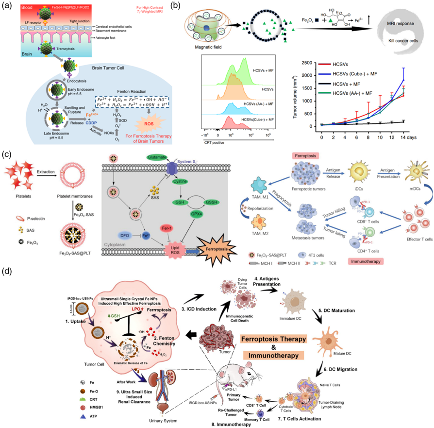
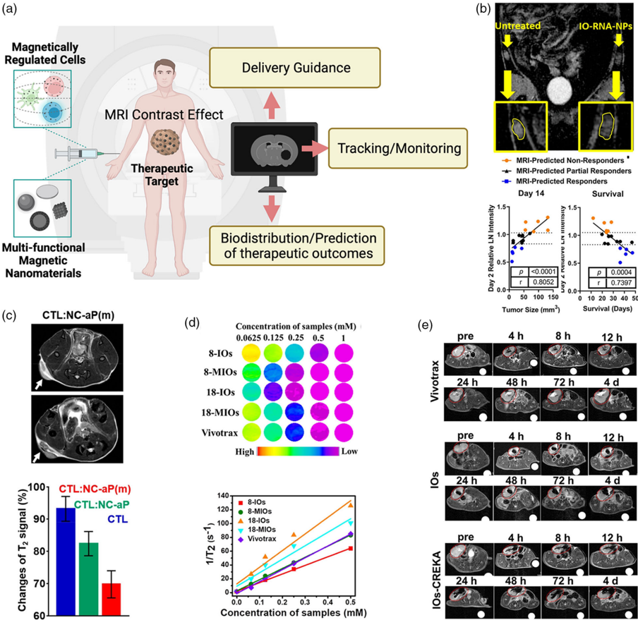
Multifunctional magnetic nanoparticles (MNPs) exhibit unique properties, such as remote motion controllability, degradability, and diagnostic imaging, which are typically not shown in nonmagnetic nanomaterials. MNPs remotely controllable via magnetic fields offer advantages of high tissue penetrability and biocompatibility. In this review, recent advances of multifunctional MNPs exhibiting unique characteristic for therapeutic applications are summarized, which utilize the “dynamic” motion, iron ion degradation, or imaging‐guided targeting of the MNPs under diverse magnetic field modes. The magnetic field‐controlled MNP motion enables spatiotemporal and reversible in situ cell regulation and mechanosensitive molecule modulation or thermal energy generation. Furthermore, the iron‐based MNPs can produce degraded ions and reactive oxygen species to enable targeted ferroptosis therapy with medical imaging‐guided approaches. The state‐of‐the‐art imaging‐guided “dynamic” therapy using the MNPs that can provide in situ feedback at each therapeutic stage is highlighted. Potential hurdles in translating the magnetic dynamic imaging and therapy toward clinical practices are also discussed. The imaging capability of the MNPs during “dynamic” magneto‐cell regulation enables noninvasive, safe, localized, and on‐demand regulation for the state‐of‐the‐art regenerative therapy, immunotherapy, and cancer treatment.
Overview
The claims and statistics of this paper haven’t been checked yet.
Run the check — free, no account needed. It reads the open-access text and checks the sources it cites.
Integrity signals
Registry
Cites
Statistics
Figures
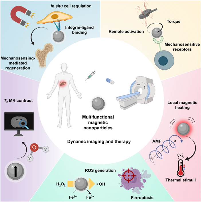
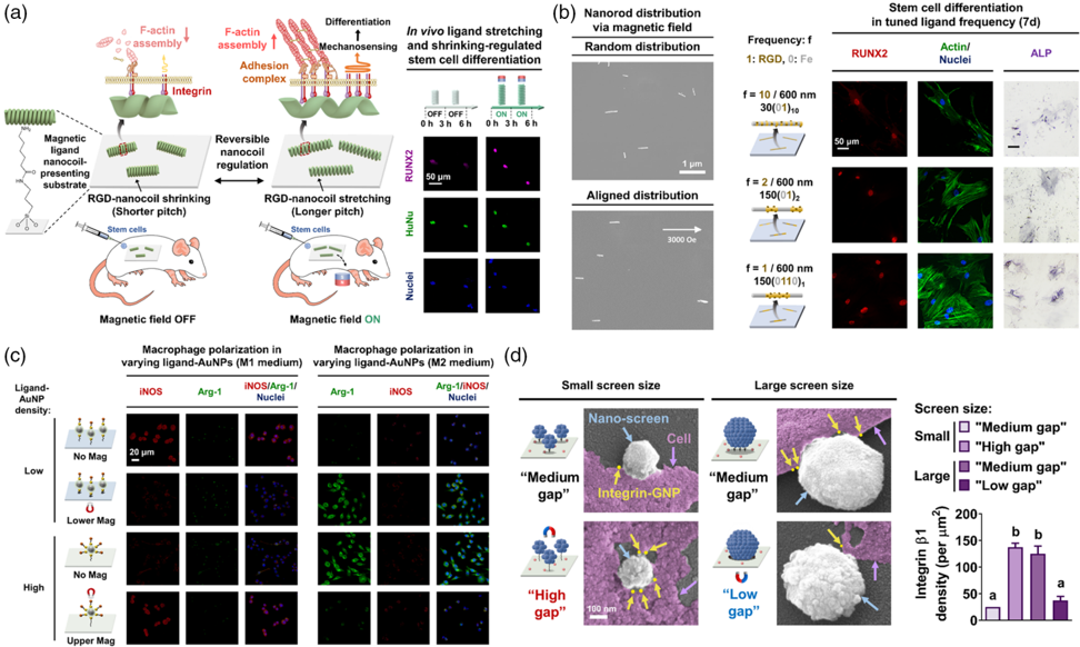
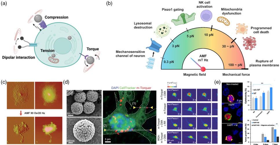
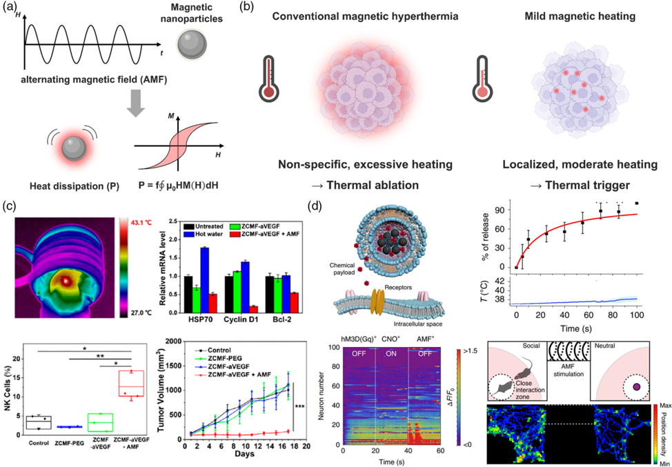




Verification
Claims the paper makes
The claims and statistics of this paper haven’t been checked yet.
Run the check — free, no account needed. It reads the open-access text and checks the sources it cites.
Claims other papers make about this one
Each is checked against this paper’s own text: does it say what the citing paper says it does?
In the citing paper
“Another advantage of MNPs lies in their imaging capability [90].”
From Advances in green-synthesized magnetic nanoparticles for targeted cancer therapy: mechanisms, applications, and future perspectives (2026) · cites this paper as [90]
Self-check
Percentages, sums and reported differences re-computed from the paper’s own numbers.
The self-check needs the paper’s full text.
Citations
Cites
Multifunctional sonosensitizers in sonodynamic cancer therapy
Son, S., Kim, J. H., Wang, X., Zhang, C., Yoon, S. A., Shin, J., Sharma, A., Lee, M. H., Cheng, L., Wu, J., et al. (2020) · Chemical Society Reviews · doi:10.1039/c9cs00648f · Cited by 1,150
The Role of Ferroptosis in Cancer Development and Treatment Response
Lu, B., Chen, X. B., Ying, M. D., He, Q. J., Cao, J., & Yang, B. (2018) · Frontiers in Pharmacology · doi:10.3389/fphar.2017.00992 · Cited by 444
Walling, C., & Goosen, A. (1973) · Journal of the American Chemical Society · doi:10.1021/ja00790a042 · Cited by 410
Research works printed but not linked: 220
- Banghart M., 2004, Nat. Neurosci., 7, 1381, 10.1038/nn1356 · DOI
- Kim D.-H., 2010, Nat. Mater., 9, 165, 10.1038/nmat2591 · DOI
- Wu C.-Y., 2015, Science, 350, aab4077, 10.1126/science.aab4077 · DOI
- Park H.-J., 2022, Nanomaterials, 12, 1377, 10.3390/nano12081377 · DOI
- Yu L., 2022, J. Am. Chem. Soc., 144, 11326, 10.1021/jacs.2c03256 · DOI
- Xie J., 2021, Chem. Soc. Rev., 50, 9152, 10.1039/D0CS01370F · DOI
- Mickle A. D., 2019, Nature, 565, 361, 10.1038/s41586-018-0823-6 · DOI · Paper page
- Dobson J., 2008, Nat. Nanotechnol., 3, 139, 10.1038/nnano.2008.39 · DOI
- Park B. C., 2020, Nat. Commun., 11, 1, 10.1038/s41467-019-13993-7 · DOI · Paper page
- Zhao Y., 2021, Exploration, 1, 20210089, 10.1002/EXP.20210089 · DOI
- Yang H. Y., 2021, Adv. NanoBiomed Res., 1, 2000043, 10.1002/anbr.202000043 · DOI
- Golovin Y. I., 2015, J. Controlled Release, 219, 43, 10.1016/j.jconrel.2015.09.038 · DOI
- Moon J., 2020, Adv. Funct. Mater., 30, 2000577, 10.1002/adfm.202000577 · DOI
- Xue W., 2018, J. Mater. Chem. B, 6, 2289, 10.1039/C7TB03206D · DOI
- Park J. S., 2017, Adv. Funct. Mater., 27, 1700396, 10.1002/adfm.201700396 · DOI
- Xia X., 2021, Exploration, 1, 9, 10.1002/EXP.20210002 · DOI
- Kang H., 2017, Biomaterials, 149, 12, 10.1016/j.biomaterials.2017.09.035 · DOI
- Chang D., 2018, Front. Pharmacol., 9, 831, 10.3389/fphar.2018.00831 · DOI
- Wydra R. J., 2015, Acta Biomater., 25, 284, 10.1016/j.actbio.2015.06.037 · DOI
- Zhu Y., 2022, Adv. Mater., 34, 2108389, 10.1002/adma.202108389 · DOI
- Züger F., 2022, Adv. NanoBiomed Res., 2, 2100108, 10.1002/anbr.202100108 · DOI
- Kim Y., 2020, View, 1, 20200029, 10.1002/VIW.20200029 · DOI
- Khatua C., 2020, Nano Lett., 20, 4188, 10.1021/acs.nanolett.0c00559 · DOI
- Liu J., 2020, Science, 367, 1372, 10.1126/science.aay4866 · DOI
- Kang H., 2018, Adv. Mater., 30, 1803591, 10.1002/adma.201803591 · DOI
- Stupp S. I., 2020, Adv. Mater., 32, 1906741, 10.1002/adma.201906741 · DOI
- Kang H., 2019, Nat. Commun., 10, 1696, 10.1038/s41467-019-09733-6 · DOI
- Kim D., 2020, Adv. Mater., 32, 1906989, 10.1002/adma.201906989 · DOI
- Kang H. M., 2018, Adv. Funct. Mater., 28, 1802642, 10.1002/adfm.201802642 · DOI
- Li W., 2018, Chem. Soc. Rev., 47, 8639, 10.1039/C8CS00053K · DOI
- Luo H., 2021, Adv. Funct. Mater., 31, 2101262, 10.1002/adfm.202101262 · DOI
- Kang H., 2018, Biomaterials, 178, 681, 10.1016/j.biomaterials.2018.03.007 · DOI
- Wu C. Y., 2018, Adv. Mater., 30, 1705673, 10.1002/adma.201705673 · DOI
- Armstrong J. P. K., 2020, Trends Biotechnol., 38, 254, 10.1016/j.tibtech.2019.07.005 · DOI
- Gaharwar A. K., 2020, Nat. Rev. Mater., 5, 686, 10.1038/s41578-020-0209-x · DOI
- Dabbagh S. R., 2022, Adv. NanoBiomed Res., 2, 2100103, 10.1002/anbr.202100103 · DOI
- Kim Y., 2022, Chem. Rev., 122, 5317, 10.1021/acs.chemrev.1c00481 · DOI
- Gu H., 2020, Nat. Commun., 11, 1, 10.1038/s41467-019-13993-7 · DOI · Paper page
- Gossuin Y., 2009, Wiley Interdiscip. Rev. Nanomed. Nanobiotechnol., 1, 299, 10.1002/wnan.36 · DOI
- Yang L., 2022, Small, 18, 2104783, 10.1002/smll.202104783 · DOI · Paper page
- Zwi-Dantsis L., 2020, Adv. Mater., 32, 1904598, 10.1002/adma.201904598 · DOI
- Ganesan V., 2019, J. Mol. Liq., 281, 315, 10.1016/j.molliq.2019.02.095 · DOI
- De Châtel P., 2009, J. Phys. Condens. Matter, 21, 124202, 10.1088/0953-8984/21/12/124202 · DOI
- Yang S. H., 2022, Nano-Micro Lett., 14, 1, 10.1007/s40820-021-00751-y · DOI
- Lisjak D., 2018, Prog. Mater Sci., 95, 286, 10.1016/j.pmatsci.2018.03.003 · DOI
- Dennis C. L., 2013, Int. J. Hyperther., 29, 715, 10.3109/02656736.2013.836758 · DOI
- Hildebrandt B., 2002, Crit. Rev. Oncol. Hematol., 43, 33, 10.1016/S1040-8428(01)00179-2 · DOI
- Liu X., 2020, Theranostics, 10, 3793, 10.7150/thno.40805 · DOI
- Kang H., 2017, ACS Nano, 11, 9636, 10.1021/acsnano.7b02857 · DOI
- Yu B., 2020, Nat. Commun., 11, 1, 10.1038/s41467-019-13993-7 · DOI · Paper page
- Shen Z., 2018, Adv. Mater., 30, 1704007, 10.1002/adma.201704007 · DOI
- Jiang X., 2021, Nat. Rev. Mol. Cell Biol., 22, 266, 10.1038/s41580-020-00324-8 · DOI
- Liang C., 2019, Adv. Mater., 31, 1904197, 10.1002/adma.201904197 · DOI
- Chen Q., 2021, Nanoscale, 13, 4855, 10.1039/D0NR08757B · DOI
- Wang X., 2020, Nano Today, 35, 100946, 10.1016/j.nantod.2020.100946 · DOI
- Wu H., 2021, Theranostics, 11, 64, 10.7150/thno.46124 · DOI
- Kwak K., 2022, Theranostics, 12, 2175, 10.7150/thno.67530 · DOI
- Hembury M., 2015, Proc. Natl. Acad. Sci. U.S.A., 112, 1959, 10.1073/pnas.1419622112 · DOI
- Kim D.-H., 2018, J. Imaging, 4, 18, 10.3390/jimaging4010018 · DOI
- Perez J. M., 2002, Nat. Biotechnol., 20, 816, 10.1038/nbt720 · DOI
- Wang Q., 2021, Exploration, 1, 20210009, 10.1002/EXP.20210009 · DOI
- Josephson L., 2001, Angew. Chem., 113, 3304, 10.1002/1521-3757(20010903)113:17<3304::AID-ANGE3304>3.0.CO;2-D · DOI
- Shasha C., 2021, Adv. Mater., 33, 1904131, 10.1002/adma.201904131 · DOI
- Hong H., 2021, Adv. Mater., 34, 2105460, 10.1002/adma.202105460 · DOI
- Min S., 2020, Adv. Mater., 32, 2004300, 10.1002/adma.202004300 · DOI
- Min S., 2021, Adv. Mater., 33, 2008353, 10.1002/adma.202008353 · DOI
- Lopez S., 2022, Nanoscale Adv., 4, 421, 10.1039/D1NA00474C · DOI
- Park W., 2021, Biomater. Sci., 9, 5497, 10.1039/D1BM00419K · DOI
- Chen M., 2020, Small, 16, 1905424, 10.1002/smll.201905424 · DOI
- Gregurec D., 2020, ACS Nano, 14, 8036, 10.1021/acsnano.0c00562 · DOI
- Sim T., 2021, ACS Nano, 15, 12780, 10.1021/acsnano.1c01889 · DOI
- Grippin A. J., 2019, ACS Nano, 13, 13884, 10.1021/acsnano.9b05037 · DOI
- Lee J.-U., 2021, Nat. Mater., 20, 1029, 10.1038/s41563-020-00896-y · DOI
- Jang J. T., 2018, Adv. Mater., 30, 1704362, 10.1002/adma.201704362 · DOI
- Pan J., 2021, J. Am. Chem. Soc., 143, 8116, 10.1021/jacs.1c02537 · DOI
- Balakrishnan P. B., 2020, Adv. Mater., 32, 2003712, 10.1002/adma.202003712 · DOI
- Chen R., 2015, Science, 347, 1477, 10.1126/science.1261821 · DOI
- Rao S., 2019, Nat. Nanotechnol., 14, 967, 10.1038/s41565-019-0521-z · DOI
- Liu X., 2020, ACS Nano, 14, 1936, 10.1021/acsnano.9b08320 · DOI
- Shen Z., 2018, ACS Nano, 12, 11355, 10.1021/acsnano.8b06201 · DOI
- Hu Y., 2018, Chem. Soc. Rev., 47, 1874, 10.1039/C7CS00657H · DOI
- Huang H.-W., 2019, Sci. Adv., 5, eaau1532, 10.1126/sciadv.aau1532 · DOI
- Huang H.-W., 2016, Nat. Commun., 7, 1
- Zhang M., 2021, Biomaterials, 268, 120543, 10.1016/j.biomaterials.2020.120543 · DOI
- Rödling L., 2018, Adv. Healthcare Mater., 7, 1701403, 10.1002/adhm.201701403 · DOI
- Islam M. S., 2021, Adv. NanoBiomed Res., 1, 2000072, 10.1002/anbr.202000072 · DOI
- Li C., 2021, Adv. NanoBiomed Res., 1, 2000081, 10.1002/anbr.202000081 · DOI
- Du V., 2017, Nat. Commun., 8, 400, 10.1038/s41467-017-00543-2 · DOI
- Kang H., 2014, J. Mater. Chem. B, 2, 5676, 10.1039/C4TB00714J · DOI
- Zhang T., 2019, Adv. Funct. Mater., 29, 1900603, 10.1002/adfm.201900603 · DOI
- Zhou D. W., 2021, Nat. Commun., 12, 1, 10.1038/s41467-020-20314-w · DOI · Paper page
- Wong D. S. H., 2017, Nano Lett., 17, 1685, 10.1021/acs.nanolett.6b04958 · DOI
- Lee S., 2021, Small, 17, 2102892, 10.1002/smll.202102892 · DOI
- Cui T., 2022, Angew. Chem., Int. Ed., 61, 202113103, 10.1002/anie.202113103 · DOI
- Li C. C., 2018, Biomaterials, 176, 24, 10.1016/j.biomaterials.2018.05.029 · DOI
- Bae G., 2022, Adv. Funct. Mater., 32, 2200828, 10.1002/adfm.202200828 · DOI
- Yan X., 2017, Sci. Rob., 2, eaaq1155, 10.1126/scirobotics.aaq1155 · DOI · Paper page
- Cho S., 2019, Adv. Funct. Mater., 29, 1901384, 10.1002/adfm.201901384 · DOI
- Thangam R., 2021, Adv. Funct. Mater., 31, 2001446
- Kang H., 2018, Acs Nano, 12, 5978, 10.1021/acsnano.8b02226 · DOI
- Wu D., 2022, ACS Appl. Mater. Interfaces, 14, 12976, 10.1021/acsami.1c22603 · DOI
- Zhu Y., 2012, Aging Cell, 11, 741, 10.1111/j.1474-9726.2012.00840.x · DOI
- Zhang L., 2020, ACS Appl. Mater. Interfaces, 12, 52467, 10.1021/acsami.0c17213 · DOI
- Kang H., 2017, Nano Lett., 17, 6415, 10.1021/acs.nanolett.7b03405 · DOI
- Kim Y., 2022, J. Am. Chem. Soc., 144, 5769, 10.1021/jacs.1c08861 · DOI
- Wong S. H. D., 2020, Nano Lett., 20, 3207, 10.1021/acs.nanolett.9b05315 · DOI
- Kim J. W., 2018, Acc. Chem. Res, 51, 839, 10.1021/acs.accounts.8b00004 · DOI
- Coste B., 2010, Science, 330, 55, 10.1126/science.1193270 · DOI · Paper page
- Shimasaki N., 2020, Nat. Rev. Drug Discovery, 19, 200, 10.1038/s41573-019-0052-1 · DOI
- Wheeler M. A., 2016, Nat. Neurosci., 19, 756, 10.1038/nn.4265 · DOI
- Xu F.-X., 2020, Nat. Neurosci., 23, 1041, 10.1038/s41593-019-0475-3 · DOI
- Shin T.-H., 2017, Acc. Chem. Res., 50, 567, 10.1021/acs.accounts.6b00559 · DOI
- Müller D. J., 2009, Nat. Chem. Biol., 5, 383, 10.1038/nchembio.181 · DOI
- Gu Z., 2019, J. Am. Chem. Soc., 141, 6122, 10.1021/jacs.8b10904 · DOI
- Thangam R., 2021, Vaccines, 9, 935, 10.3390/vaccines9080935 · DOI
- Tadijan A., 2021, Front. Cell Dev. Biol., 9, 3482, 10.3389/fcell.2021.786758 · DOI
- Kim D.-H., 2009, IEEE Trans. Magn., 45, 4821, 10.1109/TMAG.2009.2024000 · DOI
- Boyden E. S., 2005, Nat. Neurosci., 8, 1263, 10.1038/nn1525 · DOI
- Yu Y., 2021, Adv. Sci., 9, 2104194, 10.1002/advs.202104194 · DOI
- Jin Y., 2019, Nano Lett., 19, 6517, 10.1021/acs.nanolett.9b02756 · DOI
- Katz B., 1967, J. Physiol., 192, 407, 10.1113/jphysiol.1967.sp008307 · DOI
- Wen C., 2016, Drug Delivery Trans. Res., 6, 121, 10.1007/s13346-015-0242-2 · DOI
- Sadtler K., 2016, Nat. Rev. Mater., 1, 16040, 10.1038/natrevmats.2016.40 · DOI
- Rao V., 2015, Front. Bioeng. Biotechnol., 3, 1
- Bae G., 2021, Adv. Funct. Mater., 31, 2001446
- Zuo S., 2021, Theranostics, 11, 7471, 10.7150/thno.59953 · DOI
- Xu J., 2020, Adv. Sci., 7, 1903394, 10.1002/advs.201903394 · DOI
- Cheng H. W., 2021, Adv. Healthcare Mater., 10, 2001451, 10.1002/adhm.202001451 · DOI
- Kim Y., 2022, Adv. Mater., 34, 2110340, 10.1002/adma.202110340 · DOI
- Min S., 2020, Nano Lett., 20, 7272, 10.1021/acs.nanolett.0c02655 · DOI
- Kim D.-H., 2008, J. Magn. Magn. Mater., 320, 2390, 10.1016/j.jmmm.2008.05.023 · DOI
- Kim D.-H., 2010, Materials, 3, 4051, 10.3390/ma3074051 · DOI
- Ramos-Sebastian A., 2022, Adv. Sci., 9, 2103863, 10.1002/advs.202103863 · DOI
- Kang H., 2018, J. Am. Chem. Soc., 140, 5909, 10.1021/jacs.8b03001 · DOI
- Gavilán H., 2021, Chem. Soc. Rev., 50, 11614, 10.1039/D1CS00427A · DOI
- Périgo E. A., 2015, Appl. Phys. Rev., 2, 041302, 10.1063/1.4935688 · DOI
- Cabrera D., 2018, ACS Nano, 12, 2741, 10.1021/acsnano.7b08995 · DOI
- Jordan A., 1999, J. Magn. Magn. Mater., 201, 413, 10.1016/S0304-8853(99)00088-8 · DOI
- Hergt R., 2007, J. Magn. Magn. Mater., 311, 187, 10.1016/j.jmmm.2006.10.1156 · DOI
- Mo A. H., 2016, Nanoscale, 8, 11840, 10.1039/C6NR02445A · DOI
- Nemati Z., 2018, J. Phys. Chem. C, 122, 2367, 10.1021/acs.jpcc.7b10528 · DOI
- Di Corato R., 2014, Biomaterials, 35, 6400, 10.1016/j.biomaterials.2014.04.036 · DOI
- Jeon S., 2020, ACS Appl. Mater. Interfaces, 12, 33483, 10.1021/acsami.0c07419 · DOI
- Mamiya H., 2020, ACS Nano, 14, 8421, 10.1021/acsnano.0c02521 · DOI
- Zyuzin M. V., 2019, ACS Appl. Mater. Interfaces, 11, 41957, 10.1021/acsami.9b15501 · DOI
- Sanz B., 2016, Sci. Rep., 6, 1, 10.1038/s41598-016-0001-8 · DOI · Paper page
- Wilhelm S., 2016, Nat. Rev. Mater., 1, 1
- Han X., 2019, Biomaterials, 224, 119490, 10.1016/j.biomaterials.2019.119490 · DOI
- Spirou S. V., 2018, Nanomaterials, 8, 401, 10.3390/nano8060401 · DOI
- Mai B. T., 2019, ACS Appl. Mater. Interfaces, 11, 5727, 10.1021/acsami.8b16226 · DOI
- Chang M., 2021, Adv. Mater., 33, 2004788, 10.1002/adma.202004788 · DOI
- Moros M., 2019, Adv. Drug Delivery Rev., 138, 326, 10.1016/j.addr.2018.10.004 · DOI
- Liu X., 2019, ACS Nano, 13, 8811, 10.1021/acsnano.9b01979 · DOI
- Kang H., 2019, Nano Lett., 19, 1963, 10.1021/acs.nanolett.8b05150 · DOI
- Choi H., 2020, Adv. Funct. Mater., 30, 2001446, 10.1002/adfm.202001446 · DOI
- Wang L., 2022, Nano Today, 43, 101401, 10.1016/j.nantod.2022.101401 · DOI
- Kang H., 2015, Appl. Clay Sci., 112, 32, 10.1016/j.clay.2015.04.015 · DOI
- Manohar A., 2021, J. Alloys Compd., 874, 159868, 10.1016/j.jallcom.2021.159868 · DOI
- Tang G., 2021, Exploration, 1, 75, 10.1002/EXP.20210005 · DOI
- Metelitsa D. I., 1971, Russ. Chem. Rev., 40, 563, 10.1070/RC1971v040n07ABEH001939 · DOI
- Jiang Q., 2020, Small, 16, 2001704, 10.1002/smll.202001704 · DOI · Paper page
- Liang H., 2021, J. Am. Chem. Soc., 143, 15812, 10.1021/jacs.1c07471 · DOI
- Chen Q., 2019, Adv. Sci., 6, 1802134, 10.1002/advs.201802134 · DOI
- Yu G. T., 2018, Adv. Funct. Mater., 28, 1801389, 10.1002/adfm.201801389 · DOI
- Rao L., 2020, Adv. Mater., 32, 2004853, 10.1002/adma.202004853 · DOI
- Liu L., 2020, Small, 16, 2003543, 10.1002/smll.202003543 · DOI
- Proneth B., 2019, Cell Death Differ., 26, 14, 10.1038/s41418-018-0173-9 · DOI
- Massoud T. F., 2003, Genes Dev., 17, 545, 10.1101/gad.1047403 · DOI
- Thangam R., 2021, Nanomaterials, 12, 18, 10.3390/nano12010018 · DOI
- Sathuvan M., 2022, Int. J. Biol. Macromol., 194, 563, 10.1016/j.ijbiomac.2021.11.099 · DOI
- Constantine G., 2004, Lancet, 363, 2162, 10.1016/S0140-6736(04)16509-4 · DOI
- Lutz A. M., 2004, Radiology, 233, 149, 10.1148/radiol.2331031402 · DOI
- Padhani A. R., 2002, J. Magn. Reson. Imaging, 16, 407, 10.1002/jmri.10176 · DOI
- McRobbie D. W., 2017, MRI From Picture To Proton, 10.1017/9781107706958 · DOI
- Benyettou F., 2020, J. Am. Chem. Soc., 142, 18782, 10.1021/jacs.0c05381 · DOI
- Nie W., 2019, ACS Nano, 13, 1469, 10.1021/acsnano.8b07141 · DOI
- Du Y., 2019, Nano Lett., 19, 3618, 10.1021/acs.nanolett.9b00630 · DOI
- Kang H., 2018, Acta Biomater., 78, 365, 10.1016/j.actbio.2018.07.039 · DOI
- Kang H., 2017, Handbook of Intelligent Scaffolds for Tissue Engineering and Regenerative Medicine, 59
- Singh N., 2021, Chem. Soc. Rev., 50, 12883, 10.1039/D1CS00559F · DOI
- Thu M. S., 2012, Nat. Med., 18, 463, 10.1038/nm.2666 · DOI
- Lu C.-W., 2007, Nano Lett., 7, 149, 10.1021/nl0624263 · DOI
- Hoehn M., 2002, Proc. Natl. Acad. Sci., 99, 16267, 10.1073/pnas.242435499 · DOI
- Sharma B., 2021, Adv. NanoBiomed Res., 1, 2100019, 10.1002/anbr.202100019 · DOI
- Park S., 2021, Sci. Rep., 11, 1, 10.1038/s41598-020-79139-8 · DOI
- Yun S., 2018, Nano Lett., 18, 838, 10.1021/acs.nanolett.7b04089 · DOI
- Wang Q., 2020, ACS Nano, 14, 2053, 10.1021/acsnano.9b08660 · DOI
- Tu Z., 2022, Nat. Rev. Mater., 7, 1, 10.1038/s41578-021-00410-z · DOI
- Dellacherie M. O., 2019, Nat. Rev. Mater., 4, 379, 10.1038/s41578-019-0106-3 · DOI
- Kang H., 2018, Curr. Protoc. Stem Cell Biol., 44, 1F, 10.1002/cpsc.44 · DOI
- Dalby M. J., 2018, Nat. Rev. Mater., 3, 1, 10.1038/natrevmats.2017.91 · DOI
- Shih Y.-R. V., 2014, Proc. Natl. Acad. Sci., 111, 990, 10.1073/pnas.1321717111 · DOI
- Dai C., 2019, Biomaterials, 219, 119374, 10.1016/j.biomaterials.2019.119374 · DOI
- Poon W., 2019, ACS Nano, 13, 5785, 10.1021/acsnano.9b01383 · DOI
- Li X., 2016, J. Biomed. Mater. Res., Part A, 104, 1285, 10.1002/jbm.a.35654 · DOI
- Cardoso V. F., 2018, Adv. Healthcare Mater., 7, 1700845, 10.1002/adhm.201700845 · DOI
- Ottersbach A., 2018, Biomaterials, 155, 176, 10.1016/j.biomaterials.2017.11.012 · DOI
- Lee J.-R., 2020, Sci. Adv., 6, eaaz0952, 10.1126/sciadv.aaz0952 · DOI
- Shih Y.-R., 2015, Acta Biomater., 19, 1, 10.1016/j.actbio.2015.03.017 · DOI
- Xia Y., 2018, Biomaterials, 183, 151, 10.1016/j.biomaterials.2018.08.040 · DOI
200 of 220 shown
Cited by
Sorted by citations
El-khawaga, A. M., Youssef, O. I., El-dydamoni, O. A., Ragab, W. M., & Mohammed, R. A. E. (2026) · European Journal of Medical Research · doi:10.1186/s40001-025-03804-9 · Cited by 14
Metadata
- Authors
- Ko, M. J., Hong, H., Choi, H., Kang, H., & Kim, D.
- Venue
- Advanced NanoBiomed Research
- Publisher
- Wiley
- Published
- 2022-09-15
- Volume
- 2
- Issue
- 11
- ISSN
- 2699-9307
- Cited by
- 29
- References
- 3
- Language
- en
- Funders
- National Research Foundation of Korea (2020R1C1C1011038)
- National Cancer Institute (R01CA218659)
- National Institute of Biomedical Imaging and Bioengineering (R01EB026207)
About these checks
Registry, retraction, Cites and statistics results are computed, not judged, with one exception: a printed reference that no index lists is read once by a model before it counts as a citation error. Claims are extracted by a model and kept only when their quote appears verbatim in the text; each cited source is then checked, with a model’s help, for whether it says what the claim relies on. Those verdicts can be wrong — read the claim’s quote and the cited source before relying on one.
Statistics and arithmetic are recomputed, not judged.
Confidence bands are shown only for fields calibrated on our annotated test set. None is calibrated yet, so no band is shown.
What counts as a claim ›How we measure confidence ›How we check this ›
Are you an author, or do you see a problem with this page? Request a review
