

The DOI matches its Crossref record; 7 of the 7 references it cites were found in a registry or an index.
Characterization of early pathogenesis in the SOD1 G93A mouse model of ALS : part I, background and methods
Abstract
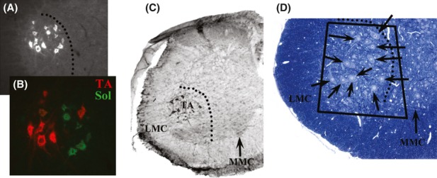
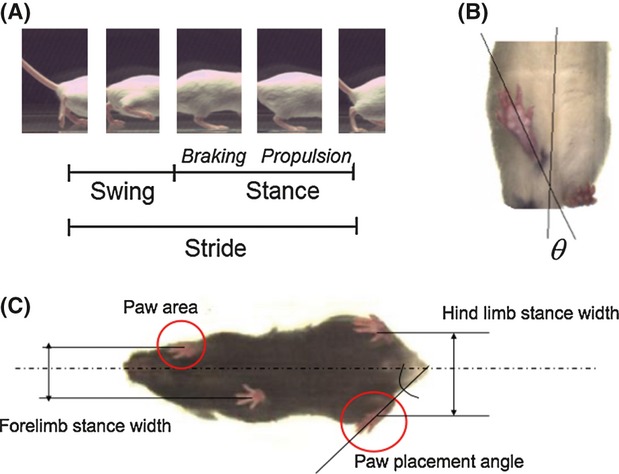
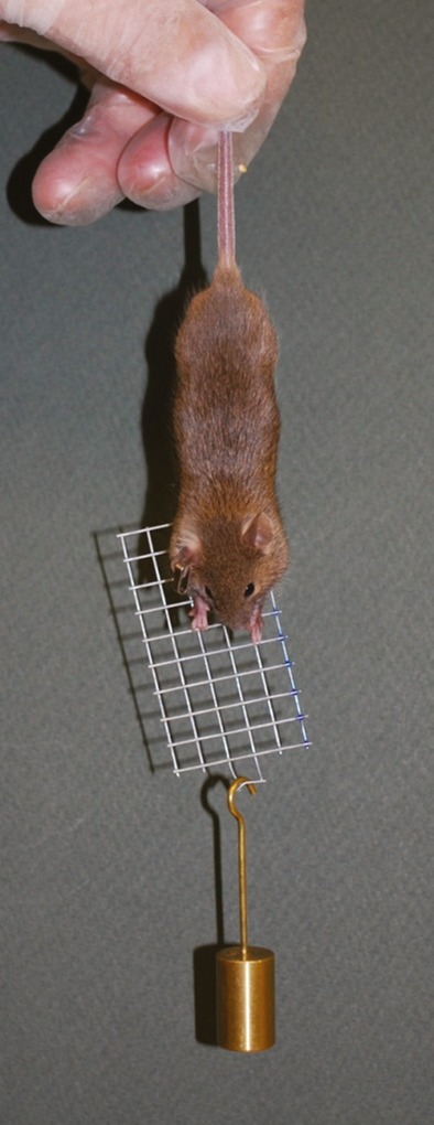
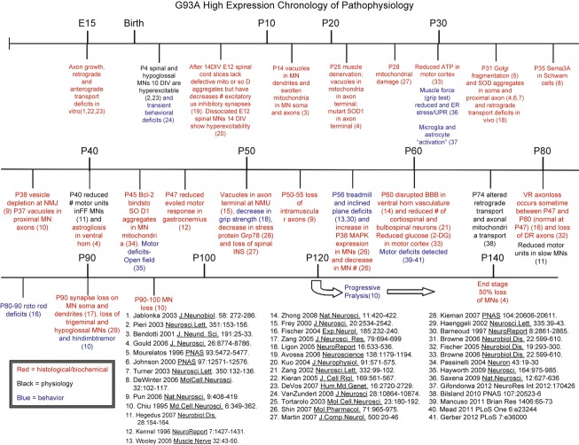
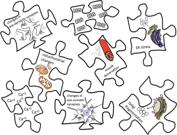
Charcot first described amyotrophic lateral sclerosis ( ALS ) in 1869; however, its causes remain largely unknown and effective, long‐term treatment strategies are not available. The first mouse model of ALS was developed after the identification of mutations in the superoxide dismutase 1 ( SOD1 ) gene in 1993, and accordingly most of our knowledge of the etiology and pathogenesis of the disease comes from studies carried out using this animal model. Although numerous preclinical trials have been conducted in the mutant SOD1 mouse models, the results have been disappointing because they did not positively translate to clinical trials. One explanation may be that current understanding of when and where pathogenesis begins is insufficient to accurately guide preclinical trials. Further characterization of these early events may provide insight into disease onset, help in the discovery of presymptomatic diagnostic disease markers, and identify novel therapeutic targets. Here, we describe the rationale, approach, and methods for our extensive analysis of early changes that included an ultrastructural examination of central and peripheral components of the neuromuscular system in the SOD1 G93A mouse and correlated these alterations with early muscle denervation, motor dysfunction, and motoneuron death. We also provide a discussion of published work to review what is known regarding early pathology in the SOD1 mouse model of ALS . The significance of this work is that we have examined early pathology simultaneously in both the spinal cord and peripheral neuromuscular system, and the results are presented in the companion paper (Part II, Results and Discussion). Our results provide evidence as to why a thorough characterization of animal models throughout the life span is critical for a strong foundation to design preclinical trials that may produce meaningful results.
Overview
The claims and statistics of this paper haven’t been checked yet.
Run the check — free, no account needed. It reads the open-access text and checks the sources it cites.
Integrity signals
Registry
Cites
Statistics
Figures
Verification
Claims the paper makes
The claims and statistics of this paper haven’t been checked yet.
Run the check — free, no account needed. It reads the open-access text and checks the sources it cites.
Claims other papers make about this one
Each is checked against this paper’s own text: does it say what the citing paper says it does?
The source backs part of the claim, not all of it — or backs it for a different population, condition or comparison.
In the citing paper
“beginning at 94 days postnatally, which corresponds to the onset of clinical disease symptoms (as evidenced by hindlimb tremor) in this model [34,35]”
From Modulation of the miR-485-3p/PGC-1α Pathway by ASO-Loaded Nanoparticles Attenuates ALS Pathogenesis (2026) · cites this paper as [34]
Experimental — accuracy not yet measured
Self-check
Percentages, sums and reported differences re-computed from the paper’s own numbers.
The self-check needs the paper’s full text.
Citations
Cites
Oligodendroglia metabolically support axons and contribute to neurodegeneration
Lee, Y., Morrison, B. M., Li, Y., Lengacher, S., Farah, M. H., Hoffman, P. N., Liu, Y., Tsingalia, A., Jin, L., Zhang, P.-W., et al. (2012) · Nature · doi:10.1038/nature11314 · Cited by 1,664
Onset and Progression in Inherited ALS Determined by Motor Neurons and Microglia
Boillée, S., Yamanaka, K., Lobsiger, C. S., Copeland, N. G., Jenkins, N. A., Kassiotis, G., Kollias, G., & Cleveland, D. W. (2006) · Science · doi:10.1126/science.1123511 · Cited by 1,623
Astrocytes as determinants of disease progression in inherited amyotrophic lateral sclerosis
Yamanaka, K., Chun, S. J., Boillee, S., Fujimori-Tonou, N., Yamashita, H., Gutmann, D. H., Takahashi, R., Misawa, H., & Cleveland, D. W. (2008) · Nature Neuroscience · doi:10.1038/nn2047 · Cited by 1,140 · cited 3×
“For example, cell-specific deletion of mutant SOD1 in genetically altered mice has implicated microglia and astrocytes as contributors to the progression but not the onset of disease (Clement et al. 2003; Boill ee et al. 2006b; Yamanaka et al. 2008a,b).”
BackgroundMolecular biology of amyotrophic lateral sclerosis: insights from genetics
Pasinelli, P., & Brown, R. H. (2006) · Nature Reviews Neuroscience · doi:10.1038/nrn1971 · Cited by 1,107
Characteristics of Excitatory and Inhibitory Synapses in the Central Nervous System of the Cat
UCHIZONO, K. (1965) · Nature · doi:10.1038/207642a0 · Cited by 917
Design, power, and interpretation of studies in the standard murine model of ALS
Scott, S., Kranz, J. E., Cole, J., Lincecum, J. M., Thompson, K., Kelly, N., Bostrom, A., Theodoss, J., Al‐Nakhala, B. M., Vieira, F. G., et al. (2008) · Amyotrophic Lateral Sclerosis · doi:10.1080/17482960701856300 · Cited by 504
Understanding the role of TDP-43 and FUS/TLS in ALS and beyond
Da Cruz, S., & Cleveland, D. W. (2011) · Current Opinion in Neurobiology · doi:10.1016/j.conb.2011.05.029 · Cited by 351
Research works printed but not linked: 106
- 10.1136/jnnp.73.2.199 · DOI
- 10.1111/j.1460-9568.2004.03745.x · DOI
- Appel S. H., 2011, The microglial‐motoneuron dialogue in ALS, Acta Myol., 30, 4
- 10.1097/00001756-199709080-00012 · DOI
- 10.1007/s10863‐011‐9392‐1 · DOI
- 10.1016/j.nbd.2006.12.015 · DOI
- 10.1016/S0022-510X(01)00627-X · DOI
- 10.1186/1471-2202-8-79 · DOI
- 10.1016/S0940-9602(98)80020-4 · DOI
- Bodian D., 1964, AN electron‐microscopic study of the monkey spinal cord. I. Fine structure of normal motor column. II. Effects of retrograde chromatolysis. III. Cytologic effects of mild and virulent poliovirus infection, Bull. Johns Hopkins Hosp., 114, 13
- 10.1126/science.151.3714.1093 · DOI
- 10.1083/jcb.44.1.115 · DOI
- Bodian D., 1972, Structure and function of synapses, 45
- 10.1172/JCI34613 · DOI
- 10.1016/j.neuron.2006.09.018 · DOI
- 10.1002/ana.410140304 · DOI
- 10.1007/BF01463929 · DOI
- 10.1586/14737175.6.3.417 · DOI
- 10.1146/annurev.neuro.27.070203.144244 · DOI · Paper page
- 10.1016/S0960-8966(97)00115-6 · DOI
- 10.1093/hmg/ddn319 · DOI
- 10.1073/pnas.0406173102 · DOI
- 10.1002/cne.901770106 · DOI
- 10.1016/S0091-679X(08)61933-0 · DOI
- 10.1126/science.1086071 · DOI
- 10.1016/S0166-2236(02)02255-5 · DOI
- 10.1038/375061a0 · DOI
- 10.1016/j.tins.2007.02.004 · DOI
- 10.1016/j.mcn.2012.06.001 · DOI
- 10.1093/hmg/ddm226 · DOI
- 10.1016/j.ymgme.2003.08.017 · DOI
- 10.1179/174313208X281235 · DOI
- 10.1152/ajpregu.00264.2003 · DOI
- 10.1016/S0960-9822(03)00206-9 · DOI · Paper page
- 10.1159/000107704 · DOI
- 10.1016/j.expneurol.2003.10.004 · DOI
- 10.1523/JNEUROSCI.20-07-02534.2000 · DOI
- 10.1073/pnas.0906809106 · DOI · Paper page
- 10.1371/journal.pone.0036000 · DOI
- 10.1523/JNEUROSCI.4057-07.2007 · DOI
- 10.1016/j.jns.2006.01.019 · DOI
- Gould T., 2012, Textbook of neural repair & rehabilitation
- 10.1016/j.neubiorev.2007.04.015 · DOI
- 10.1523/JNEUROSCI.2315-06.2006 · DOI
- 10.1172/JCI200317610 · DOI
- 10.1126/science.8209258 · DOI · Paper page
- 10.1126/science.1083129 · DOI
- 10.1080/00222890903272025 · DOI
- 10.1016/j.physbeh.2004.04.006 · DOI
- 10.1016/j.nbd.2007.07.003 · DOI
- 10.1002/cne.10648 · DOI
- 10.1097/01.ALC.0000148103.09378.81 · DOI
- 10.1083/jcb.23.2.217 · DOI
- 10.1083/jcb.200501085 · DOI
- 10.1126/science.277.5325.559 · DOI
- 10.1016/S0896-6273(02)00696-7 · DOI
- 10.1371/journal.pgen.0010004 · DOI
- 10.1097/00001756-200504250-00002 · DOI
- 10.1016/j.neures.2003.11.008 · DOI
- 10.1016/j.brainres.2011.06.033 · DOI
- 10.1073/pnas.1200658109 · DOI
- 10.1002/neu.480220707 · DOI
- 10.1371/journal.pone.0023244 · DOI
- 10.1093/brain/awg061 · DOI
- 10.1073/pnas.0609411103 · DOI
- 10.1073/pnas.93.11.5472 · DOI · Paper page
- 10.1016/B978-0-12-385870-2.00018-4 · DOI
- 10.1038/nature05289 · DOI
- 10.1093/hmg/ddq202 · DOI
- 10.1126/science.204007 · DOI
- Peters A., 1991, Fine structure of the nervous system: neurons and their supporting cells, 273
- 10.1038/ng1123 · DOI
- 10.1038/nn1653 · DOI
- 10.1038/nrneurol.2012.276 · DOI
- 10.1126/science.1068613 · DOI
- 10.1126/science.1094682 · DOI
- Robinson M. B., 2011, Neurodegeneration: theory, disorders and treatments, 45
- 10.1002/ana.21543 · DOI · Paper page
- 10.1523/JNEUROSCI.15-11-07727.1995 · DOI
- 10.1038/sj.cdd.4401560 · DOI
- 10.1007/s00401-004-0838-y · DOI
- 10.1016/S1474-4422(05)70142-3 · DOI
- 10.1016/j.pneurobio.2007.07.007 · DOI
- 10.1016/j.neuron.2011.06.031 · DOI
- 10.1038/nn.2297 · DOI · Paper page
- 10.1523/JNEUROSCI.17-15-05858.1997 · DOI · Paper page
- 10.1038/86042 · DOI · Paper page
- 10.1016/j.neuron.2011.06.003 · DOI · Paper page
- 10.1186/1749‐8104‐4‐42 · DOI
- 10.1002/ana.410390210 · DOI
- 10.1007/s004010050794 · DOI
- Suzuki M., 2007, GDNF secreting human neural progenitor cells protect dying motor neurons, but not their projection to muscle, in a rat model of familial ALS, PLoS One, 8, 1
- 10.1159/000046187 · DOI
- Truett G. E., 2000, Preparation of PCR‐quality mouse genomic DNA with hot sodium hydroxide and tris (HotSHOT), Biotechniques, 29, 52, 54, 10.2144/00291bm09 · DOI
- 10.1002/mus.880070310 · DOI
- 10.1016/j.pneurobio.2008.01.001 · DOI
- 10.1523/JNEUROSCI.21-12-04249.2001 · DOI
- 10.2170/jjphysiol.16.570 · DOI
- 10.1371/journal.pone.0034640 · DOI
- 10.1016/j.nurt.2010.05.012 · DOI
- 10.1016/S0006-8993(98)01351-1 · DOI
- 10.1016/j.nmd.2006.07.024 · DOI
- 10.1038/4553 · DOI
- 10.1073/pnas.0802556105 · DOI · Paper page
- 10.1083/jcb.139.5.1307 · DOI
- 10.1161/CIRCULATIONAHA.105.565598 · DOI
Cited by
Sorted by citations
Spatiotemporal dynamics of molecular pathology in amyotrophic lateral sclerosis
Maniatis, S., Äijö, T., Vickovic, S., Braine, C., Kang, K., Mollbrink, A., Fagegaltier, D., Andrusivová, Ž., Saarenpää, S., Saiz-Castro, G., et al. (2019) · Science · doi:10.1126/science.aav9776 · Cited by 429
Abnormalities of Mitochondrial Dynamics in Neurodegenerative Diseases
Gao, J., Wang, L., Liu, J., Xie, F., Su, B., & Wang, X. (2017) · Antioxidants · doi:10.3390/antiox6020025 · Cited by 250
Mitochondrial dynamic abnormalities in amyotrophic lateral sclerosis
Jiang, Z., Wang, W., Perry, G., Zhu, X., & Wang, X. (2015) · Translational Neurodegeneration · doi:10.1186/s40035-015-0037-x · Cited by 66
Axon Biology in ALS: Mechanisms of Axon Degeneration and Prospects for Therapy
Coleman, M. P. (2022) · Neurotherapeutics · doi:10.1007/s13311-022-01297-6 · Cited by 33
“While innervation of fast fatigable muscles is reduced by 40% by P30, motor neuron loss is limited to 20% even at P60 [24, 25].”
BackgroundModulation of the miR-485-3p/PGC-1α Pathway by ASO-Loaded Nanoparticles Attenuates ALS Pathogenesis
Ryu, I. S., Ha, D.-I., Jung, Y.-J., Lee, H. J., Kim, I., Lim, Y. N., Min, H. S., Kim, S. H., Yoon, I., Cho, H.-J., et al. (2026) · International Journal of Molecular Sciences · doi:10.3390/ijms27020615 · Cited by 2
Metadata
- Authors
- Vinsant, S., Mansfield, C., Jimenez‐Moreno, R., Moore, V. D. G., Yoshikawa, M., Hampton, T. G., Prevette, D., Caress, J., Oppenheim, R. W., & Milligan, C.
- Affiliations
- Department of Neurobiology and Anatomy The Neuroscience Program and The ALS Center Winston‐Salem North Carolina
- Department of Chemistry Elon University Elon North Carolina
- Mouse Specifics Boston Massachusetts
- Department of Neurology and the ALS Center Wake Forest University School of Medicine Winston‐Salem North Carolina
- Venue
- Brain and Behavior
- Publisher
- Wiley
- Published
- 2013-06-11
- Volume
- 3
- Issue
- 4
- Pages
- 335-350
- ISSN
- 2162-3279
- PMC
- PMC3869677
- Cited by
- 105
- References
- 7
- Language
- en
About these checks
Registry, retraction, Cites and statistics results are computed, not judged, with one exception: a printed reference that no index lists is read once by a model before it counts as a citation error. Claims are extracted by a model and kept only when their quote appears verbatim in the text; each cited source is then checked, with a model’s help, for whether it says what the claim relies on. Those verdicts can be wrong — read the claim’s quote and the cited source before relying on one.
Statistics and arithmetic are recomputed, not judged.
Confidence bands are shown only for fields calibrated on our annotated test set. None is calibrated yet, so no band is shown.
What counts as a claim ›How we measure confidence ›How we check this ›
Are you an author, or do you see a problem with this page? Request a review
How to cite
Cite CoChat for the checks on this page, and the paper for its own findings.
- This page
- CoChat. (2026). Citation check: Characterization of early pathogenesis in the SOD1 G93A mouse model of ALS : part I, background and methods. https://app.cochat.ai/papers/10.1002/brb3.143
- The paper
- Vinsant, S., Mansfield, C., Jimenez‐Moreno, R., Moore, V. D. G., Yoshikawa, M., Hampton, T. G., Prevette, D., Caress, J., Oppenheim, R. W., & Milligan, C. (2013). Characterization of early pathogenesis in the SOD1 G93A mouse model of ALS : part I, background and methods. Brain and Behavior. https://doi.org/10.1002/brb3.143
