

The DOI matches its Crossref record; 249 of the 249 references it cites were found in a registry or an index.
Electrophysiological correlates of the BOLD signal for EEG-informed fMRI
Open accessAbstract
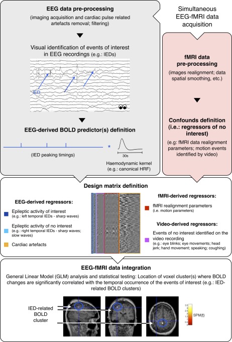
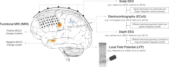
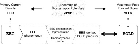
Electroencephalography (EEG) and functional magnetic resonance imaging (fMRI) are important tools in cognitive and clinical neuroscience. Combined EEG-fMRI has been shown to help to characterise brain networks involved in epileptic activity, as well as in different sensory, motor and cognitive functions. A good understanding of the electrophysiological correlates of the blood oxygen level-dependent (BOLD) signal is necessary to interpret fMRI maps, particularly when obtained in combination with EEG. We review the current understanding of electrophysiological-haemodynamic correlates, during different types of brain activity. We start by describing the basic mechanisms underlying EEG and BOLD signals and proceed by reviewing EEG-informed fMRI studies using fMRI to map specific EEG phenomena over the entire brain (EEG-fMRI mapping), or exploring a range of EEG-derived quantities to determine which best explain colocalised BOLD fluctuations (local EEG-fMRI coupling). While reviewing studies of different forms of brain activity (epileptic and nonepileptic spontaneous activity; cognitive, sensory and motor functions), a significant attention is given to epilepsy because the investigation of its haemodynamic correlates is the most common application of EEG-informed fMRI. Our review is focused on EEG-informed fMRI, an asymmetric approach of data integration. We give special attention to the invasiveness of electrophysiological measurements and the simultaneity of multimodal acquisitions because these methodological aspects determine the nature of the conclusions that can be drawn from EEG-informed fMRI studies. We emphasise the advantages of, and need for, simultaneous intracranial EEG-fMRI studies in humans, which recently became available and hold great potential to improve our understanding of the electrophysiological correlates of BOLD fluctuations.
Overview
The claims and statistics of this paper haven’t been checked yet.
Run the check — free, no account needed. It reads the open-access text and checks the sources it cites.
Integrity signals
Registry
Cites
Statistics
Figures
Verification
Claims the paper makes
The claims and statistics of this paper haven’t been checked yet.
Run the check — free, no account needed. It reads the open-access text and checks the sources it cites.
Claims other papers make about this one
Each is checked against this paper’s own text: does it say what the citing paper says it does?
The source backs part of the claim, not all of it — or backs it for a different population, condition or comparison.
In the citing paper
“In simultaneous EEG/fMRI studies, frontal theta activity is often inversely correlated with activity in DMN (Scheeringa et al. 2008;Murta et al. 2015), a brain network implicated in self-related thinking and mind wandering (Mason et al. 2007;Brewer et al. 2011).”
From Brain–Heart Interactions Underlying Traditional Tibetan Buddhist Meditation (2019) · cites this paper as Murta et al. 2015
Experimental — accuracy not yet measured
Self-check
Percentages, sums and reported differences re-computed from the paper’s own numbers.
The self-check needs the paper’s full text.
Citations
Cites
A default mode of brain function
Raichle, M. E., MacLeod, A. M., Snyder, A. Z., Powers, W. J., Gusnard, D. A., & Shulman, G. L. (2001) · Proceedings of the National Academy of Sciences · doi:10.1073/pnas.98.2.676 · Cited by 12,190
Event-related EEG/MEG synchronization and desynchronization: basic principles
Pfurtscheller, G., & Lopes da Silva, F. H. (1999) · Clinical Neurophysiology · doi:10.1016/s1388-2457(99)00141-8 · Cited by 7,328
Neuronal Oscillations in Cortical Networks
Buzsáki, G., & Draguhn, A. (2004) · Science · doi:10.1126/science.1099745 · Cited by 6,576
Neurophysiological investigation of the basis of the fMRI signal
Logothetis, N. K., Pauls, J., Augath, M., Trinath, T., & Oeltermann, A. (2001) · Nature · doi:10.1038/35084005 · Cited by 6,551
Brain magnetic resonance imaging with contrast dependent on blood oxygenation.
Ogawa, S., Lee, T. M., Kay, A. R., & Tank, D. W. (1990) · Proceedings of the National Academy of Sciences · doi:10.1073/pnas.87.24.9868 · Cited by 6,483
The origin of extracellular fields and currents — EEG, ECoG, LFP and spikes
Buzsáki, G., Anastassiou, C. A., & Koch, C. (2012) · Nature Reviews Neuroscience · doi:10.1038/nrn3241 · Cited by 4,225
Dynamic magnetic resonance imaging of human brain activity during primary sensory stimulation.
Kwong, K. K., Belliveau, J. W., Chesler, D. A., Goldberg, I. E., Weisskoff, R. M., Poncelet, B. P., Kennedy, D. N., Hoppel, B. E., Cohen, M. S., & Turner, R. (1992) · Proceedings of the National Academy of Sciences · doi:10.1073/pnas.89.12.5675 · Cited by 4,212
Interplay between Cortical Spreading Depolarization and Seizures
Kramer, D. R., Fujii, T., Ohiorhenuan, I. E., & Liu, C. Y. (2017) · Stereotactic and Functional Neurosurgery · doi:10.1159/000452841 · Cited by 3,721
Frequency Dependence of Signal Power and Spatial Reach of the Local Field Potential
Leski, S., Lindén, H., Tetzlaff, T., Pettersen, K. H., & Einevoll, G. T. (2013) · PubMed · doi:10.18154/rwth-conv-084442 · Cited by 3,667
What we can do and what we cannot do with fMRI
Logothetis, N. K. (2008) · Nature · doi:10.1038/nature06976 · Cited by 3,612
Research works printed but not linked: 3
- Ritter, 2010, EEG quality: The image acquisiton artefact. In: Mulert C, Lemieux L, editors. EEG-fMRI Physiological Basis, Technique, and Applications
- Vaughn, 1974, Bioelectric Recording Tecniques, Part B. Electroencephalography and Human Brain Potentials, 158
- Villringer, 2010, EEG-fMRI Physiological Basis, Technique, and Applications
Cited by
Sorted by citations
Electrophysiological source imaging: A noninvasive window to brain dynamics
He, B., Sohrabpour, A., Brown, E., & Liu, Z. (2018) · Annual Review of Biomedical Engineering · doi:10.1146/annurev-bioeng-062117-120853 · Cited by 297
Dynamic causal modelling revisited
Friston, K. J., Preller, K. H., Mathys, C., Cagnan, H., Heinzle, J., Razi, A., & Zeidman, P. (2019) · NeuroImage · doi:10.1016/j.neuroimage.2017.02.045 · Cited by 271
EEG-Based Estimation and Classification of Mental Fatigue
Trejo, L. J., Kubitz, K. A., Rosipal, R., Kochavi, R., & Montgomery, L. D. (2015) · Psychology · doi:10.4236/psych.2015.65055 · Cited by 251
Functional ultrasound neuroimaging: a review of the preclinical and clinical state of the art
Deffieux, T., Demené, C., Pernot, M., & Tanter, M. (2018) · Current Opinion in Neurobiology · doi:10.1016/j.conb.2018.02.001 · Cited by 201
EEG-Informed fMRI: A Review of Data Analysis Methods
Abreu, R., Leal, A., & Figueiredo, P. (2018) · Frontiers in Human Neuroscience · doi:10.3389/fnhum.2018.00029 · Cited by 171
Self-citationSimultaneous EEG-fMRI: What Have We Learned and What Does the Future Hold?
Warbrick, T. (2022) · Sensors · doi:10.3390/s22062262 · Cited by 182
Load modulates the alpha and beta oscillatory dynamics serving verbal working memory
Proskovec, A. L., Heinrichs‐Graham, E., & Wilson, T. W. (2018) · NeuroImage · doi:10.1016/j.neuroimage.2018.09.022 · Cited by 100
Fear in dreams and in wakefulness: Evidence for day/night affective homeostasis
Virginie Sterpenich, A., Perogamvros, L., Tononi, G., & Schwartz, S. (2019) · Human Brain Mapping · doi:10.1002/hbm.24843 · Cited by 110
Proskovec, A. L., Heinrichs‐Graham, E., & Wilson, T. W. (2016) · Human Brain Mapping · doi:10.1002/hbm.23178 · Cited by 99
The neural correlates of the unified percept of alcohol-related craving: a fMRI and EEG study
Huang, Y., Mohan, A., Ridder, D. D., Sunaert, S. G., & Vanneste, S. (2018) · Scientific Reports · doi:10.1038/s41598-017-18471-y · Cited by 94
Tensor Analysis and Fusion of Multimodal Brain Images
Karahan, E., Rojas-López, P. A., Bringas-Vega, M. L., Valdes-Hernandez, P. A., & Valdes-Sosa, P. A. (2015) · Proceedings of the IEEE · doi:10.1109/jproc.2015.2455028 · Cited by 88
Proskovec, A. L., Heinrichs‐Graham, E., Wiesman, A. I., McDermott, T. J., & Wilson, T. W. (2018) · Human Brain Mapping · doi:10.1002/hbm.23997 · Cited by 88
Neuroscience Information Toolbox: An Open Source Toolbox for EEG–fMRI Multimodal Fusion Analysis
Dong, L., Luo, C., Liu, X., Jiang, S., Li, F., Feng, H., Li, J., Gong, D., & Yao, D. (2018) · Frontiers in Neuroinformatics · doi:10.3389/fninf.2018.00056 · Cited by 79
Proskovec, A. L., Wiesman, A. I., Heinrichs‐Graham, E., & Wilson, T. W. (2018) · Scientific Reports · doi:10.1038/s41598-018-26863-x · Cited by 80
Yin, S., Liu, Y., & Ding, M. (2016) · Frontiers in Human Neuroscience · doi:10.3389/fnhum.2016.00364 · Cited by 75
Brain-computer interfaces in neurologic rehabilitation practice
Pichiorri, F., & Mattia, D. (2020) · Handbook of clinical neurology · doi:10.1016/b978-0-444-63934-9.00009-3 · Cited by 74
EEG Microstates Predict Concurrent fMRI Dynamic Functional Connectivity States
Abreu, R., Jorge, J., Leal, A., Koenig, T. W., & Figueiredo, P. (2020) · Brain Topography · doi:10.1007/s10548-020-00805-1 · Cited by 73
Self-citationFekete, Z., & Pongracz, A. (2016) · Sensors and Actuators B Chemical · doi:10.1016/j.snb.2016.12.096 · Cited by 76
Mesoscopic Neural Representations in Spatial Navigation
Kunz, L., Maidenbaum, S., Chen, D., Wang, L., Jacobs, J., & Axmacher, N. (2019) · Trends in Cognitive Sciences · doi:10.1016/j.tics.2019.04.011 · Cited by 71
Unimodal Versus Bimodal EEG-fMRI Neurofeedback of a Motor Imagery Task
Perronnet, L., Lécuyer, A., Mano, M., Bannier, É., Lotte, F., Clerc, M., & Barillot, C. (2017) · Frontiers in Human Neuroscience · doi:10.3389/fnhum.2017.00193 · Cited by 67
D’Hondt, F., Billieux, J., & Maurage, P. (2015) · Neuroscience & Biobehavioral Reviews · doi:10.1016/j.neubiorev.2015.10.005 · Cited by 68
Alyagon, U., Shahar, H., Hadar, A. A., Barnea‐Ygael, N., Lazarovits, A., Shalev, H., & Zangen, A. (2020) · NeuroImage Clinical · doi:10.1016/j.nicl.2020.102206 · Cited by 62
Aberrant Neuronal Dynamics during Working Memory Operations in the Aging HIV-Infected Brain
Wilson, T. W., Proskovec, A. L., Heinrichs‐Graham, E., O’Neill, J., Robertson, K. R., Fox, H. S., & Swindells, S. (2017) · Scientific Reports · doi:10.1038/srep41568 · Cited by 60
Mash, L. E., Reiter, M. A., Linke, A. C., Townsend, J., & Müller, R. (2018) · Developmental Neurobiology · doi:10.1002/dneu.22570 · Cited by 52
Methods and utility of EEG-fMRI in epilepsy.
van Graan, L. A., Lemieux, L., & Chaudhary, U. J. (2015) · Europe PMC (PubMed Central) · doi:10.3978/j.issn.2223-4292.2015.02.04 · Cited by 55
Self-citationThe strength of alpha and gamma oscillations predicts behavioral switch costs
Proskovec, A. L., Wiesman, A. I., & Wilson, T. W. (2018) · NeuroImage · doi:10.1016/j.neuroimage.2018.12.016 · Cited by 48
Murta, T., Chaudhary, U. J., Tierney, T. M., Dias, A., Leite, M., Carmichael, D. W., Figueiredo, P., & Lemieux, L. (2016) · NeuroImage · doi:10.1016/j.neuroimage.2016.08.036 · Cited by 47
Self-citationMultimodal EEG-fMRI: advancing insight into large-scale human brain dynamics
Chang, C., & Chen, J. E. (2021) · Current Opinion in Biomedical Engineering · doi:10.1016/j.cobme.2021.100279 · Cited by 45
Brain–Heart Interactions Underlying Traditional Tibetan Buddhist Meditation
Jiang, H., He, B., Guo, X., Wang, X., Guo, M., Wang, Z., Xue, T., Li, H., Xu, T., Ye, S., et al. (2019) · Cerebral Cortex · doi:10.1093/cercor/bhz095 · Cited by 40
Portnova, G. V., Tetereva, A., Balaev, V. V., Atanov, M. S., Skiteva, L. I., Ushakov, V., Ivanitsky, A. M., & Martynova, O. (2018) · Frontiers in Human Neuroscience · doi:10.3389/fnhum.2017.00654 · Cited by 41
Abreu, R., Leite, M., Jorge, J., Grouiller, F., van der Zwaag, W., Leal, A., & Figueiredo, P. (2016) · NeuroImage · doi:10.1016/j.neuroimage.2016.03.034 · Cited by 40
Self-citationGao, L., Huang, P., Dong, Z., Gao, T., Huang, S., Zhou, C., Lai, Y., Deng, G., Liu, B., Wen, G., et al. (2018) · Frontiers in Pharmacology · doi:10.3389/fphar.2018.01098 · Cited by 38
Kaur, A., Rishu Chaujar, B., & Chinnadurai, V. (2019) · Human Factors The Journal of the Human Factors and Ergonomics Society · doi:10.1177/0018720819869129 · Cited by 37
Inferring Macroscale Brain Dynamics via Fusion of Simultaneous EEG-fMRI
Philiastides, M. G., Tu, T., & Sajda, P. (2021) · Annual Review of Neuroscience · doi:10.1146/annurev-neuro-100220-093239 · Cited by 41
Two is better? combining EEG and fMRI for BCI and neurofeedback: a systematic review
Fleury, M., Figueiredo, P., Vourvopoulos, A., & Lécuyer, A. (2023) · Journal of Neural Engineering · doi:10.1088/1741-2552/ad06e1 · Cited by 40
Self-citationAbreu, R., Leal, A., & Figueiredo, P. (2019) · Scientific Reports · doi:10.1038/s41598-018-36976-y · Cited by 33
Self-citationAbreu, R., Leal, A., Lopes, F. H. D. S., & Figueiredo, P. (2018) · Clinical Neurophysiology · doi:10.1016/j.clinph.2017.12.038 · Cited by 32
Self-citationModulation of attention networks serving reorientation in healthy aging
Arif, Y., Spooner, R. K., Wiesman, A. I., Embury, C. M., Proskovec, A. L., & Wilson, T. W. (2020) · Aging · doi:10.18632/aging.103515 · Cited by 32
Fedele, T., Tzovara, A., Steiger, B. K., Hilfiker, P., Grünwald, T., Stieglitz, L. H., Jokeit, H., & Sarnthein, J. (2020) · NeuroImage · doi:10.1016/j.neuroimage.2020.116705 · Cited by 31
Multimodal Autoencoder Predicts fNIRS Resting State From EEG Signals
Sirpal, P., Damseh, R., Peng, K., Nguyen, D. K., & Lesage, F. (2021) · Neuroinformatics · doi:10.1007/s12021-021-09538-3 · Cited by 31
Abreu, R., Nunes, S., Leal, A., & Figueiredo, P. (2016) · NeuroImage · doi:10.1016/j.neuroimage.2016.08.008 · Cited by 29
Self-citationde Tommaso, M., Betti, V., Bocci, T., Bolognini, N., Russo, F. D., Fattapposta, F., Ferri, R., Invitto, S., Koch, G., Miniussi, C., et al. (2020) · Neurological Sciences · doi:10.1007/s10072-020-04420-7 · Cited by 30
Murta, T., Hu, L., Tierney, T. M., Chaudhary, U. J., Walker, M. C., Carmichael, D. W., Figueiredo, P., & Lemieux, L. (2016) · NeuroImage · doi:10.1016/j.neuroimage.2016.08.001 · Cited by 27
Self-citationThe Role of EEG-fMRI in Studying Cognitive Network Alterations in Epilepsy
Shamshiri, E., Sheybani, L., & Vulliémoz, S. (2019) · Frontiers in Neurology · doi:10.3389/fneur.2019.01033 · Cited by 26
Cognitive function: holarchy or holacracy?
Bîrle, C., Slăvoacă, D., Balea, M., Popa, L. L., Mureșanu, I. A., Ștefănescu, E., Văcăraș, V., Dina, C., Strilciuc, Ș., Popescu, B. O., et al. (2020) · Neurological Sciences · doi:10.1007/s10072-020-04737-3 · Cited by 36
Maggioni, E., Zucca, C., Reni, G., Cerutti, S., Triulzi, F. M., Bianchi, A. M., & Arrigoni, F. (2016) · Human Brain Mapping · doi:10.1002/hbm.23170 · Cited by 24
Fear in dreams and in wakefulness: Evidence for day/night affective homeostasis
Sterpenich, V, Perogamvros, L., Tononi, G., & Schwartz, S. (2019) · bioRxiv (Cold Spring Harbor Laboratory) · doi:10.1101/534099 · Cited by 99
Decroix, L., Pauw, K. D., Cutsem, J. V., Pattyn, N., Heyman, E., & Meeusen, R. (2018) · Psychopharmacology · doi:10.1007/s00213-018-4952-2 · Cited by 23
MRI magnetic compatible electrical neural interface: From materials to application
Zhang, Y., Song, L., Li, H., Ji, B., Wang, M., Tao, J., Liang, J., Zhang, X., & Kang, X. (2021) · Biosensors and Bioelectronics · doi:10.1016/j.bios.2021.113592 · Cited by 24
Multiple sclerosis-related white matter microstructural change alters the BOLD hemodynamic response
Hubbard, N. A., Turner, M. P., Hutchison, J. L., Ouyang, A., Strain, J. F., Oasay, L., Patel, S. S., Davis, S. L., Remington, G. M., Brigante, R. M., et al. (2015) · Journal of Cerebral Blood Flow & Metabolism · doi:10.1177/0271678x15615133 · Cited by 22
Clustering-Constrained ICA for Ballistocardiogram Artifacts Removal in Simultaneous EEG-fMRI
Wang, K., Li, W., Dong, L., Zou, L., & Wang, C. (2018) · Frontiers in Neuroscience · doi:10.3389/fnins.2018.00059 · Cited by 22
Markoula, S., Chaudhary, U. J., Perani, S., Ciantis, A. D., Yadee, T., Duncan, J. S., Diehl, B., McEvoy, A. W., & Lemieux, L. (2018) · Seizure · doi:10.1016/j.seizure.2018.07.016 · Cited by 21
Self-citationBelief of agency changes dynamics in sensorimotor networks
Buchholz, V. N., David, N., Sengelmann, M., & Engel, A. K. (2019) · Scientific Reports · doi:10.1038/s41598-018-37912-w · Cited by 21
Safety of Simultaneous Scalp or Intracranial EEG during MRI: A Review
Hawsawi, H. B., Carmichael, D. W., & Lemieux, L. (2017) · Frontiers in Physics · doi:10.3389/fphy.2017.00042 · Cited by 20
Self-citationLin, S.-H., Lai, H.-Y., Lo, Y., Chou, C., Chou, Y.-T., Yang, S., Sun, I., Chen, B.-W., Wang, C.-F., Liu, G.-T., et al. (2017) · Frontiers in Neuroscience · doi:10.3389/fnins.2017.00701 · Cited by 20
de Tommaso, M., Betti, V., Bocci, T., Bolognini, N., Russo, F. D., Fattapposta, F., Ferri, R., Invitto, S., Koch, G., Miniussi, C., et al. (2020) · Neurological Sciences · doi:10.1007/s10072-020-04527-x · Cited by 20
Eyndhoven, S. V., Dupont, P. J., Tousseyn, S., Vervliet, N., Paesschen, W. V., Huffel, S. V., & Hunyadi, B. (2020) · NeuroImage · doi:10.1016/j.neuroimage.2020.117652 · Cited by 20
Schulz, S., Haueisen, J., Bär, K., & Voss, A. (2018) · Physiological Measurement · doi:10.1088/1361-6579/aace9b · Cited by 19
Simões, M. A., Abreu, R., Direito, B., Sayal, A., Castelhano, J., Carvalho, P., & Castelo-Branco, M. S. (2020) · Journal of Neural Engineering · doi:10.1088/1741-2552/ab9a98 · Cited by 19
Dowdle, L. T., Ghose, G. M., Chen, C. C. C., Uǧurbil, K., Yacoub, E. S., & Vizioli, L. (2021) · Progress in Neurobiology · doi:10.1016/j.pneurobio.2021.102171 · Cited by 23
Tehrani, N., Wilson, W., Pittman, D. J., Mosher, V., Peedicail, J. S., Aghakhani, Y., Beers, C. A., Gaxiola‐Valdez, I., Singh, S., Goodyear, B. G., et al. (2021) · Epilepsia · doi:10.1111/epi.16887 · Cited by 16
Rajasilta, O., Häkkinen, S., Björnsdotter, M., Scheinin, N. M., Lehtola, S. J., Saunavaara, J., Parkkola, R., Lähdesmäki, T., Karlsson, L., Karlsson, H., et al. (2023) · European Journal of Neuroscience · doi:10.1111/ejn.15882 · Cited by 15
Meditation and the Brain in Health and Disease
Fox, K. C. R., & Cahn, B. R. (2020) · Oxford University Press eBooks · doi:10.1093/oxfordhb/9780198808640.013.23 · Cited by 16
Prokopiou, P. C., Xifra‐Porxas, A., Kassinopoulos, M., Boudrias, M., & Mitsis, G. D. (2022) · Brain Topography · doi:10.1007/s10548-022-00898-w · Cited by 15
Stable Scalp EEG Spatiospectral Patterns Across Paradigms Estimated by Group ICA
Labounek, R., Bridwell, D. A., Mareček, R., Lamoš, M., Mikl, M., Slavicek, T., Bednařík, P., Baštinec, J., Hluštı́k, P., Brázdil, M., et al. (2017) · Brain Topography · doi:10.1007/s10548-017-0585-8 · Cited by 16
Single-Trial EEG-fMRI Reveals the Generation Process of the Mismatch Negativity
Li, Q., Liu, G., Yuan, G., Wang, G., Wu, Z., & Zhao, X. (2019) · Frontiers in Human Neuroscience · doi:10.3389/fnhum.2019.00168 · Cited by 18
Measurement of the Mapping between Intracranial EEG and fMRI Recordings in the Human Brain
Carmichael, D. W., Vulliémoz, S., Murta, T., Chaudhary, U. J., Perani, S., Rodionov, R., Rosa, M. J., Friston, K. J., & Lemieux, L. (2024) · Bioengineering · doi:10.3390/bioengineering11030224 · Cited by 16
Self-citationResting state networks of the canine brain under sevoflurane anaesthesia
Beckmann, K. M., Wang‐Leandro, A., Dennler, M., Carrera, I., Richter, H., Bektas, R. N., Steiner, A. R., & Haller, S. (2020) · PLoS ONE · doi:10.1371/journal.pone.0231955 · Cited by 15
Detection of Neural Activity of Brain Functional Site Based on Microwave Scattering Principle
Wang, J.-K., Jiang, X., Peng, L. F., Li, X., An, H.-J., & Wen, B.-J. (2019) · IEEE Access · doi:10.1109/access.2019.2894128 · Cited by 15
Learning 2-in-1: Towards Integrated EEG-fMRI-NeurofeedbackPreprint
Perronnet, L., Lécuyer, A., Mano, M., Fleury, M., Lioi, G., Cury, C., Clerc, M., Lotte, F., & Barillot, C. (2018) · bioRxiv (Cold Spring Harbor Laboratory) · doi:10.1101/397729 · Cited by 15
Subject-Specific Modeling of EEG-fNIRS Neurovascular Coupling by Task-Related Tensor Decomposition
Lin, J., Lu, J., Shu, Z., Han, J., & Yu, N. (2024) · IEEE Transactions on Neural Systems and Rehabilitation Engineering · doi:10.1109/tnsre.2024.3355121 · Cited by 15
Prior Emotional Context Modulates Early Event-Related Potentials to Neutral Retrieval Cues
Bowen, H. J., Fields, E. C., & Kensinger, E. A. (2019) · Journal of Cognitive Neuroscience · doi:10.1162/jocn_a_01451 · Cited by 14
Kaur, A., Chinnadurai, V., & Rishu Chaujar, B. (2020) · Scientific Reports · doi:10.1038/s41598-020-61119-7 · Cited by 14
Mattiev, J., Sajovic, J., Drevenšek, G., & Rogelj, P. (2022) · Bioengineering · doi:10.3390/bioengineering10010042 · Cited by 14
Bjerkan, J., Lancaster, G., Meglič, B., Kobal, J., Crawford, T. J., McClintock, P. V. E., & Stefanovska, A. (2023) · Brain Research Bulletin · doi:10.1016/j.brainresbull.2023.110704 · Cited by 17
Chen, C., Yuan, K., Wang, X., Khan, A., Chu, C. W., & Tong, R. K. (2021) · Neural Plasticity · doi:10.1155/2021/8866613 · Cited by 15
Arif, Y., Embury, C. M., Spooner, R. K., Okelberry, H. J., Willett, M. P., Eastman, J. A., & Wilson, T. W. (2022) · Human Brain Mapping · doi:10.1002/hbm.25764 · Cited by 13
Direct interactions between the human insula and hippocampus during memory encoding
Huang, W., Lyu, D., Stieger, J., Gotlib, I. H., Buch, V., Wagner, A. D., & Parvizi, J. (2025) · Nature Neuroscience · doi:10.1038/s41593-025-02005-1 · Cited by 15
Quantitative Analyses Help in Choosing Between Simultaneous vs. Separate EEG and fMRI
Schrooten, M., Vandenberghe, R. R., Peeters, R. R., & Dupont, P. J. (2019) · Frontiers in Neuroscience · doi:10.3389/fnins.2018.01009 · Cited by 13
Objective selection of epilepsy-related independent components from EEG data
Abreu, R., Leite, M., Leal, A., & Figueiredo, P. (2015) · Journal of Neuroscience Methods · doi:10.1016/j.jneumeth.2015.10.003 · Cited by 13
Self-citationQuindlen, J. C., Kent, A. R., Anda, P. D., Kartha, S., Benison, A. M., & Winkelstein, B. A. (2020) · Neuromodulation Technology at the Neural Interface · doi:10.1111/ner.13116 · Cited by 13
Meditation and the brain in health and diseasePreprint
Fox, K. C. R., & Cahn, B. R. (2018) · MindRxiv (OSF Preprints) · doi:10.31231/osf.io/m2sez · Cited by 13
Söderström, P., Horne, M. A., Mannfolk, P., van Westen, D., & Roll, M. (2018) · Brain Research · doi:10.1016/j.brainres.2018.06.004 · Cited by 13
Doucette, W. T., Dwiel, L. L., Boyce, J. E., Simon, A. A., Khokhar, J. Y., & Green, A. I. (2018) · Frontiers in Psychiatry · doi:10.3389/fpsyt.2018.00336 · Cited by 11
Mann‐Krzisnik, D., & Mitsis, G. D. (2022) · Human Brain Mapping · doi:10.1002/hbm.25902 · Cited by 9
Uji, M., Cross, N. E., Pomares, F. B., Perrault, A. A., Jegou, A., Nguyen, A., Aydın, Ü., Lina, J., Dang‐Vu, T. T., & Grova, C. (2021) · Human Brain Mapping · doi:10.1002/hbm.25535 · Cited by 11
Working Memory Workload When Making Complex Decisions: A Behavioral and EEG Study
Balconi, M., Rovelli, K., Angioletti, L., & Allegretta, R. A. (2024) · Sensors · doi:10.3390/s24175754 · Cited by 14
Frequency and laminar profile of feature-specific visual activity revealed by interleaved EEG–fMRIPreprint
Clausner, T., Marques, J. P., Scheeringa, R., & Bonnefond, M. (2025) · eLife · doi:10.1101/2024.07.31.605816 · Cited by 9
A model for studying the energetics of sustained high frequency firing
Joós, B., Markham, M. R., Lewis, J. E., & Morris, C. E. (2018) · PLoS ONE · doi:10.1371/journal.pone.0196508 · Cited by 8
EEG To FMRI Synthesis: Is Deep Learning a Candidate?
Calhas, D., & Henriques, R. (2023) · Proceedings of the International Conference on Information Systems Development · doi:10.62036/isd.2023.26 · Cited by 10
Consistency of resting-state correlations between fMRI networks and EEG band power
Xavier, M., Esteves, I., Jorge, J., Abreu, R., Giraud, A., Sadaghiani, S., Wirsich, J., & Figueiredo, P. (2025) · Imaging Neuroscience · doi:10.1162/imag.a.37 · Cited by 8
Self-citationLate frontal positivity effects in Self-referential Memory: Unique to the Self?
Porter, N. A., Fields, E. C., Moore, I. L., & Gutchess, A. H. (2021) · Social Neuroscience · doi:10.1080/17470919.2021.1929460 · Cited by 8
Yue, T., Chen, Y., Zheng, Q., Xu, Z., Wang, W., & Ni, G. (2021) · Frontiers in Aging Neuroscience · doi:10.3389/fnagi.2021.677090 · Cited by 9
Rejection in Bargaining Situations: An Event-Related Potential Study in Adolescents and Adults
Zanolie, K., David De Cremer, A., Güroğlu, B., & Crone, E. A. (2015) · PLoS ONE · doi:10.1371/journal.pone.0139953 · Cited by 8
Ferreira, J. L., Wu, Y. Y., Besseling, R. M. H., Lamerichs, R. M. J. N., & Aarts, R. M. (2016) · Journal of Medical Engineering · doi:10.1155/2016/9614323 · Cited by 8
The spatiotemporal pattern of pure tone processing: A single-trial EEG-fMRI study
Li, Q., Liu, G., Wei, D., Guo, J., Yuan, G., & Wu, S. (2017) · NeuroImage · doi:10.1016/j.neuroimage.2017.11.059 · Cited by 8
BOLD signatures of sleepPreprint
Song, C., Boly, M., Tagliazucchi, E., Laufs, H., & Tononi, G. (2019) · bioRxiv (Cold Spring Harbor Laboratory) · doi:10.1101/531186 · Cited by 8
Jiříček, S., Koudelka, V., Mantini, D., Mareček, R., & Hlinka, J. (2025) · Frontiers in Neuroscience · doi:10.3389/fnins.2025.1549172 · Cited by 7
Gustatory Cortex Is Involved in Evidence Accumulation during Food Choice
Ataei, A., Amini, A., & Ghazizadeh, A. (2022) · eNeuro · doi:10.1523/eneuro.0006-22.2022 · Cited by 7
The Effect of a Prostration (Sajdeh) on the Prefrontal Brain Activity, a Pilot Study
Yousefzadeh, F., Jahromi, G. P., Manshadi, E. M., & Hatef, B. (2018) · Basic and Clinical Neuroscience Journal · doi:10.32598/bcn.9.10.195 · Cited by 7
Metadata
- Authors
- Murta, T., Leite, M., Carmichael, D. W., Figueiredo, P., & Lemieux, L.
- Affiliations
- University of Lisbon (PT)
- University College London (GB)
- National Hospital for Neurology and Neurosurgery (GB)
- Corresponding author
- Murta, Teresa
- Venue
- Human Brain Mapping
- Publisher
- Wiley
- Published
- 2014-10-03
- Volume
- 36
- Issue
- 1
- Pages
- 391-414
- ISSN
- 1065-9471 (ISSN-L)
- PMC
- PMC4280889
- OpenAlex
- W1591966483
- Semantic Scholar
- 213e8a7e4f617a884af93681624845d67264d85a
- Cited by
- 165
- References
- 249
- Citations by year
- 2026: 16 · 2025: 12 · 2024: 7 · 2023: 4 · 2022: 11 · 2021: 17 · 2020: 20 · 2019: 21 · 2018: 26 · 2017: 10 · 2016: 13 · 2015: 8
- FWCI
- 1.61
- Type
- journal article
- Language
- en
- Open access
- bronze
- doi.org
- Locations
- Human Brain Mapping (publishedVersion)
- PubMed Central (publishedVersion, cc-by)
- PubMed (publishedVersion)
- UCL Discovery (University College London) (submittedVersion)
- Funders
- Department of Health's NIHR Biomedical Research Centres funding scheme
- Portuguese Science Foundation (FCT) (SFRH/BD/80421/2011 and PTDC/SAU-ENB/112294/2009)
- Action Medical Research (SP4646)
- Topics
- Health Sciences › Medicine › Radiology, Nuclear Medicine and Imaging › Advanced MRI Techniques and Applications
- Life Sciences › Neuroscience › Cognitive Neuroscience › EEG and Brain-Computer Interfaces
- Health Sciences › Medicine › Radiology, Nuclear Medicine and Imaging › Advanced Neuroimaging Techniques and Applications
- Keywords
- BOLD signal, epilepsy, hemodynamic response, spontaneous brain activity
- MeSH terms
- Brain, Brain Mapping, Electroencephalography, Humans, Image Processing, Computer-Assisted, Magnetic Resonance Imaging, Oxygen, Brain Waves
About these checks
Registry, retraction, Cites and statistics results are computed, not judged, with one exception: a printed reference that no index lists is read once by a model before it counts as a citation error. Claims are extracted by a model and kept only when their quote appears verbatim in the text; each cited source is then checked, with a model’s help, for whether it says what the claim relies on. Those verdicts can be wrong — read the claim’s quote and the cited source before relying on one.
Statistics and arithmetic are recomputed, not judged.
Confidence bands are shown only for fields calibrated on our annotated test set. None is calibrated yet, so no band is shown.
What counts as a claim ›How we measure confidence ›How we check this ›
Are you an author, or do you see a problem with this page? Request a review
