

The DOI matches its Crossref record; 72 of the 72 references it cites were found in a registry or an index.
Ins and outs of AlphaFold2 transmembrane protein structure predictions
Open accessAbstract
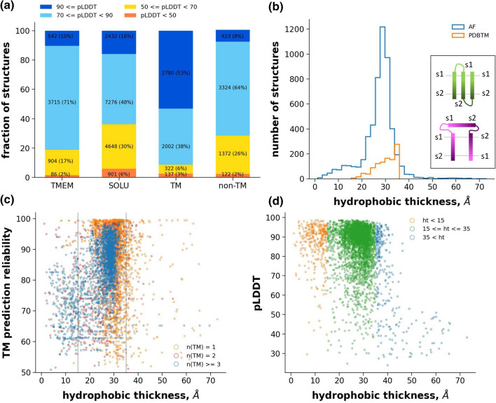
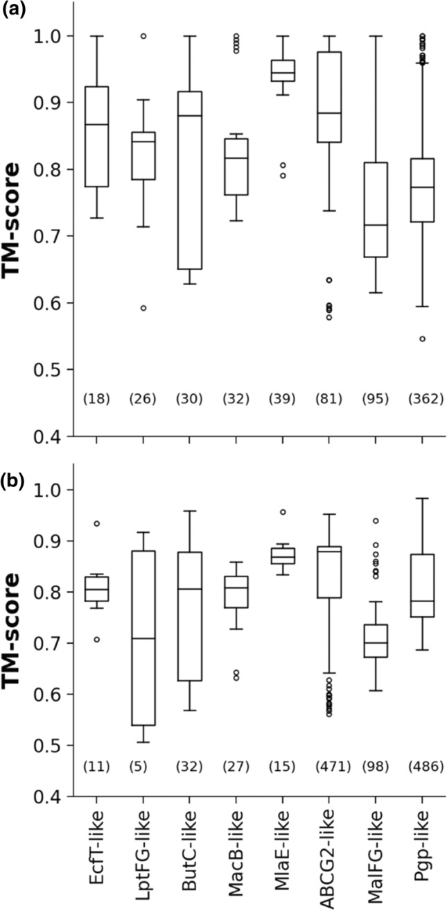
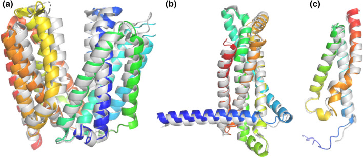
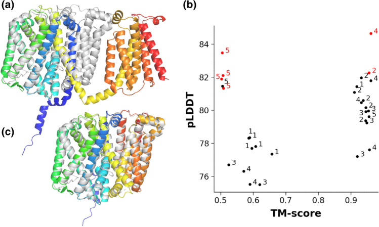
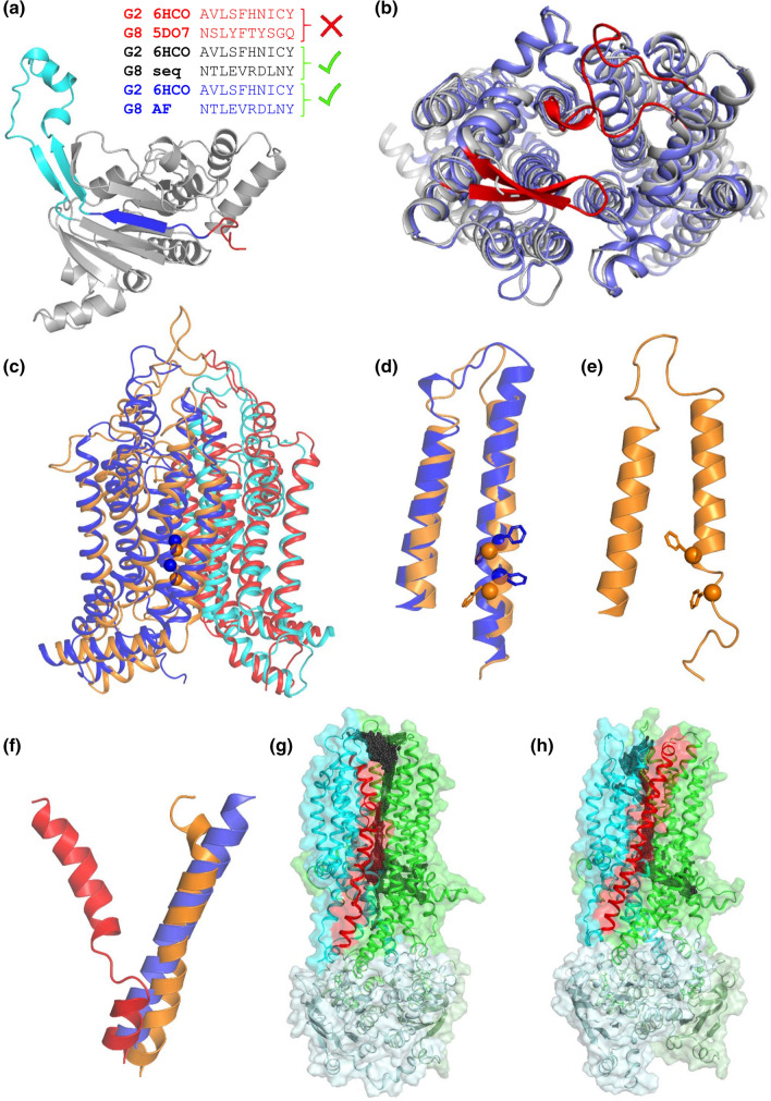
Transmembrane (TM) proteins are major drug targets, but their structure determination, a prerequisite for rational drug design, remains challenging. Recently, the DeepMind's AlphaFold2 machine learning method greatly expanded the structural coverage of sequences with high accuracy. Since the employed algorithm did not take specific properties of TM proteins into account, the reliability of the generated TM structures should be assessed. Therefore, we quantitatively investigated the quality of structures at genome scales, at the level of ABC protein superfamily folds and for specific membrane proteins (e.g. dimer modeling and stability in molecular dynamics simulations). We tested template-free structure prediction with a challenging TM CASP14 target and several TM protein structures published after AlphaFold2 training. Our results suggest that AlphaFold2 performs well in the case of TM proteins and its neural network is not overfitted. We conclude that cautious applications of AlphaFold2 structural models will advance TM protein-associated studies at an unexpected level.
Overview
The claims and statistics of this paper haven’t been checked yet.
Run the check — free, no account needed. It reads the open-access text and checks the sources it cites.
Integrity signals
Registry
Cites
Statistics
Figures
Verification
Claims the paper makes
The claims and statistics of this paper haven’t been checked yet.
Run the check — free, no account needed. It reads the open-access text and checks the sources it cites.
Claims other papers make about this one
Each is checked against this paper’s own text: does it say what the citing paper says it does?
The cited source says what the claim attributes to it.
In the citing paper
“Hegedűs et al benchmarked several membrane proteins not included in the original AF2 training set and concluded that on average AF2 predicts transmembrane proteins as well as soluble proteins38”
From The power and pitfalls of AlphaFold2 for structure prediction beyond rigid globular proteins (2024) · cites this paper as [38]
The cited source says what the claim attributes to it.
In the citing paper
“AF2 models with trans-membrane region lengths corresponding to non-physiological membrane thickness values can exhibit very high pLDDT scores (high model confidence), suggesting pLDDT scores alone are not sufficient to select native membrane protein conformations”
From The power and pitfalls of AlphaFold2 for structure prediction beyond rigid globular proteins (2024) · cites this paper as [38]
In the citing paper
“AF2 performs poorly for targets embedded in membrane thickness outside the 15–35 Å range as well as targets with novel features not commonly present in the PDB”
From The power and pitfalls of AlphaFold2 for structure prediction beyond rigid globular proteins (2024) · cites this paper as [38]
In the citing paper
“Benchmarking AF2 against membrane proteins represents a challenge since the membrane environment, which includes lipids and other proteins, is not directly considered by current versions of AlphaFold38”
From The power and pitfalls of AlphaFold2 for structure prediction beyond rigid globular proteins (2024) · cites this paper as [38]
Self-check
Percentages, sums and reported differences re-computed from the paper’s own numbers.
The self-check needs the paper’s full text.
Citations
Cites
Highly accurate protein structure prediction with AlphaFold
Jumper, J., Evans, R., Pritzel, A., Green, T., Figurnov, M., Ronneberger, O., Tunyasuvunakool, K., Bates, R., Žídek, A., Potapenko, A., et al. (2021) · Nature · doi:10.1038/s41586-021-03819-2 · Cited by 43,764
Matplotlib: a 2D graphics environment
Hunter, J. D. (2007) · Computing in Science & Engineering · doi:10.1109/mcse.2007.55 · Cited by 39,249
Particle mesh Ewald: An N⋅log(N) method for Ewald sums in large systems
Darden, T., York, D., & Pedersen, L. (1993) · The Journal of Chemical Physics · doi:10.1063/1.464397 · Cited by 29,638
Clustal W and Clustal X version 2.0
Larkin, M., Blackshields, G., Brown, N. P., Chenna, R., McGettigan, P., McWilliam, H., Valentin, F., Wallace, I. M., Wilm, A., López, R., et al. (2007) · Bioinformatics · doi:10.1093/bioinformatics/btm404 · Cited by 28,598
Abraham, M. J., Murtola, T., Schulz, R., Páll, S., Smith, J. C., Hess, B., & Lindahl, E. (2015) · SoftwareX · doi:10.1016/j.softx.2015.06.001 · Cited by 27,190
Harris, C. R., Millman, K. J., van der Walt, S. J., Gommers, R., Virtanen, P., Cournapeau, D., Wieser, E., Taylor, J., Berg, S., Smith, N. J., et al. (2020) · Nature · doi:10.1038/s41586-020-2649-2 · Cited by 23,826
LINCS: A linear constraint solver for molecular simulations
Hess, B., Bekker, H., Berendsen, H. J. C., & Fraaije, J. G. E. M. (1997) · Journal of Computational Chemistry · doi:10.1002/(sici)1096-987x(199709)18:12 · Cited by 18,479
Harris, C., Millman, K., van der Walt, S., Gommers, R., Virtanen, P., Cournapeau, D., Wieser, E., Taylor, J., Berg, S., Smith, N., et al. (2020) · RePEc: Research Papers in Economics · Cited by 17,844
ColabFold - Making protein folding accessible to all
Mirdita, M., Schütze, K., Moriwaki, Y., Heo, L., Ovchinnikov, S., & Steinegger, M. (2021) · doi:10.1101/2021.08.15.456425 · Cited by 9,391
CHARMM‐GUI: A web‐based graphical user interface for CHARMM
Jo, S., Kim, T., Iyer, V. G., & Im, W. (2008) · Journal of Computational Chemistry · doi:10.1002/jcc.20945 · Cited by 9,284
Research works printed but not linked: 1
- McCoy AJ, Sammito MD, Read RJ (2021) Possible implications of AlphaFold2 for crystallographic phasing by molecular replacement. BioRxiv. https://doi.org/10.1101/2021.05.18.444614 · DOI
Cited by
Sorted by citations
AlphaFold2 and its applications in the fields of biology and medicine
Yang, Z., Zeng, X., Zhao, Y. C., & Chen, R. (2023) · Signal Transduction and Targeted Therapy · doi:10.1038/s41392-023-01381-z · Cited by 655
The application of large language models in medicine: A scoping review
Meng, X., Yan, X., Zhang, K., Liu, D., Cui, X., Yang, Y., Zhang, M., Cao, C., Wang, J., Wang, X., et al. (2024) · iScience · doi:10.1016/j.isci.2024.109713 · Cited by 414
Using AlphaFold to predict the impact of single mutations on protein stability and function
Pak, M. A., Markhieva, K. A., Novikova, M. S., Petrov, D. S., Vorobyev, I. S., Maksimova, E. S., Kondrashov, F. A., & Ivankov, D. N. (2023) · PLOS One · doi:10.1371/journal.pone.0282689 · Cited by 322
The power and pitfalls of AlphaFold2 for structure prediction beyond rigid globular proteins
Agarwal, V., & McShan, A. C. (2024) · Nature Chemical Biology · doi:10.1038/s41589-024-01638-w · Cited by 155
“Benchmarking AF2 against membrane proteins represents a challenge, as the membrane environment, which includes lipids and other proteins, is not directly considered by current versions of AlphaFold 38 .”
BackgroundAnalysis of AlphaMissense data in different protein groups and structural context
Tordai, H., Torres, O., Csepi, M., Padányi, R., Lukács, G. L., & Hegedűs, T. (2024) · Scientific Data · doi:10.1038/s41597-024-03327-8 · Cited by 158
Self-citation
Metadata
- Authors
- Hegedűs, T., Geisler, M., Lukács, G. L., & Farkas, B.
- Affiliations
- Semmelweis University (HU)
- University of Fribourg (CH)
- McGill University (CA)
- Corresponding author
- Hegedűs, Tamás
- Venue
- Cellular and Molecular Life Sciences
- Published
- 2022-01
- Volume
- 79
- Issue
- 1
- Pages
- 73-73
- ISSN
- 1420-682X (ISSN-L)
- 1420-9071
- PMC
- PMC8761152
- OpenAlex
- W4205172550
- Cited by
- 127
- References
- 69
- Citations by year
- 2026: 15 · 2025: 19 · 2024: 23 · 2023: 41 · 2022: 28
- FWCI
- 9.06
- Type
- journal article
- Language
- en
- Open access
- hybrid
- doi.org
- Licence
- Locations
- Cellular and Molecular Life Sciences (publishedVersion, cc-by)
- PubMed Central (publishedVersion, cc-by)
- PubMed (publishedVersion)
- Funders
- Nemzeti Kutatási Fejlesztési és Innovációs Hivatal (K127961)
- Cystic Fibrosis Foundation (HEGEDU20I0)
- Cystic Fibrosis Canada (LUKACS20G0)
- Schweizerischer Nationalfonds zur Förderung der Wissenschaftlichen Forschung (310030_197563)
- Canadian Institutes of Health Research
- Canada Foundation for Innovation
- Canada Research Chairs
- Semmelweis University
- Topics
- Life Sciences › Biochemistry, Genetics and Molecular Biology › Molecular Biology › Protein Structure and Dynamics
- Life Sciences › Biochemistry, Genetics and Molecular Biology › Molecular Biology › Machine Learning in Bioinformatics
- Life Sciences › Biochemistry, Genetics and Molecular Biology › Structural Biology › Enzyme Structure and Function
- Keywords
- AlphaFold2, transmembrane proteins, protein structure prediction, prediction accuracy assessment, ABC transporters, membrane protein dimerization, CASP14, overfitting, structural genomics
- MeSH terms
- Machine Learning, Protein Domains, Algorithms, Computer Simulation, Humans, Lipids, Membrane Proteins, Protein Conformation, Software, Reproducibility of Results, Genome, Protein Structure, Secondary, Protein Folding, Computational Biology, Proteome, Proteomics, Molecular Dynamics Simulation
About these checks
Registry, retraction, Cites and statistics results are computed, not judged, with one exception: a printed reference that no index lists is read once by a model before it counts as a citation error. Claims are extracted by a model and kept only when their quote appears verbatim in the text; each cited source is then checked, with a model’s help, for whether it says what the claim relies on. Those verdicts can be wrong — read the claim’s quote and the cited source before relying on one.
Statistics and arithmetic are recomputed, not judged.
Confidence bands are shown only for fields calibrated on our annotated test set. None is calibrated yet, so no band is shown.
What counts as a claim ›How we measure confidence ›How we check this ›
Are you an author, or do you see a problem with this page? Request a review
