

The DOI matches its Crossref record; 60 of the 60 references it cites were found in a registry or an index.
Emergence and evolution of the renin-angiotensin-aldosterone system
Open accessAbstract
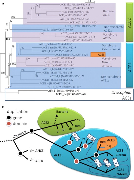
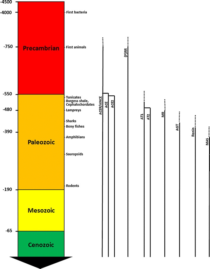
The renin-angiotensin-aldosterone system (RAAS) is not the sole, but perhaps the most important volume regulator in vertebrates. To gain insights into the function and evolution of its components, we conducted a phylogenetic analysis of its main related genes. We found that important parts of the system began to appear with primitive chordates and tunicates and that all major components were present at the divergence of bony fish, with the exception of the Mas receptor. The Mas receptor first appears after the bony-fish/tetrapod divergence. This phase of evolutionary innovation happened about 400 million years ago. We found solid evidence that angiotensinogen made its appearance in cartilage fish. The presence of several RAAS genes in organisms that lack all the components shows that these genes have had other ancestral functions outside of their current role. Our analysis underscores the utility of sequence comparisons in the study of evolution. Such analyses may provide new hypotheses as to how and why in today's population an increased activity of the RAAS frequently leads to faulty salt and volume regulation, hypertension, and cardiovascular diseases, opening up new and clinically important research areas for evolutionary medicine.
Overview
The claims and statistics of this paper haven’t been checked yet.
Run the check — free, no account needed. It reads the open-access text and checks the sources it cites.
Integrity signals
Registry
Cites
Statistics
Figures
Verification
Claims the paper makes
The claims and statistics of this paper haven’t been checked yet.
Run the check — free, no account needed. It reads the open-access text and checks the sources it cites.
Claims other papers make about this one
Each is checked against this paper’s own text: does it say what the citing paper says it does?
In the citing paper
“Because BIO101 does not interfere with AT1R like TRV-027, our positive results in COVID-19 perhaps suggest a particular importance of MasR activation in the regulation of respiratory function.28”
In the citing paper
“Our results, and those of other clinical studies, suggests that the role of the RAS in COVID-19 pathophysiology may be more complex than previously thought.9,28”
In the citing paper
“These contrasting results between the effect of BIO101 and of these other compounds may be explained by the complexity of the RAS composed of many proteases, receptors and peptidic ligands with often opposing effects.28”
Self-check
Percentages, sums and reported differences re-computed from the paper’s own numbers.
The self-check needs the paper’s full text.
Citations
Cites
Gapped BLAST and PSI-BLAST: a new generation of protein database search programs
Altschul, S. (1997) · Nucleic Acids Research · doi:10.1093/nar/25.17.3389 · Cited by 73,302
MUSCLE: multiple sequence alignment with high accuracy and high throughput
Edgar, R. C. (2004) · Nucleic Acids Research · doi:10.1093/nar/gkh340 · Cited by 44,662
Clustal W and Clustal X version 2.0
Larkin, M., Blackshields, G., Brown, N. P., Chenna, R., McGettigan, P., McWilliam, H., Valentin, F., Wallace, I. M., Wilm, A., López, R., et al. (2007) · Bioinformatics · doi:10.1093/bioinformatics/btm404 · Cited by 28,598
Global burden of hypertension: analysis of worldwide data
Kearney, P. M., Whelton, M., Reynolds, K., Muntner, P., Whelton, P. K., & He, J. (2005) · The Lancet · doi:10.1016/s0140-6736(05)17741-1 · Cited by 8,698
Handbook of Proteolytic Enzymes
(2004) · Elsevier eBooks · doi:10.1016/c2009-0-03628-9 · Cited by 2,623
Stratigraphic placement and age of modern humans from Kibish, Ethiopia
McDougall, I., Brown, F. H., & Fleagle, J. G. (2005) · Nature · doi:10.1038/nature03258 · Cited by 1,023
Fulminant hypertension in transgenic rats harbouring the mouse Ren-2 gene
Mullins, J. J., Peters, J., & Ganten, D. (1990) · Nature · doi:10.1038/344541a0 · Cited by 919
Self-citationThe serpin superfamily of proteinase inhibitors: structure, function, and regulation.
Potempa, J., Korzus, E., & Travis, J. W. (1994) · Journal of Biological Chemistry · doi:10.1016/s0021-9258(17)33954-6 · Cited by 881
The Dawn of Darwinian Medicine
Williams, G. C., & Nesse, R. M. (1991) · The Quarterly Review of Biology · doi:10.1086/417048 · Cited by 739
NCBI Reference Sequences: current status, policy and new initiatives
Pruitt, K. D., Tatusova, T., Klimke, W., & Maglott, D. R. (2009) · Nucleic Acids Research · doi:10.1093/nar/gkn721 · Cited by 736 · cited 1×
“To complement the available physiological evidence on the evolution of the RAAS, we used the information available from the gene and protein sequences in public databases [20].”
Method
Cited by
Sorted by citations
Classical Renin‐Angiotensin System in Kidney Physiology
Sparks, M. A., Crowley, S. D., Gurley, S. B., Mirotsou, M., & Coffman, T. M. (2014) · Comprehensive physiology · doi:10.1002/cphy.c130040 · Cited by 593
Rossier, B. C., Baker, M. E., & Studer, R. A. (2015) · Physiological Reviews · doi:10.1152/physrev.00011.2014 · Cited by 269
Primary aldosteronism — a multidimensional syndrome
Turcu, A. F., Yang, J., & Vaidya, A. (2022) · Nature Reviews Endocrinology · doi:10.1038/s41574-022-00730-2 · Cited by 196
The Hypertension Pandemic: An Evolutionary Perspective
Rossier, B. C., Bochud, M., & Devuyst, O. (2017) · Physiology · doi:10.1152/physiol.00026.2016 · Cited by 173
ACE2 in Brain Physiology and Pathophysiology: Evidence from Transgenic Animal Models
Alenina, N. V., & Bäder, M. (2018) · Neurochemical Research · doi:10.1007/s11064-018-2679-4 · Cited by 145
Top‐down and bottom‐up control of stress‐coping
de Kloet, E. R., de Kloet, S. F., de Kloet, C. S., & de Kloet, A. D. (2018) · Journal of Neuroendocrinology · doi:10.1111/jne.12675 · Cited by 130
Villela, D. C., Passos-Silva, D. G., & Santos, R. A. S. (2014) · Current Opinion in Nephrology & Hypertension · doi:10.1097/01.mnh.0000441052.44406.92 · Cited by 108
Renin as a Marker of Tissue-Perfusion and Prognosis in Critically Ill Patients*
Gleeson, P. J., Crippa, I. A., Mongkolpun, W., Cavicchi, F. Z., Meerhaeghe, T. V., Brimioulle, S., Taccone, F. S., Vincent, J., & Créteur, J. (2019) · Critical Care Medicine · doi:10.1097/ccm.0000000000003544 · Cited by 106
Lam, S. H., Lui, E. Y., Li, Z., Cai, S., Sung, W.-K. K., Mathavan, S., Lam, T. J., & Ip, Y. K. (2014) · BMC Genomics · doi:10.1186/1471-2164-15-921 · Cited by 102
Variants in ACE2; potential influences on virus infection and COVID-19 severity
Bakhshandeh, B., Sorboni, S. G., Javanmard, A., Mottaghi, S. S., Mehrabi, M., Sorouri, F., Abbasi, A., & Jahanafrooz, Z. (2021) · Infection Genetics and Evolution · doi:10.1016/j.meegid.2021.104773 · Cited by 101
The Angiotensin AT2 Receptor: From a Binding Site to a Novel Therapeutic Target
Steckelings, U. M., Widdop, R. E., Sturrock, E. D., Lubbe, L., Hussain, T., Kaschina, E., Unger, T., Hallberg, A., Carey, R. M., & Sumners, C. (2022) · Pharmacological Reviews · doi:10.1124/pharmrev.120.000281 · Cited by 97
Lind, A., Lai, Y. Y. Y., Mostovoy, Y., Holloway, A. K., Iannucci, A., Mak, A. C. Y., Fondi, M., Orlandini, V., Eckalbar, W. L., Milan, M., et al. (2019) · Nature Ecology & Evolution · doi:10.1038/s41559-019-0945-8 · Cited by 94
Predicting the zoonotic capacity of mammals to transmit SARS-CoV-2
Fischhoff, I. R., Castellanos, A. A., Rodrigues, J., Varsani, A., & Han, B. A. (2021) · Proceedings of the Royal Society B Biological Sciences · doi:10.1098/rspb.2021.1651 · Cited by 90
Lubbe, L., Cozier, G. E., Oosthuizen, D., Acharya, K. R., & Sturrock, E. D. (2020) · Clinical Science · doi:10.1042/cs20200899 · Cited by 93
Central nervous system neuroplasticity and the sensitization of hypertension
Johnson, A. K., & Xue, B. (2018) · Nature Reviews Nephrology · doi:10.1038/s41581-018-0068-5 · Cited by 80
Sodium and Its Role in Cardiovascular Disease – The Debate Continues
Kong, Y. W., Baqar, S., Jerums, G., & Ekinci, E. I. (2016) · Frontiers in Endocrinology · doi:10.3389/fendo.2016.00164 · Cited by 77
Renin cells in homeostasis, regeneration and immune defence mechanisms
Gómez, R. A., & Sequeira-Lόpez, M. L. S. (2018) · Nature Reviews Nephrology · doi:10.1038/nrneph.2017.186 · Cited by 74
Alternative Renin-Angiotensin System
Bäder, M., Steckelings, U. M., Alenina, N. V., Santos, R. A. S., & Ferrario, C. M. (2024) · Hypertension · doi:10.1161/hypertensionaha.123.21364 · Cited by 83
Saravi, B., Li, Z., Lang, C. N., Schmid, B., Lang, F. K., Grad, S., Alini, M., Richards, R. G., Schmal, H., Südkamp, N. P., et al. (2021) · Cells · doi:10.3390/cells10030650 · Cited by 68
Angiotensin II in Refractory Septic Shock
Antonucci, E., Gleeson, P. J., Annoni, F., Agosta, S., Orlando, S., Taccone, F. S., Velissaris, D., & Scolletta, S. (2016) · Shock · doi:10.1097/shk.0000000000000807 · Cited by 64
Aldosterone and the Risk of Hypertension
Xanthakis, V., & Vasan, R. S. (2013) · Current Hypertension Reports · doi:10.1007/s11906-013-0330-y · Cited by 60
Evolution, kidney development, and chronic kidney disease
Chevalier, R. L. (2018) · Seminars in Cell and Developmental Biology · doi:10.1016/j.semcdb.2018.05.024 · Cited by 64
Renin–Angiotensin–Aldosterone System: From History to Practice of a Secular Topic
Ksiazek, S. H., Hu, L., Andò, S., Pirklbauer, M., Säemann, M. D., Ruotolo, C., Zaza, G., Manna, G. L., Nicola, L. D., Mayer, G. J., et al. (2024) · International Journal of Molecular Sciences · doi:10.3390/ijms25074035 · Cited by 57
Mitochondrial angiotensin receptors and cardioprotective pathways
Escobales, N., Nuñez, R. E., & Javadov, S. A. (2019) · American Journal of Physiology-Heart and Circulatory Physiology · doi:10.1152/ajpheart.00772.2018 · Cited by 53
Bisphenols impact hormone levels in animals: A meta-analysis
Rubin, A. M., & Seebacher, F. (2022) · The Science of The Total Environment · doi:10.1016/j.scitotenv.2022.154533 · Cited by 48
Renin-angiotensin system in vertebrates: phylogenetic view of structure and function
Nishimura, H. (2016) · Anatomical Science International · doi:10.1007/s12565-016-0372-8 · Cited by 46
Kasal, D. A. B., Lorenzo, A. D., & Tibiriçá, E. V. (2020) · Heart Lung and Circulation · doi:10.1016/j.hlc.2020.08.010 · Cited by 41
Renin Activity in Heart Failure with Reduced Systolic Function—New Insights
Sullivan, R. D., Mehta, R. M., Tripathi, R., Reed, G. L., & Gladysheva, I. P. (2019) · International Journal of Molecular Sciences · doi:10.3390/ijms20133182 · Cited by 42
Cumpstey, A. F., Clark, A. D., Santolini, J., Jackson, A. A., & Feelisch, M. (2021) · Antioxidants and Redox Signaling · doi:10.1089/ars.2021.0017 · Cited by 40
Rider, S., Mullins, L. J., Verdon, R., MacRae, C. A., & Mullins, J. J. (2015) · American Journal of Physiology-Renal Physiology · doi:10.1152/ajprenal.00247.2015 · Cited by 39
Lomba, L., Lapeña, D., Ros, N., Aso, E., Cannavò, M., Errazquin, D., & Giner, B. (2020) · Environmental Science and Pollution Research · doi:10.1007/s11356-019-07592-8 · Cited by 39
Lumbers, E. R., Head, R. J., Smith, G. R., Delforce, S. J., Jarrott, B., Martin, J. H., & Pringle, K. G. (2022) · Pharmacology Research & Perspectives · doi:10.1002/prp2.917 · Cited by 39
Structural basis for the specificity of renin-mediated angiotensinogen cleavage
Yan, Y., Zhou, A., Carrell, R. W., & Read, R. J. (2018) · Journal of Biological Chemistry · doi:10.1074/jbc.ra118.006608 · Cited by 38
Margiotta‐Casaluci, L., Owen, S. F., Rand‐Weaver, M., & Winter, M. J. (2019) · Frontiers in Pharmacology · doi:10.3389/fphar.2019.00893 · Cited by 35
Angiotensin II: a new therapeutic option for vasodilatory shock
Bussard, R. L., & Busse, L. W. (2018) · Therapeutics and Clinical Risk Management · doi:10.2147/tcrm.s150434 · Cited by 34
Effects of bariatric surgery on cardiac remodeling: Clinical and pathophysiologic implications
Graziani, F., Leone, A. M., Cialdella, P., Basile, E., Pennestrì, F., Bona, R. D., Iaconelli, A., Liuzzo, G., Biasucci, L. M., Cardillo, M. T., et al. (2013) · International Journal of Cardiology · doi:10.1016/j.ijcard.2013.04.202 · Cited by 34
Drug discovery in renin–angiotensin system intervention: past and future
Williams, B. (2016) · Therapeutic Advances in Cardiovascular Disease · doi:10.1177/1753944716642680 · Cited by 36
Chappell, D. L., McAvoy, T., Weiss, B., WEINER, R. S., & Laterza, O. (2012) · Bioanalysis · doi:10.4155/bio.12.268 · Cited by 32
Hardenberg, J.-H., & Luft, F. C. (2020) · Acta Physiologica · doi:10.1111/apha.13539 · Cited by 32
Self-citationSharma, S. K., Ghosh, S., Geetha, A. R. S. J., Mandal, S., & Mandal, L. (2019) · Genetics · doi:10.1534/genetics.119.302209 · Cited by 30
Assessing the SARS-CoV-2 threat to wildlife: Potential risk to a broad range of mammals
Martínez‐Hernández, F., Isaak-Delgado, A. B., Alfonso-Toledo, J. A., Muñoz-García, C. I., Villalobos, G., Aréchiga-Ceballos, N., & Rendón‐Franco, E. (2020) · Perspectives in Ecology and Conservation · doi:10.1016/j.pecon.2020.09.008 · Cited by 28
Sanches, B. D. A., Tamarindo, G. H., Maldarine, J. S., da Silva, A. D. T., Santos, V. A., Góes, R. M., Taboga, S. R., & Carvalho, H. F. (2021) · Cell Biology International · doi:10.1002/cbin.11612 · Cited by 29
Fitzgerald, B. C., & Beaufrère, H. (2016) · Elsevier eBooks · doi:10.1016/b978-1-4557-4671-2.00015-x · Cited by 28
Lobo, S. M., Plantefève, G., Nair, G., Joaquim Cavalcante, A., Franzin de Moraes, N., Nunes, E., Barnum, O., Berdun Stadnik, C. M., Lima, M. P., Lins, M., et al. (2024) · eClinicalMedicine · doi:10.1016/j.eclinm.2023.102383 · Cited by 27
Venkatesh, R., Kasaboina, S., Gaikwad, H. K., Janardhan, S., Bantu, R., Nagarapu, L., Sastry, G. N., & Banerjee, S. (2015) · European Journal of Medicinal Chemistry · doi:10.1016/j.ejmech.2015.04.009 · Cited by 26
Thomas, J., Smith, H., Smith, C. A., Coward, L. U., Gorman, G. S., Luca, M. D., & Jumbo‐Lucioni, P. P. (2021) · Pathophysiology · doi:10.3390/pathophysiology28020020 · Cited by 25
Huh, T. P., Larouche‐Lebel, É., Loughran, K. A., & Oyama, M. A. (2020) · Journal of Veterinary Internal Medicine · doi:10.1111/jvim.15948 · Cited by 24
Shimizu, T., Shimizu, S., Higashi, Y., & Saito, M. (2021) · International Journal of Urology · doi:10.1111/iju.14663 · Cited by 24
Central angiotensinergic mechanisms associated with hypertension
O’Callaghan, E. L., Choong, Y., Jancovski, N., & Allen, A. M. (2013) · Autonomic Neuroscience · doi:10.1016/j.autneu.2013.01.010 · Cited by 23
Nakamura, K., Shimizu, T., Yanagita, T., Nemoto, T., Taniuchi, K., Shimizu, S., Dimitriadis, F., Yawata, T., Higashi, Y., Ueba, T., et al. (2014) · Scientific Reports · doi:10.1038/srep07248 · Cited by 22
Living the high life: Could gut microbiota matter for adaptation to high altitude?
Mazel, F. (2019) · Molecular Ecology · doi:10.1111/mec.15093 · Cited by 22
High-mobility group box 1 protein, angiotensins, ACE2, and target organ damage
Luft, F. C. (2015) · Journal of Molecular Medicine · doi:10.1007/s00109-015-1372-1 · Cited by 22
Self-citationLukens, E., & Wilcoxen, T. E. (2020) · Marine and Freshwater Behaviour and Physiology · doi:10.1080/10236244.2020.1772062 · Cited by 21
Control of aging by the renin–angiotensin system: a review of C. elegans, Drosophila, and mammals
Egan, B. M., Scharf, A., Pohl, F., & Kornfeld, K. (2022) · Frontiers in Pharmacology · doi:10.3389/fphar.2022.938650 · Cited by 21
Fate of Renin Cells During Development and Disease
Gómez, R. A. (2017) · Hypertension · doi:10.1161/hypertensionaha.116.08316 · Cited by 21
Devaux, C. A., & Camoin-Jau, L. (2022) · Frontiers in Microbiology · doi:10.3389/fmicb.2022.1042200 · Cited by 20
Cini, E., Banfi, L., Barreca, G., Carcone, L., Malpezzi, L., Manetti, F., Marras, G., Rasparini, M., Riva, R., Roseblade, S. J., et al. (2016) · Organic Process Research & Development · doi:10.1021/acs.oprd.5b00396 · Cited by 20
Too Much Salt and How We Can Get Rid of It
Ekmekçioğlu, C., Blasche, G., & Dorner, T. E. (2013) · Complementary Medicine Research · doi:10.1159/000357413 · Cited by 20
SARS‐CoV‐2 effects on the renin‐angiotensin‐aldosterone system, therapeutic implications
Ye, Q., Lai, E. Y., Luft, F. C., Persson, P. B., & Mao, J. (2020) · Acta Physiologica · doi:10.1111/apha.13608 · Cited by 18
Self-citationA primitive type of renin-expressing lymphocyte protects the organism against infections
Belyea, B., Santiago, A. E., Vasconez, W. A., Nagalakshmi, V. K., Xu, F. L., Mehalic, T. C., Sequeira-Lόpez, M. L. S., & Gómez, R. A. (2021) · Scientific Reports · doi:10.1038/s41598-021-86629-w · Cited by 16
Convergent and lineage-specific genomic changes shape adaptations in sugar-consuming birds
Osipova, E., Ko, M., Petricek, K. M., Sin, S. Y. W., Brown, T. F., Winkler, S., Pippel, M., Jarrells, J., Weiche, S., Mosbech, M., et al. (2026) · Science · doi:10.1126/science.adt1522 · Cited by 18
Rider, S., Christian, H., Mullins, L. J., Howarth, A. R., MacRae, C. A., & Mullins, J. J. (2017) · American Journal of Physiology-Renal Physiology · doi:10.1152/ajprenal.00608.2016 · Cited by 17
Deciphering the Identity of Renin Cells in Health and Disease
Guessoum, O., Martini, A. G., Sequeira-Lόpez, M. L. S., & Gómez, R. A. (2020) · Trends in Molecular Medicine · doi:10.1016/j.molmed.2020.10.003 · Cited by 16
Postlethwait, J. H., Massaquoi, M. S., Farnsworth, D. R., Yan, Y., Guillemin, K. J., & Miller, A. C. (2021) · Biology Open · doi:10.1242/bio.058172 · Cited by 16
Heavner, M. S., McCurdy, M. T., Mazzeffi, M. A., Galvagno, S. M., Tanaka, K. A., & Chow, J. H. (2020) · Journal of Intensive Care Medicine · doi:10.1177/0885066620911601 · Cited by 16
Annoni, F., Moro, F., Caruso, E., Zoerle, T., Taccone, F. S., & Zanier, E. R. (2022) · Frontiers in Immunology · doi:10.3389/fimmu.2022.841692 · Cited by 15
Lv, Y., Li, Y., Yi, Y., Zhang, L., Shi, Q., & Yang, J. (2018) · Molecules · doi:10.3390/molecules23112923 · Cited by 15
Katsu, Y., Oana, S., Lin, X., Hyodo, S., & Baker, M. E. (2021) · The Journal of Steroid Biochemistry and Molecular Biology · doi:10.1016/j.jsbmb.2021.106024 · Cited by 14
Addla, D., Jallapally, A., Kanwal, A., Sridhar, B., Banerjee, S., & Kantevari, S. (2013) · Bioorganic & Medicinal Chemistry · doi:10.1016/j.bmc.2013.05.031 · Cited by 15
Beyond the Paradigm: Novel Functions of Renin-Producing Cells
Steglich, A., Hickmann, L., Linkermann, A., Bornstein, S. R., Hugo, C., & Todorov, V. (2020) · Reviews of physiology, biochemistry and pharmacology · doi:10.1007/112_2020_27 · Cited by 14
Eje Renina Angiotensina, Enzima Convertidora de Angiotensina 2 y Coronavirus
Cano, F., Gajardo, M., & Freundlich, M. (2020) · Revista chilena de pediatría · doi:10.32641/rchped.v91i3.2548 · Cited by 14
Armesto, P., Cousin, X., Salas-Leitón, E., Asensio, E., Manchado, M., & Infante, C. (2015) · Comparative Biochemistry and Physiology Part A Molecular & Integrative Physiology · doi:10.1016/j.cbpa.2015.01.021 · Cited by 14
Aldosterone signaling and soluble adenylyl cyclase—A nexus for the kidney and vascular endothelium
Schmitz, B., Brand, S., & Brand, E. (2014) · Biochimica et Biophysica Acta (BBA) - Molecular Basis of Disease · doi:10.1016/j.bbadis.2014.05.036 · Cited by 14
Cudnoch‐Jędrzejewska, A., Czarzasta, K., Puchalska, L., Dobruch, J. T., Borowik, O., Pachucki, J., & Szczepanska‐Sadowska, E. K. (2014) · BioMed Research International · doi:10.1155/2014/385082 · Cited by 14
Class A Orphans (version 2019.5) in the IUPHAR/BPS Guide to Pharmacology Database
Alexander, S. P. H., Battey, J., Benson, H. E., Benya, R. V., Bonner, T. I., Davenport, A. P., Eguchi, S., Harmar, A. J., Holliday, N. D., Jensen, R. T., et al. (2019) · IUPHAR/BPS Guide to Pharmacology CITE · doi:10.2218/gtopdb/f16/2019.5 · Cited by 12
Tornabene, B. J., Breuner, C. W., Hossack, B. R., & Crespi, E. J. (2021) · General and Comparative Endocrinology · doi:10.1016/j.ygcen.2021.113972 · Cited by 13
Faulkner, P. C., Hala, D. N., Rahman, M. S., & Petersen, L. H. (2019) · Comparative Biochemistry and Physiology Part A Molecular & Integrative Physiology · doi:10.1016/j.cbpa.2019.110531 · Cited by 13
No evidence for a local renin-angiotensin system in liver mitochondria
Astin, R., Bentham, R. B., Djafarzadeh, S., Horscroft, J. A., Kuc, R. E., Leung, P. S., Skipworth, J. R. A., Vicencio, J. M., Davenport, A. P., Murray, A. J., et al. (2013) · Scientific Reports · doi:10.1038/srep02467 · Cited by 12
Wong, M. K. (2015) · Elsevier eBooks · doi:10.1016/b978-0-12-801028-0.00029-5 · Cited by 11
The role of renin-angiotensin aldosterone system related micro-ribonucleic acids in hypertension
Wang, H., & Yang, J. (2015) · Saudi Medical Journal · doi:10.15537/smj.2015.10.12458 · Cited by 11
GPR15–C10ORF99 functional pairing initiates colonic Treg homing in amniotes
Song, J., Zheng, H., Xue, J., Liu, J., Sun, Q., Yang, W., Liu, F., Xiang, X., He, K., Chen, Y., et al. (2021) · EMBO Reports · doi:10.15252/embr.202153246 · Cited by 11
Vlassi, M., Brauns, K., & Andrade-Navarro, M. A. (2013) · BMC Structural Biology · doi:10.1186/1472-6807-13-17 · Cited by 11
Self-citationRossier, B. C. (2016) · The Nephron journals/Nephron journals · doi:10.1159/000444307 · Cited by 11
Ghalayini, J., & Boulianne, G. L. (2023) · Frontiers in Neuroscience · doi:10.3389/fnins.2023.1166973 · Cited by 10
Heparin Binds Lamprey Angiotensinogen and Promotes Thrombin Inhibition through a Template Mechanism
Wei, H., Cai, H., Wu, J., Wei, Z., Zhang, F., Huang, X., Ma, L., Feng, L., Zhang, R., Wang, Y., et al. (2016) · Journal of Biological Chemistry · doi:10.1074/jbc.m116.725895 · Cited by 9
Investigating the RAS can be a fishy business: interdisciplinary opportunities using Zebrafish
Hoffmann, S., Mullins, L. J., Buckley, C., Rider, S., & Mullins, J. J. (2018) · Clinical Science · doi:10.1042/cs20180721 · Cited by 9
Comparative Studies of Renin-Null Zebrafish and Mice Provide New Functional Insights
Hoffmann, S., Mullins, L., Rider, S., Brown, C., Buckley, C. B., Assmus, A. M., Li, Z., Beltran, M. S., Henderson, N., del Pozo, J., et al. (2022) · Hypertension · doi:10.1161/hypertensionaha.121.18600 · Cited by 9
Patney, V., Chaudhary, K. R., & Whaley‐Connell, A. T. (2018) · Advances in Chronic Kidney Disease · doi:10.1053/j.ackd.2017.11.002 · Cited by 9
The evolution of evolutionary molecular medicine
Ganten, D., & Nesse, R. M. (2012) · Journal of Molecular Medicine · doi:10.1007/s00109-012-0903-2 · Cited by 9
Self-citationMello, W. C. D. (2014) · Peptides · doi:10.1016/j.peptides.2014.06.003 · Cited by 8
Faulkner, P. C., Burleson, M. L., Simonitis, L. E., Marshall, C. D., Hala, D. N., & Petersen, L. H. (2018) · Journal of Experimental Biology · doi:10.1242/jeb.181172 · Cited by 8
Class A Orphans (version 2020.5) in the IUPHAR/BPS Guide to Pharmacology Database
Alexander, S. P. H., Battey, J., Benson, H. E., Benya, R. V., Bonner, T. I., Davenport, A. P., Singh, K. D., Eguchi, S., Harmar, A. J., Holliday, N. D., et al. (2020) · IUPHAR/BPS Guide to Pharmacology CITE · doi:10.2218/gtopdb/f16/2020.5 · Cited by 8
The Possible Influence of Vitamin D Levels on the Development of Atrial Fibrillation—An Update
Graczyk, S., Grzeczka, A., Pasławska, U., & Kordowitzki, P. (2023) · Nutrients · doi:10.3390/nu15122725 · Cited by 10
Wang, X., Wang, W., Zhang, W., Li, J., Cui, F., & Qiao, L. (2019) · Virology · doi:10.1016/j.virol.2019.05.007 · Cited by 8
Czick, M. E., Shapter, C., & Shapter, R. (2020) · Diabetes Metabolic Syndrome and Obesity · doi:10.2147/dmso.s265518 · Cited by 8
Tseng, M., Huang, S., Konrad, M., Huang, J., Shaw, S. W., Tian, Y.-C., Chueh, H., Fan, W., Wu, T.-W., Ding, J.-J., et al. (2021) · Cells · doi:10.3390/cells10040782 · Cited by 7
Mineralocorticoid replacement therapy in salt‐wasting congenital adrenal hyperplasia
Lang, K., Quinkler, M. O., & Kienitz, T. (2023) · Clinical Endocrinology · doi:10.1111/cen.14959 · Cited by 7
Filice, M., Mazza, R., Imbrogno, S., Mileti, O., Baldino, N., Barca, A., Vecchio, G. D., Verri, T., Gattuso, A., & Cerra, M. C. (2022) · Antioxidants · doi:10.3390/antiox11040764 · Cited by 7
Bigiani, A. (2020) · Elsevier eBooks · doi:10.1016/b978-0-12-809324-5.23910-2 · Cited by 8
Fenrich, M., Habjanović, K., Kajan, J., & Heffer, M. (2020) · BioEssays · doi:10.1002/bies.202000115 · Cited by 6
Metadata
- Authors
- Fournier, D., Luft, F. C., Bader, M., Ganten, D., & Andrade-Navarro, M. A.
- Affiliations
- Max Delbrück Center (DE)
- Charité – Universitätsmedizin Berlin (DE)
- Corresponding author
- Andrade-Navarro, Miguel A.
- Venue
- Journal of Molecular Medicine
- Published
- 2012-04-14
- Volume
- 90
- Issue
- 5
- Pages
- 495-508
- ISSN
- 0946-2716 (ISSN-L)
- 1432-1440
- PMC
- PMC3354321
- OpenAlex
- W2086052108
- Cited by
- 165
- References
- 60
- Citations by year
- 2026: 6 · 2025: 3 · 2024: 12 · 2023: 11 · 2022: 15 · 2021: 18 · 2020: 23 · 2019: 12 · 2018: 15 · 2017: 4 · 2016: 11 · 2015: 9 · 2014: 12 · 2013: 12 · 2012: 2
- FWCI
- 1.89
- Type
- journal article
- Language
- en
- Open access
- hybrid
- doi.org
- Licence
- cc-by
- Locations
- Journal of Molecular Medicine (publishedVersion, cc-by)
- PubMed Central (publishedVersion, cc-by)
- PubMed (publishedVersion)
- MDC Repository (Max-Delbrueck-Center for Molecular Medicine) (acceptedVersion)
- Topics
- Health Sciences › Medicine › Cardiology and Cardiovascular Medicine › Renin-Angiotensin System Studies
- Health Sciences › Medicine › Endocrinology, Diabetes and Metabolism › Hormonal Regulation and Hypertension
- Life Sciences › Biochemistry, Genetics and Molecular Biology › Molecular Biology › Genomics and Phylogenetic Studies
- Keywords
- renin-angiotensin-aldosterone system, phylogenetic analysis, angiotensinogen, Mas receptor, angiotensin II receptor, volume regulation, water-salt balance, Chordata, tunicates, Chondrichthyes, bony fishes, tetrapods, evolutionary innovation, hypertension, cardiovascular disease, evolutionary medicine
- MeSH terms
- Animals, Humans, Phylogeny, Renin-Angiotensin System, Time Factors, Sequence Homology, Amino Acid, Sequence Analysis, DNA, Evolution, Molecular
About these checks
Registry, retraction, Cites and statistics results are computed, not judged, with one exception: a printed reference that no index lists is read once by a model before it counts as a citation error. Claims are extracted by a model and kept only when their quote appears verbatim in the text; each cited source is then checked, with a model’s help, for whether it says what the claim relies on. Those verdicts can be wrong — read the claim’s quote and the cited source before relying on one.
Statistics and arithmetic are recomputed, not judged.
Confidence bands are shown only for fields calibrated on our annotated test set. None is calibrated yet, so no band is shown.
What counts as a claim ›How we measure confidence ›How we check this ›
Are you an author, or do you see a problem with this page? Request a review
