

The DOI matches its Crossref record; 52 of the 52 references it cites were found in a registry or an index.
Metformin activates AMP-activated protein kinase in primary human hepatocytes by decreasing cellular energy status
Open accessAbstract
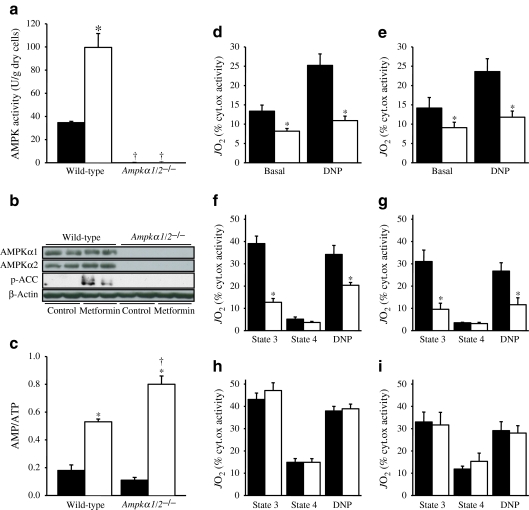
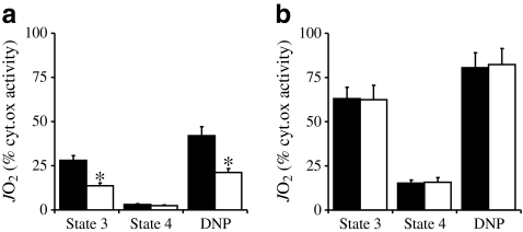
AIM/HYPOTHESIS: The glucose-lowering drug metformin has been shown to activate hepatic AMP-activated protein kinase (AMPK), a master kinase regulating cellular energy homeostasis. However, the underlying mechanisms remain controversial and have never been investigated in primary human hepatocytes. METHODS: Hepatocytes isolated from rat, mouse and human livers were treated with various concentrations of metformin. Isoform-specific AMPKα abundance and activity, as well as intracellular adenine nucleotide levels and mitochondrial oxygen consumption rates were determined at different time points. RESULTS: Metformin dose- and time-dependently increased AMPK activity in rat and human hepatocytes, an effect associated with a significant rise in cellular AMP:ATP ratio. Surprisingly, we found that AMPKα2 activity was undetectable in human compared with rat hepatocytes, while AMPKα1 activities were comparable. Accordingly, metformin only increased AMPKα1 activity in human hepatocytes, although both AMPKα isoforms were activated in rat hepatocytes. Analysis of mRNA expression and protein levels confirmed that only AMPKα1 is present in human hepatocytes; it also showed that the distribution of β and γ regulatory subunits differed between species. Finally, we demonstrated that the increase in AMP:ATP ratio in hepatocytes from liver-specific Ampkα1/2 (also known as Prkaa1/2) knockout mice and humans is due to a similar and specific inhibition of the mitochondrial respiratory-chain complex 1 by metformin. CONCLUSIONS/INTERPRETATION: Activation of hepatic AMPK by metformin results from a decrease in cellular energy status owing to metformin's AMPK-independent inhibition of the mitochondrial respiratory-chain complex 1. The unique profile of AMPK subunits found in human hepatocytes should be considered when developing new pharmacological agents to target the kinase.
Overview
The claims and statistics of this paper haven’t been checked yet.
Run the check — free, no account needed. It reads the open-access text and checks the sources it cites.
Integrity signals
Registry
Cites
Statistics
Figures
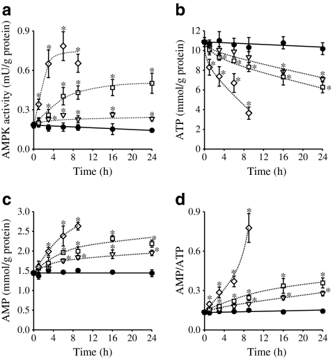
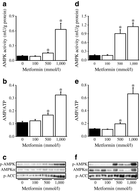
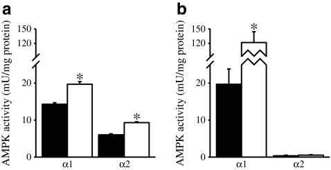
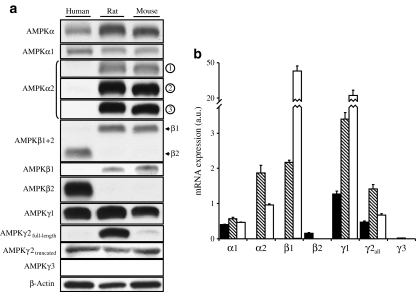
Verification
Claims the paper makes
The claims and statistics of this paper haven’t been checked yet.
Run the check — free, no account needed. It reads the open-access text and checks the sources it cites.
Claims other papers make about this one
Each is checked against this paper’s own text: does it say what the citing paper says it does?
In the citing paper
“Evidence that metformin targets mitochondrial complex 1 is supported by data from several studies (112).”
From A Critical Review of the Evidence That Metformin Is a Putative Anti-Aging Drug That Enhances Healthspan and Extends Lifespan (2021) · cites this paper as [112]
Self-check
Percentages, sums and reported differences re-computed from the paper’s own numbers.
The self-check needs the paper’s full text.
Citations
Cites
Role of AMP-Activated Protein Kinase in Mechanism of Metformin Action
Zhou, G., Myers, R., Li, Y., Chen, Y., Shen, X., Fenyk-Melody, J., Wu, M., Ventre, J., Doebber, T., Fujii, N., et al. (2001) · Journal of Clinical Investigation · doi:10.1172/jci13505 · Cited by 5,652
HIGH-YIELD PREPARATION OF ISOLATED RAT LIVER PARENCHYMAL CELLS
Berry, M. N., & Friend, D. S. (1969) · The Journal of Cell Biology · doi:10.1083/jcb.43.3.506 · Cited by 4,407
AMP-activated/SNF1 protein kinases: conserved guardians of cellular energy
Hardie, D. G. (2007) · Nature Reviews Molecular Cell Biology · doi:10.1038/nrm2249 · Cited by 2,261
OWEN, M. R., DORAN, E., & HALESTRAP, A. P. (2000) · Biochemical Journal · doi:10.1042/bj3480607 · Cited by 2,157
The Kinase LKB1 Mediates Glucose Homeostasis in Liver and Therapeutic Effects of Metformin
Shaw, R. J., Lamia, K. A., Vasquez, D., Koo, S.-H., Bardeesy, N., DePinho, R. A., Montminy, M., & Cantley, L. C. (2005) · Science · doi:10.1126/science.1120781 · Cited by 1,950
Steinberg, G. R., & Kemp, B. E. (2009) · Physiological Reviews · doi:10.1152/physrev.00011.2008 · Cited by 1,672
El-Mir, M.-Y., Nogueira, V., Fontaine, E., Avéret, N., Rigoulet, M., & Leverve, X. (2000) · Journal of Biological Chemistry · doi:10.1074/jbc.275.1.223 · Cited by 1,399
Clinical pharmacokinetics of metformin
Graham, G. G., Punt, J., Arora, M., Day, R. O., Doogue, M. P., Duong, J. K., Furlong, T. J., Greenfield, J. R., Greenup, L. C., Kirkpatrick, C. M., et al. (2011) · Clinical Pharmacokinetics · doi:10.2165/11534750-000000000-00000 · Cited by 1,318
Cited by
Sorted by citations
Cellular and molecular mechanisms of metformin: an overview
Viollet, B., Guigas, B., Garcia, N. S., Leclerc, J., Foretz, M., & Andreelli, F. (2012) · Clinical Science · doi:10.1042/cs20110386 · Cited by 1,720
Metformin: from mechanisms of action to therapies
Foretz, M., Guigas, B., Bertrand, L., Pollak, M., & Viollet, B. (2014) · Cell Metabolism · doi:10.1016/j.cmet.2014.09.018 · Cited by 1,501
Metformin inhibits mitochondrial complex I of cancer cells to reduce tumorigenesis
Wheaton, W. W., Weinberg, S. E., Hamanaka, R. B., Soberanes, S., Sullivan, L. B., Anso, E., Glasauer, A., Dufour, E., Mutlu, G. M., Budigner, G. S., et al. (2014) · eLife · doi:10.7554/elife.02242 · Cited by 1,105
AMP-activated protein kinase: the current landscape for drug development
Steinberg, G. R., & Carling, D. (2019) · Nature Reviews Drug Discovery · doi:10.1038/s41573-019-0019-2 · Cited by 785
Understanding the glucoregulatory mechanisms of metformin in type 2 diabetes mellitus
Foretz, M., Guigas, B., & Viollet, B. (2019) · Nature Reviews Endocrinology · doi:10.1038/s41574-019-0242-2 · Cited by 686
Self-citation
Metadata
- Authors
- Stephenne, X., Foretz, M., Taleux, N., van der Zon, G. C., Sokal, E., Hue, L., Viollet, B., & Guigas, B.
- Affiliations
- Centre National de la Recherche Scientifique (FR)
- Inserm (FR)
- Cliniques Universitaires Saint-Luc (BE)
- de Duve Institute (BE)
- Leiden University Medical Center (NL)
- Corresponding author
- Guigas, B.
- Venue
- Diabetologia
- Published
- 2011-09-23
- Volume
- 54
- Issue
- 12
- Pages
- 3101-3110
- ISSN
- 0012-186X (ISSN-L)
- 1432-0428
- PMC
- PMC3210354
- OpenAlex
- W2042871909
- Cited by
- 287
- References
- 52
- Citations by year
- 2026: 14 · 2025: 12 · 2024: 20 · 2023: 10 · 2022: 22 · 2021: 14 · 2020: 24 · 2019: 30 · 2018: 25 · 2017: 26 · 2016: 19 · 2015: 16 · 2014: 22 · 2013: 19 · 2012: 12
- FWCI
- 6.97
- Type
- journal article
- Language
- en
- Open access
- hybrid
- doi.org
- Licence
- cc-by-nc
- Locations
- Diabetologia (publishedVersion, cc-by-nc)
- Europe PMC (PubMed Central) (publishedVersion, cc-by-nc)
- PubMed Central (publishedVersion, cc-by-nc)
- PubMed (publishedVersion)
- Data Archiving and Networked Services (DANS) (submittedVersion)
- Leiden Repository (Leiden University) (submittedVersion)
- DIAL (Catholic University of Leuven) (submittedVersion)
- Funders
- European Commission (LSHMCT-2004-005272)
- Agence Nationale de la Recherche (ANR-10-BLAN-1123, ANR-10)
- Fonds De La Recherche Scientifique - FNRS
- Topics
- Life Sciences › Biochemistry, Genetics and Molecular Biology › Molecular Biology › Metabolism, Diabetes, and Cancer
- Life Sciences › Biochemistry, Genetics and Molecular Biology › Molecular Biology › Biochemical and Molecular Research
- Life Sciences › Biochemistry, Genetics and Molecular Biology › Molecular Biology › Peroxisome Proliferator-Activated Receptors
- Keywords
- metformin, AMPK, AMPKα1, AMPKα2, human hepatocytes, cellular energy status, AMP/ATP ratio, mitochondrial complex I inhibition
- MeSH terms
- Adenine Nucleotides, Animals, Cells, Cultured, Humans, Hypoglycemic Agents, Isoenzymes, Male, Metformin, Mitochondria, Liver, Oxygen Consumption, Mice, Knockout, Hepatocytes, Mice, Rats, AMP-Activated Protein Kinases
About these checks
Registry, retraction, Cites and statistics results are computed, not judged, with one exception: a printed reference that no index lists is read once by a model before it counts as a citation error. Claims are extracted by a model and kept only when their quote appears verbatim in the text; each cited source is then checked, with a model’s help, for whether it says what the claim relies on. Those verdicts can be wrong — read the claim’s quote and the cited source before relying on one.
Statistics and arithmetic are recomputed, not judged.
Confidence bands are shown only for fields calibrated on our annotated test set. None is calibrated yet, so no band is shown.
What counts as a claim ›How we measure confidence ›How we check this ›
Are you an author, or do you see a problem with this page? Request a review
